

The DOI matches its Crossref record; 756 of the 760 references it cites were found in a registry or an index.
FGF/FGFR signaling in health and disease
Open accessAbstract
Growing evidences suggest that the fibroblast growth factor/FGF receptor (FGF/FGFR) signaling has crucial roles in a multitude of processes during embryonic development and adult homeostasis by regulating cellular lineage commitment, differentiation, proliferation, and apoptosis of various types of cells. In this review, we provide a comprehensive overview of the current understanding of FGF signaling and its roles in organ development, injury repair, and the pathophysiology of spectrum of diseases, which is a consequence of FGF signaling dysregulation, including cancers and chronic kidney disease (CKD). In this context, the agonists and antagonists for FGF-FGFRs might have therapeutic benefits in multiple systems.
Overview
The claims and statistics of this paper haven’t been checked yet.
Run the check — free, no account needed. It reads the open-access text and checks the sources it cites.
Integrity signals
Registry
Cites
Statistics
Figures
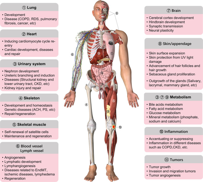
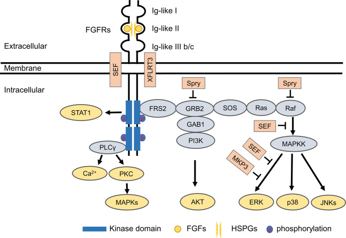
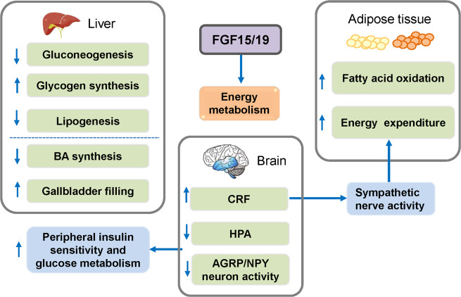
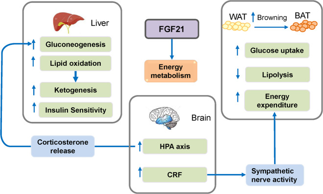
Verification
Claims the paper makes
The claims and statistics of this paper haven’t been checked yet.
Run the check — free, no account needed. It reads the open-access text and checks the sources it cites.
Claims other papers make about this one
Each is checked against this paper’s own text: does it say what the citing paper says it does?
The source backs part of the claim, not all of it — or backs it for a different population, condition or comparison.
In the citing paper
“We then assessed the expression of known FGFR3 ligands56,57 and found concomitant enrichment of FGFR3 and its ligands FGF20 and FGF23 in eGFP+ Paneth-like cells, accompanied by elevated p-FGFR3 levels (Figures 5D, S6D, and S6E).”
From Paneth-like transition drives resistance to dual targeting of KRAS and EGFR in colorectal cancer (2026) · cites this paper as [57]
Experimental — accuracy not yet measured
Self-check
Percentages, sums and reported differences re-computed from the paper’s own numbers.
The self-check needs the paper’s full text.
Citations
Cites
5 retracted
Yan, D., Chen, D., Cool, S. M., van Wijnen, A. J., Mikecz, K., Murphy, G., & Im, H. (2011) · Arthritis Research & Therapy · doi:10.1186/ar3441 · Cited by 146
RetractedIm, H., Li, X., Muddasani, P., Kim, G., Davis, F. J., Rangan, J., Forsyth, C. B., Ellman, M. B., & Thonar, E. J. ‐. A. (2007) · Journal of Cellular Physiology · doi:10.1002/jcp.21317 · Cited by 98
RetractedBasu, M., Mukhopadhyay, S., Chatterjee, U., & Roy, S. S. (2013) · Journal of Biological Chemistry · doi:10.1074/jbc.m113.535427 · Cited by 42
RetractedFgfr2 is integral for bladder mesenchyme patterning and function
Walker, K. A., Ikeda, Y., Zabbarova, I. V., Schaefer, C., Bushnell, D. S., de Groat, W. C., Kanai, A., & Bates, C. M. (2015) · American Journal of Physiology-Renal Physiology · doi:10.1152/ajprenal.00624.2014 · Cited by 16
RetractedTang, Z.-F., & Li, H.-Y. (2018) · PubMed · doi:10.26355/eurrev_201804_14838 · Cited by 11
RetractedThe metabolic phenotype in obesity: Fat mass, body fat distribution, and adipose tissue function
Goossens, G. H. (2017) · Obesity Facts · doi:10.1159/000471488 · Cited by 8,481
Origin and physiological roles of inflammation
Medzhitov, R. (2008) · Nature · doi:10.1038/nature07201 · Cited by 6,618
Fibroblast growth factor signalling: from development to cancer
Turner, N. C., & Grose, R. P. (2010) · Nature reviews. Cancer · doi:10.1038/nrc2780 · Cited by 2,768
Role of platelet-derived growth factors in physiology and medicinePreprint
Andræ, J., Gallini, R., & Betsholtz, C. (2008) · Genes & Development · doi:10.1101/gad.1653708 · Cited by 2,534
What we talk about when we talk about fat
Rosen, E. D., & Spiegelman, B. M. (2014) · Cell · doi:10.1016/j.cell.2013.12.012 · Cited by 2,373
Research works printed but not linked: 1
- Wang, Y. & Becker, D. Antisense targeting of basic fibroblast growth factor and fibroblast growth factor receptor-1 in human melanomas blocks intratumoral angiogenesis and tumor growth. Nat. Med. 3, 887–893 (1997). · DOI
Cited by
Sorted by citations
Items 501–600 of 906
Two sisters with RSPRY1‐related spondyloepimetaphyseal dysplasia
Singh, S., Shah, H. H., Dalal, A. B., Shukla, A., Bhavani, G. S., & Girisha, K. M. (2024) · American Journal of Medical Genetics Part A · doi:10.1002/ajmg.a.63601 · Cited by 4
Zhang, X., Meng, F., Lyu, W., He, J., Wei, R., Du, Z., Zhang, C., Guan, Y., Huang, X., Lyu, G., et al. (2023) · bioRxiv (Cold Spring Harbor Laboratory) · doi:10.1101/2023.05.26.542348 · Cited by 4
Salas-Bastos, A., Bardet, C., Kopper, K., Jauze, L., Collaud, F., Francois, A., Moor, M. B., Chen, G., Mohammadi, M., Stockmann, C., et al. (2024) · bioRxiv (Cold Spring Harbor Laboratory) · doi:10.1101/2024.01.18.576116 · Cited by 4
Aebisher, D., Woźnicki, P., Czarnecka–Czapczyńska, M., Dynarowicz, K., Szliszka, E., Kawczyk‐Krupka, A., & Bartusik‐Aebisher, D. (2024) · International Journal of Molecular Sciences · doi:10.3390/ijms25168708 · Cited by 3
Phenotyping of FGF12AV52H mutation in mouse implies a complex FGF12 network
Huang, J., Sun, C., Zhu, Q., Wu, G., Cao, Y., Shi, J., He, S., Jiang, L., Liao, J., Li, L., et al. (2024) · Neurobiology of Disease · doi:10.1016/j.nbd.2024.106637 · Cited by 3
Wang, J., Liu, H., Liu, M., Shen, T., Wen, T., He, F., & Wang, X. (2025) · International Journal of Biological Macromolecules · doi:10.1016/j.ijbiomac.2025.141831 · Cited by 3
Fibroblast growth factor 18 attenuates renal fibrosis via AMPK/NOX4 pathway in mice
Zhou, J., Shou, Y., Shi, Q., Ye, J., Li, X., Zhu, Z., & Wang, X. (2025) · Biochemical and Biophysical Research Communications · doi:10.1016/j.bbrc.2025.151913 · Cited by 3
An FGFR-p53 developmental signaling axis drives salivary cancer progression
Musicant, A. M., Billington, J. M. R., Damrauer, J., Modliszewski, J. L., Landau, L. J. B., Tsai, Y.-H., Mehta, J. H., Powers, J. J., Betancourt, R., Sekhri, R., et al. (2025) · Oncogene · doi:10.1038/s41388-025-03444-7 · Cited by 4
Targeting neurotrophic dysregulation in diabetic retinopathy: a novel therapeutic avenue
Rana, D. B., Dhankhar, S., Chauhan, R., Saini, M., Singh, R., Kumar, P., Singh, T. G., Chauhan, S., & Devi, S. (2025) · Molecular Biology Reports · doi:10.1007/s11033-025-10671-4 · Cited by 3
FGFR1 agonists alleviate pathology and cognitive impairment in an Alzheimer's disease mouse model
Hung, W.-C., Sun, W., Su, T., Lee, Y.-J., & Cheng, I. H.-J. (2025) · Experimental Neurology · doi:10.1016/j.expneurol.2025.115357 · Cited by 6
Liu, Z., Sun, M., Lu, L., Wu, X., Wang, H., Gao, Y., Liu, A., Qian, Y., Hu, H., & Jiang, H. (2025) · Computers in Biology and Medicine · doi:10.1016/j.compbiomed.2025.110828 · Cited by 3
Starrett, J. H., Allen, E. L., Neal, M., Iyer, S., Ye, Q., Hudkins, R. L., Degaonkar, V., Lihou, C. F., Swanson, R. V., Boni, V., et al. (2025) · Molecular Cancer Therapeutics · doi:10.1158/1535-7163.mct-25-0652 · Cited by 3
Soczyńska, J., Gawełczyk, W., Majcherczyk, K., Rydzek, J., Muzyka, A., Żołyniak, M., & Woźniak, S. (2025) · Cells · doi:10.3390/cells14211674 · Cited by 3
Salvati, M., Tiberio, F., Giacon, N., Augello, A., Tamburrini, G., Pietro, L. D., Vita, A., Tosi, D. D., Perini, G., Canini, G., et al. (2025) · Regenerative Biomaterials · doi:10.1093/rb/rbaf115 · Cited by 3
Liu, H., Jiang, Z., Guo, X., Liu, X., Lu, C., & Lu, D. (2025) · Journal of Orthopaedic Surgery and Research · doi:10.1186/s13018-025-06431-6 · Cited by 3
Li, X., Qi, Y., Wu, Y., Bi, X., Wang, Y., Wang, J., Wang, J., Zhang, L., Xiao, H., & Wang, J. (2025) · Advanced Science · doi:10.1002/advs.202511943 · Cited by 3
Lei, Y.-Q., Huo, J.-Y., Li, S.-J., Yang, B., Wang, L.-N., Gao, Y., Ma, L.-L., Dong, S.-J., Wu, J., & Li, S.-G. (2025) · Food Chemistry · doi:10.1016/j.foodchem.2025.147508 · Cited by 3
Hu, J., Xu, Y., Zhou, D., Chen, K., Jiang, J., Lin, M. Q., Chen, W., Wu, J., Jiang, H., Zhu, M., et al. (2025) · Biomaterials Research · doi:10.34133/bmr.0309 · Cited by 4
Self-citationPalollathil, A., Mahin, A., Gopalakrishnan, A. P., Poojari, T. R., Sambreena, A., Shivamurthy, P. B., & Raju, R. (2026) · Proteomes · doi:10.3390/proteomes14010008 · Cited by 3
Bukke, S. P. N., Thalluri, C., Medhi, J., Vasam, M., Gupta, N., Dey, P., Dennison, M. S., & Mundu, M. M. (2026) · Journal of Materials Chemistry B · doi:10.1039/d5tb02812d · Cited by 3
S100A6 promotes liver metastasis by activating FGFR3 signaling in BAP1-deficient uveal melanoma
Li, Y., Zheng, C., Liang, Y., Liu, Y., Zhou, F., Zhu, X., Fang, J., Gai, R., Zhuang, Y., Wang, K., et al. (2026) · Oncogene · doi:10.1038/s41388-026-03766-0 · Cited by 3
Yuan, D., Hao, X., Luo, J., Liu, Y., Du, P., & Yang, Z. (2026) · Cell Biochemistry and Function · doi:10.1002/cbf.70248 · Cited by 3
FGF13A interacts with NPM1 and UBF and inhibits the invasion of bladder cancer cells
Han, D., Guan, L., Zhang, Y., Yang, H., Si, L., Jia, T., Wu, Y., Lv, K., Song, T., & Yang, G. (2023) · Biochemical and Biophysical Research Communications · doi:10.1016/j.bbrc.2023.08.040 · Cited by 3
Circadian Regulation of Endocrine Fibroblast Growth Factors on Systemic Energy Metabolism
Yang, Z., Zarbl, H., & Guo, G. L. (2024) · Molecular Pharmacology · doi:10.1124/molpharm.123.000831 · Cited by 3
Carrillo-Dávila, I. A., Garibaldi-Ríos, A. F., Figuera, L. E., Gómez‐Meda, B. C., Zúñiga‐González, G. M., Puebla‐Pérez, A. M., García-Verdín, P. M., Castro-García, P. B., Gutiérrez-Hurtado, I. A., Torres-Mendoza, B. M., et al. (2024) · Biomedicines · doi:10.3390/biomedicines12030602 · Cited by 3
Genetic factors underlying Mandibular prognathism: insights from recent human and animal studies
Fang, H., Li, P., Zhu, S., & Bi, R. (2024) · Mammalian Genome · doi:10.1007/s00335-024-10084-x · Cited by 3
Sticky Polyelectrolyte Shield for Enhancing Biological Half-Life of Growth Factors
Kim, Y., Jeon, S., Kim, B., Jeong, Y. J., Kim, T. H., Jeong, S., Kim, I., Oh, J., Jung, Y., Lee, K., et al. (2024) · ACS Applied Materials & Interfaces · doi:10.1021/acsami.4c16261 · Cited by 3
The effect of combined oral contraceptive pills on angiogenesis in endometriotic lesions
Antonis, S., Papakonstantinou, Ε., Keramida, M., Panteris, E., Kalogeropoulos, S., Georgopoulos, N. A., Taniguchi, F., George, A., Tasuku, H., & Kaponis, A. I. (2025) · HORMONES · doi:10.1007/s42000-025-00636-4 · Cited by 3
Current Understanding of Crouzon Syndrome Pathophysiology and New Therapeutic Approaches
Tiberio, F., Polito, L., Salvati, M., Pietro, L. D., Massimi, L., Parolini, O., Tamburrini, G., & Lattanzi, W. (2025) · Journal of Craniofacial Surgery · doi:10.1097/scs.0000000000011376 · Cited by 4
Tsimafeyeu, I. V. (2025) · Exploration of Targeted Anti-tumor Therapy · doi:10.37349/etat.2025.1002327 · Cited by 3
Lian, K., Yang, W. H., Yang, R., Xu, X., & Guan, J. (2025) · Molecular Neurobiology · doi:10.1007/s12035-025-05221-9 · Cited by 4
Long-Lasting Proteinaceous Nanoformulation for Tumor Imaging and Therapy
Zhao, L., Gu, X., Jiang, F., Li, B., Lü, S., Wang, F., Sun, Y., Liu, K., & Li, J. (2022) · ACS Omega · doi:10.1021/acsomega.2c03561 · Cited by 3
FGF signalling is involved in cumulus migration in the common house spider Parasteatoda tepidariorum
Wang, R., Leite, D. J., Karadas, L., Schiffer, P. H., & Pechmann, M. (2022) · Developmental Biology · doi:10.1016/j.ydbio.2022.11.009 · Cited by 3
Sanggenol B, a plant bioactive, as a safer alternative to tackle cancer by antagonising human FGFR
Nagaraj, A., Raghavan, S. S., Niraikulam, A., Gautham, N., & Gunasekaran, K. (2023) · Journal of Biomolecular Structure and Dynamics · doi:10.1080/07391102.2023.2245047 · Cited by 3
Wu, J., Chen, J., Yu, X., & You, Y. (2023) · Toxicology Mechanisms and Methods · doi:10.1080/15376516.2023.2253305 · Cited by 3
Lenvatinib-Associated Fulminant Type 1 Diabetes Mellitus
Liao, P., Peng, T., Wu, T., & Hu, Y. (2022) · American Journal of Therapeutics · doi:10.1097/mjt.0000000000001495 · Cited by 3
Greenen, J. D. V., & Hockman, D. (2023) · Differentiation · doi:10.1016/j.diff.2023.10.005 · Cited by 3
Hioki, T., Tachi, J., Ueda, K., Matsushima‐Nishiwaki, R., Iida, H., Kozawa, O., & Tokuda, H. (2023) · Experimental and Therapeutic Medicine · doi:10.3892/etm.2023.12322 · Cited by 3
Shimpi, A. A., Williams, E. D., Lu, L., Tamir, T. Y., White, F. M., & Fischbach, C. (2023) · ACS Biomaterials Science & Engineering · doi:10.1021/acsbiomaterials.3c01034 · Cited by 3
Yang, Y.-F., Ma, H., Wang, X., Nie, M., Mao, J., & Wu, X. (2024) · Asian Journal of Andrology · doi:10.4103/aja202366 · Cited by 3
Schmidt, J., Kaulfuß, S., Ott, H., Gaubert, M., Reintjes, N., Bremmer, F., Dreha‐Kulaczewski, S. F., Ströbel, P., Yigit, G., & Wollnik, B. (2024) · Human Genetics · doi:10.1007/s00439-023-02634-1 · Cited by 3
Nature’s Elixir for Cancer Treatment: Targeting Tumor-induced Neovascularization
Kumari, R., Syeda, S., & Shrivastava, A. (2024) · Current Medicinal Chemistry · doi:10.2174/0109298673282525240222050051 · Cited by 3
Liang, R., Tan, B., Lei, K., Xu, K., Liang, J., Huang, J., Liang, Y., Huang, J., Zhang, L., Shi, X., et al. (2024) · Translational Oncology · doi:10.1016/j.tranon.2024.101974 · Cited by 3
Guardiola‐Diaz, H. M., DiBenedictis, B. T., Prendaj, E., & Bansal, R. (2024) · ASN NEURO · doi:10.1080/17590914.2024.2371163 · Cited by 4
Analysis of cell signaling profiles induced by DNA aptamer-based FGFR1 agonist
Hoshiyama, J., Hayata, Y., Eguchi, A., Morimoto, J., Ueki, R., & Sando, S. (2024) · Analytical Sciences · doi:10.1007/s44211-024-00660-1 · Cited by 3
Micale, L., Vourlia, A., Fusco, C., Pracella, R., Karagiannis, D., Nardella, G., Vaccaro, L., Leone, M. P., Gramazio, A., Dentici, M. L., et al. (2024) · European Journal of Human Genetics · doi:10.1038/s41431-024-01721-9 · Cited by 3
Role of FGF2 in Promoting Osteogenic Differentiation for Craniofacial Bone Regeneration
Yang, X., & Ma, P. X. (2025) · Regenerative Engineering and Translational Medicine · doi:10.1007/s40883-025-00447-4 · Cited by 3
Paeoniflorin and depression: a comprehensive review of underlying molecular mechanisms
Sahani, P., & Singh, L. (2025) · Metabolic Brain Disease · doi:10.1007/s11011-025-01671-1 · Cited by 4
Docosahexaenoic Acid Ester of Phloridzin Reduces Inflammation and Insulin Resistance via AMPK
Chen, J., Wu, Z., Wang, J., Si, X., Zhang, R., Sun, T., Dong, Q., Wu, W., & Qiu, Y. (2022) · Current Pharmaceutical Design · doi:10.2174/1381612828666220518102440 · Cited by 5
Gong, X., Akil, A., Ndi, A., Ji, T., Liu, X., Lovern, M., & Chen, X. (2023) · CPT Pharmacometrics & Systems Pharmacology · doi:10.1002/psp4.13064 · Cited by 3
A high-sensitivity ELISA for detection of human FGF18 in culture supernatants from tumor cell lines
Tsuchiya, Y., Komazawa‐Sakon, S., Tanaka, M., Kanokogi, T., Moriwaki, K., Akiba, H., Yagita∥, H., Okumura, K., Entzminger, K. C., Okumura, C. J., et al. (2023) · Biochemical and Biophysical Research Communications · doi:10.1016/j.bbrc.2023.06.070 · Cited by 3
Letizia, A., Espinàs, M., & Llimargas, M. (2023) · bioRxiv · doi:10.1101/2023.01.26.525651 · Cited by 2
An Insight on Protein Kinases and Their Therapeutic Perspective
Dhal, A. K., & Ritaparna, P. (2024) · Biochemistry · doi:10.5772/intechopen.113285 · Cited by 2
FGFR2 variants in Schimmelpenning–Feuerstein–Mims syndrome
Wang, Y., Zhao, L., Chen, Y., Pan, L., Huang, N., Zhou, R., & Li, L. (2025) · British Journal of Dermatology · doi:10.1093/bjd/ljaf025 · Cited by 2
Preparation and Characterization of an Engineered FGF1 Conjugated to 161Tb for Targeting of FGFRs
Song, L., Kostas, M., Laerdahl, J. K., Skálová, M., Janská, T., Juzeniene, A., Ræstad, S., Krivokapić, A., Kalantzopoulos, G. N., Šoltés, J., et al. (2025) · ACS Omega · doi:10.1021/acsomega.4c09179 · Cited by 2
FGFR4 inhibition augments paclitaxel-induced cell death in ovarian cancer
Zeng, R., Chen, X., Chen, Y., & Dong, J. (2025) · International Immunopharmacology · doi:10.1016/j.intimp.2025.114626 · Cited by 2
Li, I., Huo, Y., Yang, T., Gunawan, H., Alexandrov, L. B., & Zage, P. E. (2025) · Cancer Drug Resistance · doi:10.20517/cdr.2024.208 · Cited by 4
Voukali, E., Cabral, J. V., Smorodinova, N., Kolín, V., Netuková, M., Vacı́k, T., & Jirsová, K. (2025) · Bioengineering · doi:10.3390/bioengineering12070688 · Cited by 2
Luo, H., Wang, L., Gao, H., Zhou, D., Qiu, Y., Yang, L., Li, J., Du, D., Huang, X., Zhao, Y. X., et al. (2025) · Biomedicines · doi:10.3390/biomedicines13071667 · Cited by 2
Karaosmanoğlu, O., Berber, H., Sivas, H., & Uysal, Ü. D. (2025) · Cell Biochemistry and Biophysics · doi:10.1007/s12013-025-01826-y · Cited by 3
Exploring the Neurobiological Mechanisms of Cancer Growth
Hussain, M. S., Maqbool, M., Agrawal, M., Rana, A. J., Sultana, A., Sulthana, N., Bisht, A. S., & Khan, G. (2025) · Current Pharmaceutical Design · doi:10.2174/0113816128402718250806151308 · Cited by 2
Fibroblast growth factor 2 in the hotbed of rheumatoid arthritis pathogenesis
Illanad, G. H., Rithvik, A., & Rasool, M. (2025) · International Immunopharmacology · doi:10.1016/j.intimp.2025.115778 · Cited by 2
Blood Derivatives in the Therapy of Ocular Surface Diseases
Stępień, P. W., Żarnowski, T., & Wróbel‐Dudzińska, D. (2025) · International Journal of Molecular Sciences · doi:10.3390/ijms262211097 · Cited by 2
Breaking the Toxicity-Efficacy Dilemma of Polymyxins: Advances in Synergists
Cai, Z., Zhang, Y., Xin, Y., Han, L., Xu, B., Du, M., & Zhao, Y. (2025) · ACS Infectious Diseases · doi:10.1021/acsinfecdis.5c00782 · Cited by 2
Savyon, G., Korol, R., Abdel-Haq, M., Saadon, S. B., Bistritz, D., Lankry, H., Peled, Y., Rak, R., Levavi‐Sivan, B., Benny, O., et al. (2025) · Frontiers in Nutrition · doi:10.3389/fnut.2025.1669909 · Cited by 2
The alternative splicing landscape of hepatocellular carcinoma and its potential for HCC detection
Hershberger, C. E., Daniels, N. J., Patterson, W. M., & Rotroff, D. M. (2025) · Hepatology Communications · doi:10.1097/hc9.0000000000000838 · Cited by 2
Recent advances in the treatment of FGFR-altered cholangiocarcinoma
Kawashima, J., Akabane, M., Endo, I., & Pawlik, T. M. (2025) · Expert Opinion on Pharmacotherapy · doi:10.1080/14656566.2025.2605200 · Cited by 3
Udeabor, S. E., Ishfaq, M., Shah, S. J., Khalid, I., Baig, F. A. H., Hamid, M. M. M., Elfadeel, A. S. A., Onwuka, C. I., Ali, S. A. A., & Mustafa, M. M. (2025) · BMC Cancer · doi:10.1186/s12885-025-15471-4 · Cited by 2
Kong, Q., Wang, X., & Ding, W. (2026) · Frontiers in Molecular Biosciences · doi:10.3389/fmolb.2025.1728698 · Cited by 2
Thymosin beta 4: An emerging therapeutic candidate for kidney diseases
Di, H., Huang, J., Zhang, D., Ni, F., Zheng, R., & Geng, H. (2026) · Peptides · doi:10.1016/j.peptides.2026.171467 · Cited by 2
Self-citationA paracrine-to-autocrine shunt of GREM1 fuels colorectal cancer metastasis via ACVR1C
Zhou, H., Jin, Q., Fu, Z., Yang, Y., Gao, Y., Wang, N., Zhao, B., Gui, L., Li, J., Zhu, Z., et al. (2026) · Molecular Cancer · doi:10.1186/s12943-025-02554-w · Cited by 2
Exome findings in children with short stature evaluated by growth hormone stimulation testing
de la Piscina, I. M., Zürcher, M., Zingg, T., Janner, M., Saner, C. C. S., Boettcher, C., Zweier, C., & Flück, C. E. (2026) · European Journal of Endocrinology · doi:10.1093/ejendo/lvag038 · Cited by 2
The antiaging protein Klotho is a key factor in susceptibility to cerebral ischemia
Aguilera, P., Hernandez‐Cruz, A., Franco‐Pérez, J., Peralta‐Arrieta, I., & Alquisiras-Burgos, I. (2026) · Neuroscience · doi:10.1016/j.neuroscience.2026.03.005 · Cited by 2
RNA Splicing of the FGFR2 Gene Promotes Endothelial Cell Senescence and Atherosclerosis Development
Ni, Q., Qi, H., Chu, Y., Cao, R., Wang, Y., Yang, S., Yao, Y., Wang, H., Yuan, K., Lv, L., et al. (2026) · Arteriosclerosis Thrombosis and Vascular Biology · doi:10.1161/atvbaha.125.323834 · Cited by 2
Self-citationAlfaraj, G. A., Alfaraj, H. A., Adel, M., & AlGadeeb, R. B. (2026) · European Journal of Pediatrics · doi:10.1007/s00431-026-06970-y · Cited by 2
Comprehending fibroblast growth factor receptor like 1: Oncogene or tumor suppressor?
Aprajita, & Sharma, R. (2021) · Cancer Treatment and Research Communications · doi:10.1016/j.ctarc.2021.100472 · Cited by 3
Research progress of fibroblast growth factor in nervous system diseases
Huang, W., Wan-hua, Q., Chen, K., Ye, S., Wang, D., HU, J. L., Xu, H., Lin, L., & Xiaokun, L. I. (2022) · Journal of Zhejiang University (Medical Sciences) · doi:10.3724/zdxbyxb-2022-0180 · Cited by 2
Anaya, D. M., Juárez-Velázquez, R., Ruvalcaba, S. R., Navarrete-Meneses, M. D. P., Labadía, C. S., Lieberman, E., & Pérez‐Vera, P. (2023) · Molecular Syndromology · doi:10.1159/000528472 · Cited by 2
Villazón, F. G., Pacheco‐Tena, C., Anchondo-López, A., Ordoñez-Solorio, L. A., Martínez, B. C., Muñoz-Cobos, A., Luévano-González, A., & González‐Chávez, S. A. (2023) · European Spine Journal · doi:10.1007/s00586-023-07721-5 · Cited by 2
Fibrogenic Factors and Molecular Mechanisms
Kondaiah, P. (2023) · Textbooks in contemporary dentistry · doi:10.1007/978-3-031-12855-4_12 · Cited by 2
Boraka, Ö., Klintman, M., Vallon‐Christersson, J., Zackrisson, S., Hall, P. N., Borgquist, S., & Rosendahl, A. H. (2023) · Frontiers in Oncology · doi:10.3389/fonc.2023.1230821 · Cited by 2
Magalhães, C. G., Mansueli, C. P., Manieri, T. M., Quintilio, W., Garbuio, A., Marinho, J. D. J., de Moraes, J. Z., Tsuruta, L. R., & Moro, A. M. (2023) · Bioengineered · doi:10.1080/21655979.2023.2252667 · Cited by 2
Potential roles of FGF5 as a candidate therapeutic target in prostate cancer.
Stangis, M. M., Colah, A. N., McLean, D. T., Halberg, R. B., Collier, L. S., & Ricke, W. A. (2023) · PubMed Central · Cited by 2
Najeeb, S., Manekia, F. A., Sadiq, M. S. K., Adanir, N., Khurshid, Z., Zafar, M. S., & Heboyan, A. G. (2024) · Science Progress · doi:10.1177/00368504241228964 · Cited by 2
[Role of Fibroblast Growth Factor 7 in Craniomaxillofacial Development].
Wang, R., An, K., Xie, J., & Zou, S. (2024) · PubMed Central · doi:10.12182/20240360505 · Cited by 2
The potential of lenvatinib in breast cancer therapy
Shang, Y., Liu, Y., & Wang, W. (2024) · Medical Oncology · doi:10.1007/s12032-024-02477-4 · Cited by 2
Li, X., Pan, Z., Luan, T., Xiao, Q., Li, L., Wu, Q., Yao, G., Zhang, X., & Song, D. (2024) · Frontiers in Immunology · doi:10.3389/fimmu.2024.1493673 · Cited by 2
FGF Signaling: A Key Pathway During Skeletal Development
Fayad, C., & Legeai‐Mallet, L. (2024) · Biology of extracellular matrix · doi:10.1007/978-3-031-70835-0_8 · Cited by 2
Vozár, J., Hudáková, N., Nosálová, N., Huniadi, M., Marcinčáková, D., Horňák, S., Horňáková, Ľ., Majerová, P., & Cizkova, D. (2025) · Frontiers in Veterinary Science · doi:10.3389/fvets.2024.1517349 · Cited by 2
Demirdjian, L., Triantos, S., Standish, K., Thomas, S., Xia, Q., Zhang, J., Greshock, J. D., Paone, J., Sheridan, P. E., Pant, S., et al. (2025) · Cancer Medicine · doi:10.1002/cam4.70546 · Cited by 2
E3 ligase MIB1 regulates STAT1/P21 signaling via regulation of FGFR1 in colorectal cancer
Jung, C., Park, J. E., & Seo, S. (2025) · Genes & Genomics · doi:10.1007/s13258-025-01629-8 · Cited by 2
Dai, J., Xia, B., Liu, N., & Shui, P. (2025) · Shock · doi:10.1097/shk.0000000000002565 · Cited by 2
The C-Terminal Kinase Domain–Binding and Suppression Motif Prevents Constitutive Activation of FGFR2
Zingg, D., Lin, C., Yemelyanenko, J., Wieteska, Ł., Kas, S. M., Bleijerveld, O. B., Chao, X., Bhin, J., Lutz, C., Wientjens, E., et al. (2025) · Cancer Research · doi:10.1158/0008-5472.can-24-3349 · Cited by 2
Wei, Z., Zhang, S., Bai, K., Liu, Y., Luan, Y., Hu, Z., Li, Y., Qu, Z., Hu, X., Ding, X., et al. (2025) · Development · doi:10.1242/dev.204532 · Cited by 2
Jiao, Y., Liu, J., Li, C., Hu, Y., & Zhao, S. (2025) · Journal of Developmental Biology · doi:10.3390/jdb13030033 · Cited by 2
A higher proportion of craniosynostosis genes are cancer driver genes
Misra, S., Shih, A., Yan, X., & Li, W. (2025) · Human Gene · doi:10.1016/j.humgen.2025.201378 · Cited by 2
Li, M. (2021) · Aging Pathobiology and Therapeutics · doi:10.31491/apt.2021.09.065 · Cited by 2
Unmet needs in the treatment of intrahepatic cholangiocarcinoma harboring FGFR2 gene rearrangements.
Beri, N. (2022) · Future Oncology · doi:10.2217/fon-2021-1089 · Cited by 3
Hallmarks of Cancer: Molecular Underpinnings
Chitale, D. A. (2022) · Springer eBooks · doi:10.1007/978-3-030-93084-4_1 · Cited by 2
Non-inositol 1,4,5-trisphosphate (IP3) receptor IP3-binding proteins
Mackrill, J. J. (2023) · Biochimica et Biophysica Acta (BBA) - Molecular Cell Research · doi:10.1016/j.bbamcr.2023.119470 · Cited by 2
Metadata
- Authors
- Xie, Y., Su, N., Yang, J., Tan, Q., Huang, S., Jin, M., Ni, Z., Zhang, B., Zhang, D., Luo, F., Chen, H., Sun, X., Feng, J. Q., Qi, H., & Chen, L.
- Affiliations
- Army Medical University (CN)
- Texas A&M University (US)
- Corresponding author
- Xie, Yangli
- ORCID
- Venue
- Signal Transduction and Targeted Therapy
- Published
- 2020-09-02
- Volume
- 5
- Issue
- 1
- Pages
- 181-181
- ISSN
- 2059-3635 (ISSN-L)
- PMC
- PMC7468161
- OpenAlex
- W3082979654
- Cited by
- 904
- References
- 760
- Citations by year
- 2026: 148 · 2025: 190 · 2024: 205 · 2023: 151 · 2022: 124 · 2021: 76 · 2020: 9
- FWCI
- 8.62
- Type
- journal article
- Language
- en
- Open access
- gold
- doi.org
- Locations
- Signal Transduction and Targeted Therapy (publishedVersion, cc-by)
- PubMed Central (publishedVersion, cc-by)
- PubMed (publishedVersion)
- Funders
- National Natural Science Foundation of China (81772306, IRT1216, 81991513, 81530071, 2018YFA0800802, 81721001)
- National Key Research and Development Program of China (2018YFA0800802)
- Topics
- Life Sciences › Biochemistry, Genetics and Molecular Biology › Molecular Biology › Fibroblast Growth Factor Research
- Life Sciences › Biochemistry, Genetics and Molecular Biology › Molecular Biology › Connective Tissue Growth Factor Research
- Life Sciences › Biochemistry, Genetics and Molecular Biology › Developmental Biology › Congenital limb and hand anomalies
- Keywords
- fibroblast growth factor, fibroblast growth factor receptor, FGF/FGFR signaling, organogenesis, tissue repair, cancer, chronic kidney disease, therapeutic target, cell differentiation, apoptosis
- MeSH terms
- Cell Differentiation, Fibroblast Growth Factors, Homeostasis, Humans, Neoplasms, Signal Transduction, Apoptosis, Receptors, Fibroblast Growth Factor, Embryonic Development, Cell Proliferation
About these checks
Registry, retraction, Cites and statistics results are computed, not judged, with one exception: a printed reference that no index lists is read once by a model before it counts as a citation error. Claims are extracted by a model and kept only when their quote appears verbatim in the text; each cited source is then checked, with a model’s help, for whether it says what the claim relies on. Those verdicts can be wrong — read the claim’s quote and the cited source before relying on one.
Statistics and arithmetic are recomputed, not judged.
Confidence bands are shown only for fields calibrated on our annotated test set. None is calibrated yet, so no band is shown.
What counts as a claim ›How we measure confidence ›How we check this ›
Are you an author, or do you see a problem with this page? Request a review
