

The DOI matches its Crossref record; 833 of the 833 references it cites were found in a registry or an index.
Molecular mechanisms in liver repair and regeneration: from physiology to therapeutics
Open accessAbstract
Liver repair and regeneration are crucial physiological responses to hepatic injury and are orchestrated through intricate cellular and molecular networks. This review systematically delineates advancements in the field, emphasizing the essential roles played by diverse liver cell types. Their coordinated actions, supported by complex crosstalk within the liver microenvironment, are pivotal to enhancing regenerative outcomes. Recent molecular investigations have elucidated key signaling pathways involved in liver injury and regeneration. Viewed through the lens of metabolic reprogramming, these pathways highlight how shifts in glucose, lipid, and amino acid metabolism support the cellular functions essential for liver repair and regeneration. An analysis of regenerative variability across pathological states reveals how disease conditions influence these dynamics, guiding the development of novel therapeutic strategies and advanced techniques to enhance liver repair and regeneration. Bridging laboratory findings with practical applications, recent clinical trials highlight the potential of optimizing liver regeneration strategies. These trials offer valuable insights into the effectiveness of novel therapies and underscore significant progress in translational research. In conclusion, this review intricately links molecular insights to therapeutic frontiers, systematically charting the trajectory from fundamental physiological mechanisms to innovative clinical applications in liver repair and regeneration.
Overview
The claims and statistics of this paper haven’t been checked yet.
Run the check — free, no account needed. It reads the open-access text and checks the sources it cites.
Integrity signals
Registry
Cites
Statistics
Figures
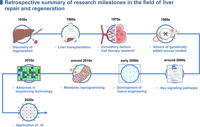
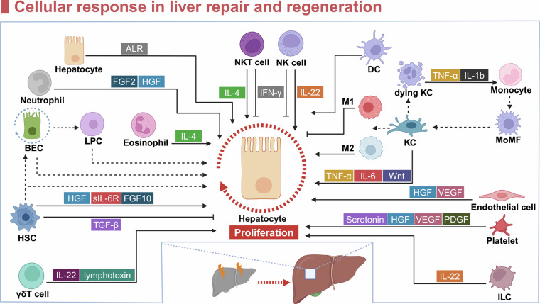
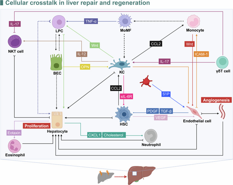
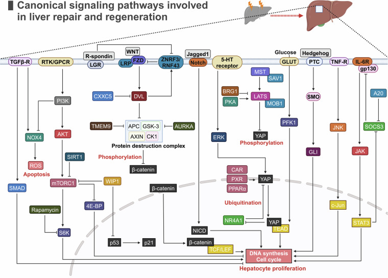
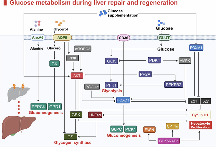
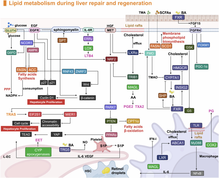
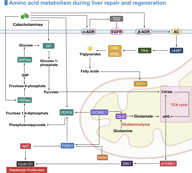
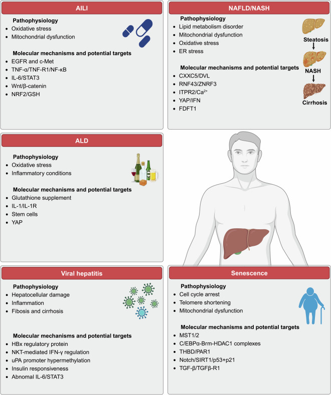
Verification
Claims the paper makes
The claims and statistics of this paper haven’t been checked yet.
Run the check — free, no account needed. It reads the open-access text and checks the sources it cites.
Claims other papers make about this one
Each is checked against this paper’s own text: does it say what the citing paper says it does?
The cited source says what the claim attributes to it.
In the citing paper
“Consistent with prior reports of transient hypoglycemia,34,35 hepatic steatosis,34,53 and BA overload4,34 occurring after PHx”
From Constitutive Androstane Receptor Regulates Bile Acid Homeostasis and Metabolism During Liver Regeneration (2026) · cites this paper as [34]
Self-check
Percentages, sums and reported differences re-computed from the paper’s own numbers.
The self-check needs the paper’s full text.
Citations
Cites
Items 401–600 of 833
The development and hepatotoxicity of acetaminophen: reviewing over a century of progress
McGill, M. R., & Hinson, J. (2020) · Drug Metabolism Reviews · doi:10.1080/03602532.2020.1832112 · Cited by 109
Liver Injury and Regeneration: Current Understanding, New Approaches, and Future Perspectives
Hora, S., & Wüestefeld, T. (2023) · Cells · doi:10.3390/cells12172129 · Cited by 109
Chaturantabut, S., Shwartz, A., Evason, K. J., Cox, A. G., LaBella, K. A., Schepers, A. G., Yang, S. P., Acuña, M., Houvras, Y. J., Mâncio-Silva, L., et al. (2019) · Gastroenterology · doi:10.1053/j.gastro.2019.01.010 · Cited by 108
Haga, S., Ogawa, W., Inoue, H., Terui, K., Ogino, T., Igarashi, R., Takeda, K., Akira, S., Enosawa, S., Furukawa, H., et al. (2005) · Journal of Hepatology · doi:10.1016/j.jhep.2005.03.027 · Cited by 108
Sympathetic Nervous System Inhibition Increases Hepatic Progenitors and Reduces Liver Injury
Oben, J. A., Roskams, T., Yang, S., Lin, H., Sinelli, N., Li, Z., Torbenson, M. S., Huang, J., Guarino, P., Kafrouni, M. I., et al. (2003) · Hepatology · doi:10.1053/jhep.2003.50371 · Cited by 108
Self-citationPI3K/Akt activation is critical for early hepatic regeneration after partial hepatectomy
Jackson, L. N., Larson, S. D., Silva, S. R., Rychahou, P. G., Chen, L. A., Qiu, S., Rajaraman, S., & Evers, B. M. (2008) · American Journal of Physiology-Gastrointestinal and Liver Physiology · doi:10.1152/ajpgi.00062.2008 · Cited by 108
Incompatibility of the circadian protein BMAL1 and HNF4α in hepatocellular carcinoma
Fekry, B., Ribas‐Latre, A., Baumgartner, C., Deans, J., Kwok, C., Patel, P., Fu, L., Berdeaux, R., Sun, K., Kolonin, M. G., et al. (2018) · Nature Communications · doi:10.1038/s41467-018-06648-6 · Cited by 108
Self-citationJin, J., Iakova, P. A., Jiang, Y., Medrano, E. E., & Timchenko, N. A. (2011) · Hepatology · doi:10.1002/hep.24471 · Cited by 107
McGettigan, B. M., McMahan, R. H., Orlicky, D. J., Burchill, M. A., Danhorn, T., Francis, P., Cheng, L. L., Golden‐Mason, L., Jakubzick, C., & Rosen, H. R. (2018) · Hepatology · doi:10.1002/hep.30401 · Cited by 107
Qing, J., Ren, Y., Zhang, Y., Yan, M., Zhang, H., Wu, D., Ma, Y., Chen, Y., Huang, X., Wu, Q., et al. (2021) · Journal of Hepatology · doi:10.1016/j.jhep.2021.09.032 · Cited by 107
Thevananther, S., Sun, H., Li, D., Arjunan, V., Awad, S. S., Wyllie, S., Zimmerman, T. L., Goss, J. A., & Karpen, S. J. (2004) · Hepatology · doi:10.1002/hep.20075 · Cited by 107
Abshagen, K., Eipel, C., Kalff, J. C., Menger, M. D., & Vollmar, B. (2007) · American Journal of Physiology-Gastrointestinal and Liver Physiology · doi:10.1152/ajpgi.00399.2006 · Cited by 107
Understanding the heterogeneity of resident liver macrophages
Blériot, C., & Ginhoux, F. (2019) · Frontiers in Immunology · doi:10.3389/fimmu.2019.02694 · Cited by 107
Torre, C., Perret, C., & Colnot, S. (2009) · The International Journal of Biochemistry & Cell Biology · doi:10.1016/j.biocel.2009.11.004 · Cited by 106
Ritschka, B., Knauer-Meyer, T., Gonçalves, D. S., Mas, A., Plassat, J.-L., Durik, M., Jacobs, H., Pedone, E., Di Vicino, U., Cosma, M. P., et al. (2020) · Genes & Development · doi:10.1101/gad.332643.119 · Cited by 105
The LATS2 tumor suppressor inhibits SREBP and suppresses hepatic cholesterol accumulation
Aylon, Y., Gershoni, A., Rotkopf, R., Biton, I. E., Porat, Z., Koh, A. P., Sun, X., Lee, Y., Fiel, M.-I., Hoshida, Y., et al. (2016) · European Journal of Cancer · doi:10.1016/s0959-8049(16)61580-0 · Cited by 105
Zhao, S., Jiang, J., Jing, Y., Liu, W., Yang, X. F., Hou, X., Gao, L., & Wei, L. (2020) · Cell Death and Disease · doi:10.1038/s41419-020-2264-z · Cited by 105
Immune cells in liver regeneration
Li, N., & Hua, J. (2017) · Oncotarget · doi:10.18632/oncotarget.12275 · Cited by 104
Jiang, Y., Feng, D., Ma, X., Fan, S., Gao, Y., Fu, K., Wang, Y., Sun, J., Yao, X., Liu, C., et al. (2019) · Hepatology · doi:10.1002/hep.30131 · Cited by 103
Self-citationContribution of cyclooxygenase‐2 to liver regeneration after partial hepatectomy
Casado, M., Callejas, N. A., Rodrigo, J., Zhao, X., Dey, S. K., Boscá, L., & Martín‐Sanz, P. (2001) · The FASEB Journal · doi:10.1096/fj.01-0158fje · Cited by 103
Two Faces of Neutrophils in Liver Disease Development and Progression
Cho, Y., & Szabó, G. (2020) · Hepatology · doi:10.1002/hep.31680 · Cited by 103
Schaedler, S., Krause, J., Himmelsbach, K., Carvajal-Yepes, M., Lieder, F., Klingel, K., Nassal, M., Weiß, T. S., Werner, S., & Hildt, E. (2010) · Journal of Biological Chemistry · doi:10.1074/jbc.m110.145862 · Cited by 103
INTRAPORTAL INSULIN PROTECTS FROM THE LIVER INJURY OF PORTACAVAL SHUNT IN DOGS
Starzl, T. E., Porter, K. A., & Putnam, C. W. (1975) · The Lancet · doi:10.1016/s0140-6736(75)92076-0 · Cited by 103
Gandhi, C. R., Chaillet, J. R., Nalesnik, M. A., Kumar, S., Dangi, A., Demetris, A. J., Ferrell, R. E., Wu, T., Divanovic, S., Stankeiwicz, T., et al. (2014) · Gastroenterology · doi:10.1053/j.gastro.2014.10.008 · Cited by 102
Regeneration of kidney parenchyma under normal and pathological conditions.
Wd, H., & Stöcker, E. (1971) · PubMed · Cited by 102
Kamimoto, K., Kaneko, K., Kok, C., Okada, H., Miyajima, A., & Itoh, T. (2016) · eLife · doi:10.7554/elife.15034 · Cited by 102
Ang, C. H., Hsu, S. H., Guo, F., Tan, C. T., Yu, V. C., Visvader, J. E., Chow, P. K. H., & Fu, N. Y. (2019) · Proceedings of the National Academy of Sciences · doi:10.1073/pnas.1908099116 · Cited by 102
Feder, L. S., Todaro, J. A., & Laskin, D. L. (1993) · Journal of Leukocyte Biology · doi:10.1002/jlb.53.2.126 · Cited by 101
Novel Mechanism of Foxo1 Phosphorylation in Glucagon Signaling in Control of Glucose Homeostasis
Wu, Y., Pan, Q., Yan, H., Zhang, K., Guo, X., Xu, Z., Yang, W., Qi, Y., Guo, C., Hornsby, C., et al. (2018) · Diabetes · doi:10.2337/db18-0674 · Cited by 101
Valdecantos, M. P., Pardo, V., Ruiz, L., Castro‐Sánchez, L., Lanzón, B., Fernández‐Millán, E., García‐Monzón, C., Arroba, A. I., González‐Rodríguez, Á., Escrivá, F., et al. (2017) · Hepatology · doi:10.1002/hep.28962 · Cited by 100
Kawasaki, T., Murata, S., Takahashi, K., Nozaki, R., Ohshiro, Y., Ikeda, N., Pak, S., Myronovych, A., Hisakura, K., Fukunaga, K., et al. (2010) · Journal of Hepatology · doi:10.1016/j.jhep.2010.04.021 · Cited by 100
Emerging role of aging in the progression of NAFLD to HCC
He, Y., Su, Y., Duan, C., Wang, S., He, W. G., Zhang, Y., An, X., & He, M. (2022) · Ageing Research Reviews · doi:10.1016/j.arr.2022.101833 · Cited by 100
Wu, Z., Jiao, P., Huang, X., Feng, B., Feng, Y., Yang, S., Hwang, P. W., Du, J., Nie, Y., Xiao, G., et al. (2010) · Journal of Clinical Investigation · doi:10.1172/jci43250 · Cited by 99
Zhang, C., Feng, J., Du, J., Zhuo, Z., Yang, S., Zhang, W., Wang, W., Zhang, S., Iwakura, Y., Meng, G., et al. (2018) · Cellular & Molecular Immunology · doi:10.1038/cmi.2017.22 · Cited by 99
Shrestha, N., Chand, L., Han, M., Lee, S. O., Kim, C. Y., & Jeong, Y. J. (2016) · Food and Chemical Toxicology · doi:10.1016/j.fct.2016.04.024 · Cited by 97
Meyer, J., Lejmi, E., Fontana, P., Morel, P., Gonelle‐Gispert, C., & Bühler, L. H. (2015) · Journal of Hepatology · doi:10.1016/j.jhep.2015.07.002 · Cited by 96
HDAC1 Cooperates with C/EBPα in the Inhibition of Liver Proliferation in Old Mice
Wang, G.-L., Salisbury, E. A., Shi, X., Timchenko, L., Medrano, E. E., & Timchenko, N. A. (2008) · Journal of Biological Chemistry · doi:10.1074/jbc.m803544200 · Cited by 96
p21 is required for dextrose-mediated inhibition of mouse liver regeneration #
Weymann, A., Hartman, E., Gazit, V. A., Wang, C., Glauber, M., Turmelle, Y. P., & Rudnick, D. A. (2009) · Hepatology · doi:10.1002/hep.22979 · Cited by 94
Fernandez-Rojo, M. A., Restall, C. M., Ferguson, C., Martel, N., Martin, S., Bosch, M., Kassan, A., Leong, G. M., Martin, S. D., McGee, S. L., et al. (2011) · Hepatology · doi:10.1002/hep.24810 · Cited by 94
Soto–Gutiérrez, A., Gough, A., Vernetti, L. A., Taylor, D. L., & Monga, S. P. (2017) · Experimental Biology and Medicine · doi:10.1177/1535370217707731 · Cited by 94
Chiu, H.-J., Gardner, C. R., Dambach, D. M., Durham, S. K., Brittingham, J. A., Laskin, J. D., & Laskin, D. L. (2003) · Toxicology and Applied Pharmacology · doi:10.1016/j.taap.2003.07.003 · Cited by 94
Notch signaling regulates tubular morphogenesis during repair from biliary damage in mice
Fiorotto, R., Raizner, A., Morell, C. M., Torsello, B., Scirpo, R., Fabris, L., Spirlı̀, C., & Strazzabosco, M. (2013) · Journal of Hepatology · doi:10.1016/j.jhep.2013.02.025 · Cited by 94
Organs-on-a-chip: a union of tissue engineering and microfabrication
Zhao, Y., Wang, E. Y., Lai, F. B. L., Cheung, K., & Radisic, M. (2023) · Trends in Biotechnology · doi:10.1016/j.tibtech.2022.12.018 · Cited by 93
Liver Regeneration is Impaired in Lipodystrophic Fatty Liver Dystrophy Mice
Gazit, V. A., Weymann, A., Hartman, E., Finck, B. N., Hruz, P. W., Tzekov, A., & Rudnick, D. A. (2010) · Hepatology · doi:10.1002/hep.23920 · Cited by 93
Walesky, C. M., Gunewardena, S., Terwilliger, E. F., Edwards, G., Borude, P. C., & Apte, U. M. (2012) · American Journal of Physiology-Gastrointestinal and Liver Physiology · doi:10.1152/ajpgi.00064.2012 · Cited by 93
Pre‐treatments enhance the therapeutic effects of mesenchymal stem cells in liver diseases
Hu, C., Wu, Z., & Li, L. (2019) · Journal of Cellular and Molecular Medicine · doi:10.1111/jcmm.14788 · Cited by 93
Feng, D., Xiang, X., Guan, Y., Guillot, A., Lu, H., Chang, C., He, Y., Wang, H., Pan, H., Ju, C., et al. (2023) · Journal of Clinical Investigation · doi:10.1172/jci166954 · Cited by 93
Aging Reprograms the Hematopoietic-Vascular Niche to Impede Regeneration and Promote Fibrosis
Chen, Y., Pu, Q., Ma, Y., Zhang, H., Ye, T., Zhao, C., Huang, X., Ren, Y., Qiao, L., Liu, H., et al. (2020) · Cell Metabolism · doi:10.1016/j.cmet.2020.11.019 · Cited by 92
He, P., Zhang, Y., Ye, Z., Li, H., Liu, M., Zhou, C., Yang, S., Gan, X., Zhang, Y., & Qin, X. (2023) · Metabolism · doi:10.1016/j.metabol.2023.155643 · Cited by 92
Changes in glutathione homeostasis during liver regeneration in the rat
Huang, Z., Li, H., Cai, J., Kuhlenkamp, J., Kaplowitz, N., & Lu, S. C.-L. (1998) · Hepatology · doi:10.1002/hep.510270123 · Cited by 92
Murata, S., Hashimoto, I., Nakano, Y., Myronovych, A., Watanabe, M., & Ohkohchi, N. (2008) · Annals of Surgery · doi:10.1097/sla.0b013e31818584c7 · Cited by 92
Wang, L., Scott, I., Zhu, L., Wu, K., Han, K., Chen, Y., Guček, M., & Sack, M. N. (2017) · Nature Communications · doi:10.1038/s41467-017-00521-8 · Cited by 91
Marot, A., Singal, A. K., Moreno, C., & Deltenre, P. (2020) · JHEP Reports · doi:10.1016/j.jhepr.2020.100139 · Cited by 91
Aguilar‐Bravo, B., Rodrigo‐Torres, D., Ariño, S., Coll, M., Pose, E., Blaya, D., Graupera, I., Perea, L., Vallverdú, J., Rubio‐Tomás, T., et al. (2018) · Hepatology · doi:10.1002/hep.30472 · Cited by 90
Angiocrine signaling in sinusoidal homeostasis and liver diseases
Gao, J., Lan, T., Kostallari, E., Guo, Y., Lai, E., Guillot, A., Ding, B., Tacke, F., Tang, C., & Shah, V. H. (2024) · Journal of Hepatology · doi:10.1016/j.jhep.2024.05.014 · Cited by 90
Butyrate Protects Rat Liver against Total Hepatic Ischemia Reperfusion Injury with Bowel Congestion
Liu, B., Qian, J., Wang, Q., Wang, F., Ma, Z., & Qiao, Y. (2014) · PLoS ONE · doi:10.1371/journal.pone.0106184 · Cited by 89
Hepatic Stellate Cells Support Hematopoiesis and are Liver-Resident Mesenchymal Stem Cells
Kordes, C., Sawitza, I., Götze, S., & Häussinger, D. (2013) · Cellular Physiology and Biochemistry · doi:10.1159/000343368 · Cited by 89
ChatGPT: The transformative influence of generative AI on science and healthcare
Varghese, J., & Chapiro, J. (2024) · Journal of Hepatology · doi:10.1016/j.jhep.2023.07.028 · Cited by 89
Liver sinusoidal endothelial cell: An important yet often overlooked player in the liver fibrosis
Qu, J., Wang, L., Li, Y., & Li, X. (2024) · Clinical and Molecular Hepatology · doi:10.3350/cmh.2024.0022 · Cited by 89
Dayoub, R., Vogel, A., Schuett, J., Lupke, M., Spieker, S. M., Kettern, N., Hildt, E., Melter, M., & Weiß, T. S. (2013) · Molecular Medicine · doi:10.2119/molmed.2013.00027 · Cited by 89
T cell-derived lymphotoxin regulates liver regeneration
Tumanov, A. V., Koroleva, E. P., Christiansen, P. A., Khan, M. A., Ruddy, M. J., Burnette, B., Papa, S., Franzoso, G., Nedospasov, S. A., Fu, Y., et al. (2009) · Gastroenterology · doi:10.1053/j.gastro.2008.09.015 · Cited by 89
Wang, L., Wang, X., Wang, L., Chiu, J. D., van de Ven, G., Gaarde, W. A., & DeLeve, L. D. (2012) · Gastroenterology · doi:10.1053/j.gastro.2012.08.008 · Cited by 88
Senescent hepatic stellate cells promote liver regeneration through IL-6 and ligands of CXCR2
Cheng, N., Kim, K.-H., & Lau, L. F. (2022) · JCI Insight · doi:10.1172/jci.insight.158207 · Cited by 87
Ko, S., Russell, J. O., Tian, J., Gao, C., Kobayashi, M., Feng, R., Yuan, X., Shao, C., Ding, H., Poddar, M., et al. (2018) · Gastroenterology · doi:10.1053/j.gastro.2018.09.039 · Cited by 87
Doignon, I., Julien, B., Serrière-Lanneau, V., Garcin, I., Alonso, G., Nicou, A., Monnet, F., Gigou, M., Humbert, L., Rainteau, D., et al. (2011) · Journal of Hepatology · doi:10.1016/j.jhep.2010.07.012 · Cited by 87
Verboven, E., Moya, I. M., Sansores-Garcia, L., Xie, J., Hillen, H., Kowalczyk, W., Vella, G., Verhulst, S., Castaldo, S. A., Algueró-Nadal, A., et al. (2021) · Gastroenterology · doi:10.1053/j.gastro.2020.10.035 · Cited by 87
Rapamycin-insensitive Regulation of 4E-BP1 in Regenerating Rat Liver
Jiang, Y., Ballou, L. M., & Lin, R. Z. (2001) · Journal of Biological Chemistry · doi:10.1074/jbc.m007758200 · Cited by 86
Melgar‐Lesmes, P., & Edelman, E. R. (2015) · Journal of Hepatology · doi:10.1016/j.jhep.2015.05.011 · Cited by 84
Hayashi, H., Sakai, K., Baba, H., & Sakai, T. (2011) · Hepatology · doi:10.1002/hep.24800 · Cited by 84
Sasso, G. L., Celli, N., Caboni, M., Murzilli, S., Salvatore, L., Morgano, A., Vacca, M., Pagliani, T., Parini, P., & Moschetta, A. (2009) · Hepatology · doi:10.1002/hep.23436 · Cited by 84
S6 kinase 1 is required for rapamycin-sensitive liver proliferation after mouse hepatectomy
Espeillac, C., Mitchell, C., Celton‐Morizur, S., Chauvin, C., Koka, V., Gillet, C., Albrecht, J. H., Desdouets, C., & Pende, M. (2011) · Journal of Clinical Investigation · doi:10.1172/jci44203 · Cited by 84
Zheng, J., Li, H., He, L., Huang, Y., Cai, J., Chen, L., Zhou, C., Fu, H., Lu, T., Zhang, Y., et al. (2018) · Cell Proliferation · doi:10.1111/cpr.12546 · Cited by 84
ZNRF3 and RNF43 cooperate to safeguard metabolic liver zonation and hepatocyte proliferation
Sun, T., Annunziato, S., Bergling, S., Sheng, C., Orsini, V., Forcella, P., Pikiolek, M., Kancherla, V., Holwerda, S., Imanci, D., et al. (2021) · Cell Stem Cell · doi:10.1016/j.stem.2021.05.013 · Cited by 84
Bou Saleh, M., Louvet, A., Ntandja-Wandji, L. C., Boleslawski, E., Gnemmi, V., Lassailly, G., Truant, S., Maggiotto, F., Ningarhari, M., Artru, F., et al. (2021) · Journal of Hepatology · doi:10.1016/j.jhep.2021.05.041 · Cited by 83
Carmona‐Cuenca, I., Herrera, B. M., Ventura, J., Roncero, C., Fernández, M., & Fabregat, I. (2005) · Journal of Cellular Physiology · doi:10.1002/jcp.20568 · Cited by 82
Nishiyama, K., Nakashima, H., Ikarashi, M., Kinoshita, M., Nakashima, M., Aosasa, S., Seki, S., & Yamamoto, J. (2015) · PLOS ONE · doi:10.1371/journal.pone.0136774 · Cited by 82
Interleukin 17–Producing γδT Cells Promote Hepatic Regeneration in Mice
Rao, R., Graffeo, C. S., Gulati, R., Jamal, M., Narayan, S., Zambirinis, C. P., Barilla, R., Deutsch, M., Greco, S. H., Ochi, A., et al. (2014) · Gastroenterology · doi:10.1053/j.gastro.2014.04.042 · Cited by 81
NASH drug treatment development: challenges and lessons
Tilg, H., Byrne, C. D., & Targher, G. (2023) · The Lancet Gastroenterology & Hepatology · doi:10.1016/s2468-1253(23)00159-0 · Cited by 81
TIGIT safeguards liver regeneration through regulating natural killer cell-hepatocyte crosstalk
Bi, J., Zheng, X., Chen, Y., Wei, H., Sun, R., & Tian, Z. (2014) · Hepatology · doi:10.1002/hep.27245 · Cited by 80
Jung, K., Kim, M., So, J., Lee, S., Ko, S., & Shin, D. (2020) · Hepatology · doi:10.1002/hep.31679 · Cited by 80
Nutritional modulation of liver regeneration by carbohydrates, lipids, and amino acids: a review
Holeček, M. (1999) · Nutrition · doi:10.1016/s0899-9007(99)00158-6 · Cited by 79
Beldi, G., Wu, Y., Sun, X., Imai, M., Enjyoji, K., Csizmadia, E., Candinas, D., Erb, L., & Robson, S. C. (2008) · Gastroenterology · doi:10.1053/j.gastro.2008.07.025 · Cited by 79
β-Adrenergic induction of lipolysis in hepatocytes is inhibited by ethanol exposure
Schott, M. B., Rasineni, K., Weller, S. G., Schulze, R. J., Sletten, A. C., Casey, C. A., & McNiven, M. A. (2017) · Journal of Biological Chemistry · doi:10.1074/jbc.m117.777748 · Cited by 79
Chen, T., Oh, S., Gregory, S., Shen, X., & Diehl, A. M. (2020) · JCI Insight · doi:10.1172/jci.insight.141024 · Cited by 79
Gene therapy for liver diseases — progress and challenges
Zabaleta, N., Unzu, C., Weber, N. D., & González‐Aseguinolaza, G. (2023) · Nature Reviews Gastroenterology & Hepatology · doi:10.1038/s41575-022-00729-0 · Cited by 78
Changes of liver-resident NK cells during liver regeneration in rats.
Vujanović, N. L., Polimeno, L., Azzarone, A., Francavilla, A., Chambers, W. H., Starzl, T. E., Herberman, R. B., & Whiteside, T. L. (1995) · The Journal of Immunology · doi:10.4049/jimmunol.154.12.6324 · Cited by 78
Mesenchymal Stem Cells for the Treatment of Liver Disease: Present and Perspectives
Kang, S. H., Kim, M. Y., Eom, Y. W., & Baik, S. K. (2020) · Gut and Liver · doi:10.5009/gnl18412 · Cited by 78
da Silva, C. G., Studer, P., Skroch, M., Mahiou, J., Minussi, D. C., Peterson, C. R., Wilson, S. W., Patel, V. I., Ma, A., Csizmadia, E., et al. (2013) · Hepatology · doi:10.1002/hep.26197 · Cited by 77
Suzuki, H., Iyomasa, S., Nimura, Y., & Yoshida, S. (1994) · Hepatology · doi:10.1002/hep.1840200532 · Cited by 77
Albright, C. D., Salganik, R. I., Craciunescu, C. N., Mar, M.-H. H., & Zeisel, S. H. (2003) · Journal of Cellular Biochemistry · doi:10.1002/jcb.10498 · Cited by 77
Bhushan, B., Chavan, H. D., Borude, P. C., Xie, Y., Du, K., McGill, M. R., Lebofsky, M., Jaeschke, H., Krishnamurthy, P. C., & Apte, U. M. (2016) · Toxicological Sciences · doi:10.1093/toxsci/kfw213 · Cited by 76
New Therapeutics for Hepatitis B: The Road to Cure
Naggie, S., & Lok, A. S.-F. (2020) · Annual Review of Medicine · doi:10.1146/annurev-med-080119-103356 · Cited by 76
Platelets and protease-activated receptor-4 contribute to acetaminophen-induced liver injury in mice
Miyakawa, K., Joshi, N., Sullivan, B. P., Albee, R., Brandenberger, C., Jaeschke, H., McGill, M. R., Scott, M. A., Ganey, P. E., Luyendyk, J. P., et al. (2015) · Blood · doi:10.1182/blood-2014-09-598656 · Cited by 75
Chébaro, A., Buc, E., Durin, T., Chiche, L., Brustia, R., Alexandre, D., Pruvot, F., Kitano, Y., Muscari, F., Lécolle, K., et al. (2021) · Annals of Surgery · doi:10.1097/sla.0000000000005121 · Cited by 75
Wen, Y., Feng, D., Wu, H., Liu, W., Li, H., Wang, F., Xia, Q., Gao, W., & Kong, X. (2015) · International Journal of Biological Sciences · doi:10.7150/ijbs.12118 · Cited by 75
Ding, B., Liu, C. H., Sun, Y., Chen, Y., Swendeman, S. L., Jung, B., Chavez, D., Cao, Z., Christoffersen, C., Nielsen, L. B., et al. (2016) · JCI Insight · doi:10.1172/jci.insight.87058 · Cited by 74
WNT and β-Catenin in Cancer: Genes and Therapy
Jackstadt, R., Hodder, M. C., & Sansom, O. J. (2019) · Annual Review of Cancer Biology · doi:10.1146/annurev-cancerbio-030419-033628 · Cited by 74
Bhushan, B., Banerjee, S., Paranjpe, S., Koral, K., Mars, W. M., Stoops, J. W., Orr, A. V., Bowen, W. C., Locker, J. D., & Michalopoulos, G. K. (2019) · Hepatology · doi:10.1002/hep.30696 · Cited by 74
Crumm, S. E., Cofán, M., Juskeviciute, E., & Hoek, J. B. (2008) · Hepatology · doi:10.1002/hep.22421 · Cited by 73
External biliary drainage and liver regeneration after major hepatectomy
Otao, R., Beppu, T., Isiko, T., Mima, K., Okabe, H., Hayashi, H., Masuda, T., Chikamoto, A., Takamori, H., & Baba, H. (2012) · British journal of surgery · doi:10.1002/bjs.8906 · Cited by 73
The Role of Catecholamines in Pathophysiological Liver Processes
Lelou, E., Corlu, A., Nesseler, N., Rauch, C., Mallédant, Y., Séguin, P., & Aninat, C. (2022) · Cells · doi:10.3390/cells11061021 · Cited by 73
A hyper-quiescent chromatin state formed during aging is reversed by regeneration
Yang, N., Occean, J. R., Melters, D. P., Shi, C., Wang, L., Stransky, S., Doyle, M. E., Cui, C., Delannoy, M. R., Fan, J., et al. (2023) · Molecular Cell · doi:10.1016/j.molcel.2023.04.005 · Cited by 73
Contribution of the Lymphotoxin β Receptor to Liver Regeneration
Anders, R. A., Subudhi, S. K., Wang, J. H., Pfeffer, K., & Fu, Y. (2005) · The Journal of Immunology · doi:10.4049/jimmunol.175.2.1295 · Cited by 73
Invading macrophages play a major role in the liver progenitor cell response to chronic liver injury
Viebahn, C. S., Benseler, V., Holz, L. E., Elsegood, C. L., Vo, M., Bertolino, P., Ganß, R., & Yeoh, G. C. (2010) · Journal of Hepatology · doi:10.1016/j.jhep.2010.04.010 · Cited by 72
Akt‐mediated foxo1 inhibition is required for liver regeneration
Pauta, M., Rotllán, N., Fernández‐Hernando, A., Langhi, C., Ribera, J., Lu, M., Boix, L., Bruix, J., Jiménez, W., Suárez, Y., et al. (2015) · Hepatology · doi:10.1002/hep.28286 · Cited by 72
Metabolic hallmarks of liver regeneration
Solhi, R., Lotfinia, M., Gramignoli, R., Najimi, M., & Vosough, M. (2021) · Trends in Endocrinology and Metabolism · doi:10.1016/j.tem.2021.06.002 · Cited by 72
Jin, J., Wang, G., Shi, X., Darlington, G., & Timchenko, N. A. (2009) · Molecular and Cellular Biology · doi:10.1128/mcb.00456-09 · Cited by 72
Tralhão, J. G., Roudier, J., Morosan, S., Giannini, C., Tu, H., Goulenok, C., Carnot, F., Zavala, F., Joulin, V., Kremsdorf, D., et al. (2002) · Proceedings of the National Academy of Sciences · doi:10.1073/pnas.092657699 · Cited by 72
The insulin receptor translocates to the nucleus to regulate cell proliferation in liver
Amaya, M. J., de Oliveira, A. G., Guimarães, E. S., Casteluber, M. C. D. F., Carvalho, S. M., de Andrade, L. M., Pinto, M. C. X., Mennone, A., Oliveira, C. A., Resende, R. R., et al. (2013) · Hepatology · doi:10.1002/hep.26609 · Cited by 71
Wu, X., Sun, R., Chen, Y., Zheng, X., Bai, L., Lian, Z., Wei, H., & Tian, Z. (2015) · Hepatology · doi:10.1002/hep.27791 · Cited by 71
Belenguer, G., Mastrogiovanni, G., Pacini, C., Hall, Z., Dowbaj, A. M., Arnes-Benito, R., Sljukic, A., Prior, N., Kakava, S., Bradshaw, C. R., et al. (2022) · Nature Communications · doi:10.1038/s41467-021-27923-z · Cited by 71
Hamano, M., Ezaki, H., Kiso, S., Furuta, K., Egawa, M., Kizu, T., Chatani, N., Kamada, Y., Yoshida, Y., & Takehara, T. (2013) · Journal of Gastroenterology · doi:10.1007/s00535-013-0780-7 · Cited by 70
Von Willebrand factor delays liver repair after acetaminophen-induced acute liver injury in mice
Groeneveld, D. J., Cline-Fedewa, H. M., Baker, K. S., Williams, K. J., Roth, R. A., Mittermeier, K., Lisman, T., Palumbo, J. S., & Luyendyk, J. P. (2019) · Journal of Hepatology · doi:10.1016/j.jhep.2019.09.030 · Cited by 70
Role of Vα 14 Nkt Cells in the Development of Impaired Liver Regeneration In Vivo
Ito, H., Ando, K., Nakayama, T., Taniguchi, M., Ezaki, T., Saito, K., Takemura, M., Sekikawa, K., Imawari, M., Seishima, M., et al. (2003) · Hepatology · doi:10.1053/jhep.2003.50471 · Cited by 70
Groeneveld, D. J., Pereyra, D., Veldhuis, Z. J., Adelmeijer, J., Ottens, P. J., Kopec, A. K., Starlinger, P. P., Lisman, T., & Luyendyk, J. P. (2019) · Blood · doi:10.1182/blood-2018-08-869057 · Cited by 70
He, J., Chen, J., Wei, X., Leng, H., Mu, H., Cai, P., & Luo, L. (2019) · Hepatology · doi:10.1002/hep.30790 · Cited by 69
Liu, K., Jin, H., & Zhou, B. (2020) · Journal of Biological Chemistry · doi:10.1074/jbc.rev120.011631 · Cited by 69
Bavachinin protects the liver in NAFLD by promoting regeneration via targeting PCNA
Dong, X., Lu, S., Tian, Y. T., Ma, H., Wang, Y., Zhang, X., Sun, G., Luo, Y., & Sun, X. (2023) · Journal of Advanced Research · doi:10.1016/j.jare.2023.02.007 · Cited by 69
STAT1 contributes to dsRNA inhibition of liver regeneration after partial hepatectomy in mice†
Sun, R., Park, O., Horiguchi, N., Kulkarni, S., Jeong, W., Sun, H., Radaeva, S., & Gao, B. (2006) · Hepatology · doi:10.1002/hep.21344 · Cited by 68
CXC chemokine signaling in the liver: Impact on repair and regeneration
Sweringen, H. L. V., Sakai, N., Tevar, A. D., Burns, J. M., Edwards, M. J., & Lentsch, A. B. (2011) · Hepatology · doi:10.1002/hep.24457 · Cited by 68
López‐Luque, J., Caballero‐Díaz, D., Martinez‐Palacián, A., Roncero, C., Moreno‐Càceres, J., García‐Bravo, M., Grueso, E., Fernández, A., Crosas‐Molist, E., García‐Álvaro, M., et al. (2016) · Hepatology · doi:10.1002/hep.28134 · Cited by 68
YAP suppresses gluconeogenic gene expression through PGC1α
Hu, Y. H., Shin, D., Pan, H., Lin, Z., Dreyfuss, J. M., Camargo, F. D., Miao, J., & Biddinger, S. B. (2017) · Hepatology · doi:10.1002/hep.29373 · Cited by 67
Mechanisms and biomarkers of liver regeneration after drug-induced liver injury
Clemens, M. M., McGill, M. R., & Apte, U. M. (2019) · Advances in pharmacology · doi:10.1016/bs.apha.2019.03.001 · Cited by 67
Cellular and molecular basis of liver regeneration
Bangru, S., & Kalsotra, A. (2020) · Seminars in Cell and Developmental Biology · doi:10.1016/j.semcdb.2019.12.004 · Cited by 67
Dysfunction of liver regeneration in aged liver after partial hepatectomy
Enkhbold, C., Morine, Y., Utsunomiya, T., Imura, S., Ikemoto, T., Arakawa, Y., Saito, Y., Yamada, S., Ishikawa, D., & Shimada, M. (2015) · Journal of Gastroenterology and Hepatology · doi:10.1111/jgh.12930 · Cited by 66
Kachaylo, E., Tschuor, C., Calo, N., Borgeaud, N., Ungethüm, U., Limani, P., Piguet, A., Dufour, J., Foti, M., Graf, R., et al. (2017) · Hepatology · doi:10.1002/hep.29226 · Cited by 66
Xu, D., Wang, Y., Wu, J., Zhang, Z., Chen, J., Xie, M., Tang, R., Chen, C., Chen, L., Lin, S., et al. (2021) · Cell Death and Disease · doi:10.1038/s41419-021-03450-z · Cited by 66
Duan, J., Liu, J., Ruan, B., Ding, J. H., Fang, Z.-Q., Xu, H., Song, P., Chen, X., Li, Z., Du, W., et al. (2022) · Nature Aging · doi:10.1038/s43587-022-00348-z · Cited by 66
Self-citationMatsuzaki, E., Hiratsuka, S., Hamachi, T., Takahashi‐Yanaga, F., Hashimoto, Y., Higashi, K., Kobayashi, M., Hirofuji, T., Hirata, M., & Maeda, K. (2013) · Bone · doi:10.1016/j.bone.2013.04.008 · Cited by 66
Bhushan, B., & Michalopoulos, G. K. (2020) · Chemico-Biological Interactions · doi:10.1016/j.cbi.2020.109090 · Cited by 66
NLRP3 inflammasome in hepatic diseases: A pharmacological target
Ramos‐Tovar, E., & Muriel, P. (2023) · Biochemical Pharmacology · doi:10.1016/j.bcp.2023.115861 · Cited by 66
First-in-class MKK4 inhibitors enhance liver regeneration and prevent liver failure
Zwirner, S., Abu Rmilah, A. A., Klotz, S., Pfaffenroth, B., Kloevekorn, P., Moschopoulou, A. A., Schuette, S., Haag, M., Selig, R., Li, K., et al. (2024) · Cell · doi:10.1016/j.cell.2024.02.023 · Cited by 65
Bile Acid Flux Is Necessary for Normal Liver Regeneration
Naugler, W. E. (2014) · PLoS ONE · doi:10.1371/journal.pone.0097426 · Cited by 65
Diagnosis and Management of Nonalcoholic Fatty Liver Disease
Paul, S., & Davis, A. M. (2018) · JAMA · doi:10.1001/jama.2018.17365 · Cited by 65
Mechanisms of enterohepatic fibroblast growth factor 15/19 signaling in health and disease
Jahn, D., Rau, M., Hermanns, H. M., & Geier, A. (2015) · Cytokine & Growth Factor Reviews · doi:10.1016/j.cytogfr.2015.07.016 · Cited by 65
Zhang, H., Ma, Y., Cheng, X., Wu, D., Huang, X., Chen, B., Ren, Y., Jiang, W., Tang, X., Bai, T., et al. (2021) · Science Translational Medicine · doi:10.1126/scitranslmed.abd1206 · Cited by 65
Iracheta‐Vellve, A., Petrášek, J., Gyogyosi, B., Bala, S., Csák, T., Kodys, K., & Szabó, G. (2017) · Liver International · doi:10.1111/liv.13430 · Cited by 64
Jung, Y., Stratton, S. A., Lee, S. H., Kim, M. J., Jun, S., Zhang, J., Zheng, B., Cervantes, C., Cha, J., Barton, M. C., et al. (2020) · Hepatology · doi:10.1002/hep.31305 · Cited by 64
Goikoetxea‐Usandizaga, N., Serrano‐Maciá, M., Delgado, T. C., Simón, J., Fernández‐Ramos, D., Barriales, D., Cornide, M. E., Jiménez, M., Redondo, M. P., Lachiondo‐Ortega, S., et al. (2021) · Hepatology · doi:10.1002/hep.32149 · Cited by 64
Impairment of liver regeneration correlates with activated hepatic NKT cells in HBV transgenic mice
Dong, Z., Zhang, J., Sun, R., Wei, H., & Tian, Z. (2007) · Hepatology · doi:10.1002/hep.21597 · Cited by 64
Khamphaya, T., Chukijrungroat, N., Saengsirisuwan, V., Mitchell-Richards, K., Robert, M. E., Mennone, A., Ananthanarayanan, M., Nathanson, M. H., & Weerachayaphorn, J. (2017) · Hepatology · doi:10.1002/hep.29588 · Cited by 64
Wnt signaling in liver regeneration, disease, and cancer
Zou, G., & Park, J.-I. (2023) · Clinical and Molecular Hepatology · doi:10.3350/cmh.2022.0058 · Cited by 64
Thyroid Hormones, Thyromimetics and Their Metabolites in the Treatment of Liver Disease
Kowalik, M. A., Columbano, A., & Perra, A. (2018) · Frontiers in Endocrinology · doi:10.3389/fendo.2018.00382 · Cited by 63
The roles of signaling pathways in liver repair and regeneration
Valizadeh, A., Majidinia, M., Kafil, H. S., Yousefi, M., & Yousefi, B. (2019) · Journal of Cellular Physiology · doi:10.1002/jcp.28336 · Cited by 63
The role of neutrophils in alcohol-related hepatitis
Khan, R. S., Lalor, P. F., Thursz, M. R., & Newsome, P. N. (2023) · Journal of Hepatology · doi:10.1016/j.jhep.2023.05.017 · Cited by 63
Lack of mitochondrial topoisomerase I ( TOP1mt ) impairs liver regeneration
Khiati, S., Baechler, S. A., Factor, V. M., Zhang, H., Huang, S.-Y. N., Rosa, I. D., Sourbier, C., Neckers, L. Μ., Thorgeirsson, S. S., & Pommier, Y. G. (2015) · Proceedings of the National Academy of Sciences · doi:10.1073/pnas.1511016112 · Cited by 62
Luz, J. H. M., Gomes, F. V., Costa, N. V., Vasco, I. C., Coimbra, É., Luz, P. M., Marques, H. P., Coelho, J. S., Mega, R., Ribeiro, V., et al. (2021) · Radiology · doi:10.1148/radiol.2021204055 · Cited by 62
Brandel, V., Schimek, V., Göber, S., Hammond, T. G., Brunnthaler, L., Schrottmaier, W. C., Mußbacher, M., Sachet, M., Liang, Y. Y., Reipert, S., et al. (2022) · Journal of Hepatology · doi:10.1016/j.jhep.2022.07.027 · Cited by 62
Kim, A., Wu, X., Allende, D. S., & Nagy, L. E. (2021) · Hepatology · doi:10.1002/hep.31759 · Cited by 61
Identification of α-taxilin as an essential factor for the life cycle of hepatitis B virus
Hoffmann, J., Boehm, C., Himmelsbach, K., Donnerhak, C., Roettger, H., Weiß, T. S., Ploen, D., & Hildt, E. (2013) · Journal of Hepatology · doi:10.1016/j.jhep.2013.06.020 · Cited by 61
A Complement–IL-4 Regulatory Circuit Controls Liver Regeneration
DeAngelis, R. A., Markiewski, M. M., Kourtzelis, I., Rafail, S., Syriga, M., Sandor, A., Maurya, M. R., Gupta, S., Subramaniam, S., & Lambris, J. D. (2011) · The Journal of Immunology · doi:10.4049/jimmunol.1101925 · Cited by 60
Quétier, I., Brezillon, N., Duriez, M., Massinet, H., Giang, E., Ahodantin, J., Lamant, C., Brunelle-Navas, M.-N., Soussan, P., & Kremsdorf, D. (2013) · Journal of Hepatology · doi:10.1016/j.jhep.2013.03.021 · Cited by 60
Liver sinusoidal endothelial cell modulation upon resection and shear stress in vitro
Braet, F., Shleper, M., Paizi, M., Brodsky, S. V., Kopeiko, N., Resnick, N., & Spira, G. (2004) · Comparative Hepatology · doi:10.1186/1476-5926-3-7 · Cited by 60
Torre, C., Benhamouche, S., Mitchell, C., Godard, C., Veber, P., Letourneur, F., Cagnard, N., Jacques, S., Finzi, L., Perret, C., et al. (2011) · Journal of Hepatology · doi:10.1016/j.jhep.2010.10.021 · Cited by 60
Bidault, V., Merlen, G., Glénisson, M., Doignon, I., Garcin, I., Pean, N., Boisgard, R., Bedoya, J. U., Sérino, M., Ullmer, C., et al. (2020) · JHEP Reports · doi:10.1016/j.jhepr.2020.100214 · Cited by 60
Purpose-driven biomaterials research in liver-tissue engineering
Ananthanarayanan, A., Narmada, B. C., Mo, X., McMillian, M., & Yu, H. (2010) · Trends in biotechnology · doi:10.1016/j.tibtech.2010.10.006 · Cited by 59
Lu, J., Zhou, Y., Hu, T., Zhang, H., Shen, M., Cheng, P., Dai, W., Wang, F., Chen, K., Zhang, Y., et al. (2016) · Scientific Reports · doi:10.1038/srep22754 · Cited by 59
Augmenter of liver regeneration: Essential for growth and beyond
Ibrahim, S., & Weiß, T. S. (2018) · Cytokine & Growth Factor Reviews · doi:10.1016/j.cytogfr.2018.12.003 · Cited by 59
The hippo pathway: A master regulator of liver metabolism, regeneration, and disease
Nguyen‐Lefebvre, A. T., Selzner, N., Wrana, J. L., & Bhat, M. (2021) · The FASEB Journal · doi:10.1096/fj.202002284rr · Cited by 58
Opposing effects of prednisolone treatment on T/NKT cell- and hepatotoxin-mediated hepatitis in mice
Kwon, H., Won, Y.-S., Park, O., Feng, D., & Gao, B. (2013) · Hepatology · doi:10.1002/hep.26748 · Cited by 57
Role of eicosanoids in liver repair, regeneration and cancer
Álvarez, M. D. L., & Lorenzetti, F. (2021) · Biochemical Pharmacology · doi:10.1016/j.bcp.2021.114732 · Cited by 57
Liu, N., Liu, Q., Yang, X., Zhang, F., Li, X., Ma, Y., Guan, F., Zhao, X., Li, Z., Zhang, L., et al. (2018) · Hepatology · doi:10.1002/hep.30098 · Cited by 57
Targeting nonalcoholic fatty liver disease via gut microbiome-centered therapies
Koning, M., Herrema, H., Nieuwdorp, M., & Meijnikman, A. S. (2023) · Gut Microbes · doi:10.1080/19490976.2023.2226922 · Cited by 56
Augmenter of liver regeneration: Mitochondrial function and steatohepatitis
Verma, A. K., Sharma, A., Nithyananthan, S., & Gandhi, C. R. (2022) · Journal of Hepatology · doi:10.1016/j.jhep.2022.06.019 · Cited by 56
Morais, A. D. S., Vieira, S., Zhao, X., Mao, Z., Gao, C., Oliveira, J. M., & Reis, R. L. (2020) · Advanced Healthcare Materials · doi:10.1002/adhm.201901435 · Cited by 56
Kohjima, M., Tsai, T.-H., Tackett, B. C., Thevananther, S., Li, L., Chang, B. H.-J., & Chan, L. (2013) · Journal of Hepatology · doi:10.1016/j.jhep.2013.07.025 · Cited by 55
Kalinina, O. A., Kalinin, S., Polack, E., Mikaelian, I., Panda, S., Costa, R. H., & Adami, G. R. (2003) · Oncogene · doi:10.1038/sj.onc.1206640 · Cited by 55
Regulation of Hepatic Insig-2 by the Farnesoid X Receptor
Hubbert, M. L., Zhang, Y., Lee, F. Y., & Edwards, P. A. (2007) · Molecular Endocrinology · doi:10.1210/me.2007-0089 · Cited by 55
NF-κB activation in Kupffer cells after partial hepatectomy
Yang, L., Magness, S. T., Bataller, R. A., Rippe, R. A., & Brenner, D. A. (2005) · American Journal of Physiology-Gastrointestinal and Liver Physiology · doi:10.1152/ajpgi.00526.2004 · Cited by 55
LXR directly regulates glycosphingolipid synthesis and affects human CD4+ T cell function
Waddington, K. E., Robinson, G., Rubio-Cuesta, B., Chrifi-Alaoui, E., Andreone, S., Poon, K., Ivanova, I., Martin‐Gutierrez, L., Owen, D. M., Jury, E. C., et al. (2021) · Proceedings of the National Academy of Sciences · doi:10.1073/pnas.2017394118 · Cited by 55
Activation of Mouse Natural Killer T Cells Accelerates Liver Regeneration After Partial Hepatectomy
Nakashima, H., Inui, T., Habu, Y., Kinoshita, M., Nagao, S., Kawaguchi, A., Miura, S., Shinomiya, N., Yagita∥, H., & Seki, S. (2006) · Gastroenterology · doi:10.1053/j.gastro.2006.08.028 · Cited by 55
Choi, T., Khaliq, M., Tsurusaki, S., Ninov, N., Stainier, D. Y. R., Tanaka, M., & Shin, D. (2017) · Hepatology · doi:10.1002/hep.29309 · Cited by 55
Deshayes, E., Piron, L., Bouvier, A. A., Lapuyade, B., Lermite, É., Vervueren, L., Laurent, C., Pinaquy, J., Chevallier, P., Dohan, A. J. J., et al. (2020) · BMC Cancer · doi:10.1186/s12885-020-07065-z · Cited by 55
Cancer immunometabolism: advent, challenges, and perspective
Dang, Q., Li, B., Jin, B., Zeng, Y., Lou, X., Wang, T., Wang, Y., Pan, X., Hu, Q., Li, Z., et al. (2024) · Molecular Cancer · doi:10.1186/s12943-024-01981-5 · Cited by 55
Self-citationBile acids, FGF15/19 and liver regeneration: From mechanisms to clinical applications
Álvarez‐Sola, G., Uriarte, I., Latasa, M. U., Jiménez, M., Bárcena‐Varela, M., Santamaría, E., Urtasun, R., Rodríguez‐Ortigosa, C. M., Prìeto, J., Berraondo, P., et al. (2017) · Biochimica et Biophysica Acta (BBA) - Molecular Basis of Disease · doi:10.1016/j.bbadis.2017.06.025 · Cited by 54
Ishikawa, J., Takeo, M., Iwadate, A., Koya, J., Kihira, M., Oshima, M., Suzuki, Y., Taniguchi, K., Kobayashi, A., & Tsuji, T. (2021) · Communications Biology · doi:10.1038/s42003-021-01936-2 · Cited by 54
Hepatocyte γ-catenin compensates for conditionally deleted β-catenin at adherens junctions
Wickline, E. D., Awuah, P. K., Behari, J., Ross, M., Stolz, D. B., & Monga, S. P. (2011) · Journal of Hepatology · doi:10.1016/j.jhep.2011.03.014 · Cited by 54
Aoyama, T., Ikejima, K., Kon, K., Okumura, K., Arai, K., & Watanabe, S. (2009) · Hepatology · doi:10.1002/hep.22828 · Cited by 53
Oliva‐Vilarnau, N., Vorrink, S. U., Ingelman‐Sundberg, M., & Lauschke, V. M. (2020) · Advanced Science · doi:10.1002/advs.202000248 · Cited by 53
Wu, H., Reizel, Y., Wang, Y. J., Lapiro, J. L., Kren, B. T., Schug, J., Rao, S. J., Morgan, A. R., Herman, A., Shekels, L. L., et al. (2020) · Proceedings of the National Academy of Sciences · doi:10.1073/pnas.2002898117 · Cited by 53
Yan, K., Da, T.-T., Bian, Z.-H., He, Y., Liu, M.-C., Liu, Q., Long, J., Li, L., Gao, C., Yang, S., et al. (2020) · Cell Death and Disease · doi:10.1038/s41419-020-02982-0 · Cited by 53
Cellular crosstalk during liver regeneration: unity in diversity
Shu, W., Yang, M., Yang, J., Lin, S., Wei, X., & Xu, X. (2022) · Cell Communication and Signaling · doi:10.1186/s12964-022-00918-z · Cited by 53
Li, S., Zhu, Z., Xue, M., Pan, X., Tong, G., Yi, X., Fan, J., Li, Y., Li, W., Dong, Y., et al. (2021) · Redox Biology · doi:10.1016/j.redox.2021.101859 · Cited by 52
Hepatic stellate cells and astrocytes
Schachtrup, K., Moan, N. L., Passino, M. A., & Akassoglou, K. (2011) · Cell Cycle · doi:10.4161/cc.10.11.15828 · Cited by 52
Starlinger, P. P., Pereyra, D., Haegele, S., Braeuer, P., Oehlberger, L., Primavesi, F., Kohler, A., Offensperger, F., Reiberger, T., Ferlitsch, A., et al. (2017) · Hepatology · doi:10.1002/hep.29651 · Cited by 52
MSC-derived extracellular vesicles as nanotherapeutics for promoting aged liver regeneration
Zhang, J., Lu, T., Xiao, J., Du, C., Chen, H., Li, R., Sui, X., Pan, Z., Xiao, C., Zhao, X., et al. (2023) · Journal of Controlled Release · doi:10.1016/j.jconrel.2023.02.032 · Cited by 52
Chronic hyperinsulinemia promotes human hepatocyte senescence
Baboota, R. K., Spinelli, R., Erlandsson, M. C., Brandão, B. B., Lino, M., Yang, H., Mardinoğlu, A., Bokarewa, M. I., Boucher, J., Kahn, C. R., et al. (2022) · Molecular Metabolism · doi:10.1016/j.molmet.2022.101558 · Cited by 52
Lv, J., Hu, Y., Li, L., He, Y., Wang, J., Guo, N., Fang, Y., Chen, Q., Cai, C., Tong, J., et al. (2023) · Metabolism · doi:10.1016/j.metabol.2023.155528 · Cited by 51
Self-citationGraubardt, N., Fahrner, R., Trochsler, M., Keogh, A., Breu, K., Furer, C., Stroka, D., Robson, S. C., Slack, E. M. C., Candinas, D., et al. (2012) · Hepatology · doi:10.1002/hep.26008 · Cited by 51
TGFβ Signaling in Liver Regeneration
Karkampouna, S., ten Dijke, P., Dooley, S., & Julio, M. K. (2012) · Current Pharmaceutical Design · doi:10.2174/138161212802430521 · Cited by 51
Notch Antagonists: Potential Modulators of Cancer and Inflammatory Diseases
Kumar, R., Juillerat‐Jeanneret, L., & Golshayan, D. (2016) · Journal of Medicinal Chemistry · doi:10.1021/acs.jmedchem.5b01516 · Cited by 50
Zhao, Y., Tran, M., Wang, L., Shin, D., & Wu, J. (2020) · Hepatology Communications · doi:10.1002/hep4.1484 · Cited by 50
Huang, M., Jiao, J., Cai, H., Zhang, Y., Xia, Y., Lin, J., Shang, Z., Qian, Y., Wang, F., Wu, H., et al. (2022) · Hepatology · doi:10.1002/hep.32458 · Cited by 50
P2X1-regulated IL-22 secretion by innate lymphoid cells is required for efficient liver regeneration
Kudira, R., Malinka, T., Kohler, A., Dosch, M., de Agüero, M. G., Melin, N., Haegele, S., Starlinger, P., Maharjan, N., Saxena, S., et al. (2016) · Hepatology · doi:10.1002/hep.28492 · Cited by 50
Tissue-targeted R-spondin mimetics for liver regeneration
Zhang, Z., Broderick, C., Nishimoto, M., Yamaguchi, T., Lee, S.-J., Zhang, H., Chen, H., Patel, M., Ye, J., Ponce, A., et al. (2020) · Scientific Reports · doi:10.1038/s41598-020-70912-3 · Cited by 50
Dirscherl, K., Schläpfer, M., Z’graggen, B. R., Wenger, R. H., Booy, C., Flury-Frei, R., Fatzer, R., Aloman, C., Bartosch, B., Parent, R., et al. (2020) · Scientific Reports · doi:10.1038/s41598-020-60709-9 · Cited by 49
Park, E., Park, Y. K., Shin, C. Y., Park, S. H., Ahn, S. H., Kim, D. H., Lim, K.-H., Kwon, S. Y., Kim, K. P., Yang, S.-I., et al. (2013) · Hepatology · doi:10.1002/hep.26379 · Cited by 49
Cholesterol: recapitulation of its active role during liver regeneration
Delgado‐Coello, B., Briones-Orta, M. A., Macías‐Silva, M., & Mas‐Oliva, J. (2011) · Liver International · doi:10.1111/j.1478-3231.2011.02542.x · Cited by 48
Bile acids and their receptors during liver regeneration: “Dangerous protectors”
Merlen, G., Bedoya, J. U., Bidault, V., Kahale, N., Glénisson, M., Doignon, I., Rainteau, D. P., & Tordjmann, T. (2017) · Molecular Aspects of Medicine · doi:10.1016/j.mam.2017.03.002 · Cited by 48
JNK1 induces hedgehog signaling from stellate cells to accelerate liver regeneration in mice
Langiewicz, M., Graf, R., Humar, B., & Clavien, P. (2018) · Journal of Hepatology · doi:10.1016/j.jhep.2018.04.017 · Cited by 48
Research works printed but not linked: 1
- Higgins, G. M. & Anderson, R. M. Experimental pathology of the liver. I. Restoration of the liver of the white rat following partial surgical removal. Arch. Pathol. 12, 186–202 (1931).
Cited by
Sorted by citations
Felli, É., Nulan, Y., Ortega‐Ribera, M., Fernández‐Iglesias, A., & Gracia‐Sancho, J. (2025) · Journal of Hepatology · doi:10.1016/j.jhep.2025.10.017 · Cited by 33
Beyond M1/M2: The role of reactive oxygen species in liver fibrosis and immune modulation
Lu, H., Huang, J., Wang, Y.-C., Casals, E., Casals, G., & Zeng, M. (2025) · Redox Biology · doi:10.1016/j.redox.2025.103933 · Cited by 19
Self-citationWidowati, W., Sabrina, A. H. N., Sutendi, A. F., Zahiroh, F. H., Nurjamil, A. M., Wargasetia, T. L., Nainggolan, I. M., Azis, R., Rismani, E., & Vosough, M. (2026) · Journal of Immunology Research · doi:10.1155/jimr/2656395 · Cited by 17
Macrophage heterogeneity and plasticity in liver injury and repair mechanisms
Zhang, T., Horn, P., Peiseler, M., & Tacke, F. (2025) · Stem Cells · doi:10.1093/stmcls/sxaf072 · Cited by 14
Llorens‐Giralt, P., Ruiz‐Romero, M., Nurtdinov, R., Herranz‐Itúrbide, M., Vicent, G. P., Serras, F., Fabregat, I., & Corominas, M. (2025) · Cell Genomics · doi:10.1016/j.xgen.2025.100887 · Cited by 10
Metadata
- Authors
- Ma, X., Huang, T., Chen, X., Li, Q., Liao, M., Fu, L., Huang, J., Yuan, K., Wang, Z., & Zeng, Y.
- Affiliations
- Sichuan University (CN)
- Corresponding author
- Wang, Zhen
- Zeng, Yong
- Venue
- Signal Transduction and Targeted Therapy
- Published
- 2025-02-08
- Volume
- 10
- Issue
- 1
- Pages
- 63-63
- ISSN
- 2059-3635 (ISSN-L)
- PMC
- PMC11806117
- OpenAlex
- W4407274978
- Cited by
- 133
- References
- 832
- Citations by year
- 2026: 83 · 2025: 50
- FWCI
- 19.43
- Type
- journal article
- Language
- en
- Open access
- gold
- doi.org
- Locations
- Signal Transduction and Targeted Therapy (publishedVersion, cc-by)
- PubMed Central (publishedVersion, cc-by)
- PubMed (publishedVersion)
- DOAJ (DOAJ: Directory of Open Access Journals) (submittedVersion, cc-by-sa)
- Funders
- National Natural Science Foundation of China (ZYJC18008, 82270643, 82170,621, 82070644)
- Sichuan University (ZYJC18008)
- West China Hospital, Sichuan University (ZYJC18008, ZYGD22006, 1.3.5)
- Topics
- Health Sciences › Medicine › Hepatology › Liver physiology and pathology
- Health Sciences › Medicine › Surgery › Tissue Engineering and Regenerative Medicine
- Health Sciences › Medicine › Surgery › Organ Transplantation Techniques and Outcomes
- Keywords
- liver regeneration, liver repair, liver injury, liver microenvironment, cell-cell crosstalk, signaling pathways, metabolic reprogramming, glucose metabolism, lipid metabolism, amino acid metabolism, clinical trials
- MeSH terms
- Animals, Humans, Liver, Liver Diseases, Liver Regeneration, Signal Transduction
About these checks
Registry, retraction, Cites and statistics results are computed, not judged, with one exception: a printed reference that no index lists is read once by a model before it counts as a citation error. Claims are extracted by a model and kept only when their quote appears verbatim in the text; each cited source is then checked, with a model’s help, for whether it says what the claim relies on. Those verdicts can be wrong — read the claim’s quote and the cited source before relying on one.
Statistics and arithmetic are recomputed, not judged.
Confidence bands are shown only for fields calibrated on our annotated test set. None is calibrated yet, so no band is shown.
What counts as a claim ›How we measure confidence ›How we check this ›
Are you an author, or do you see a problem with this page? Request a review
