

The DOI matches its Crossref record; 226 of the 226 references it cites were found in a registry or an index.
Immunopathogenic mechanisms and immunoregulatory therapies in MASLD
Open accessAbstract
Metabolic dysfunction-associated steatotic liver disease (MASLD), previously known as nonalcoholic fatty liver disease (NAFLD), is the most prevalent chronic liver disease worldwide, with an estimated global prevalence of approximately 30%; however, effective pharmacotherapies are still limited due to its complex pathogenesis and etiology. Therefore, a more thorough understanding of disease pathogenesis is urgently needed. An increasing number of studies suggest that MASLD and its progressive form, metabolic dysfunction-associated steatohepatitis (MASH), are driven by chronic overnutrition, multiple genetic susceptibility factors, and pathogenic consequences, including hepatocyte damage and liver inflammation. Hepatic inflammation is the key event fueling the conversion from simple steatosis to steatohepatitis and fibrosis. Current therapies for MASH, including the recently approved thyroid hormone receptor-beta agonist resmetirom or the available incretin mimetics, mainly target metabolic injury to the liver but not inflammation directly. In this review, we provide an in-depth discussion of current data related to the immunological mechanisms of MASLD and summarize the effects of current and experimental therapies on immunoregulation in MASLD.
Overview
The claims and statistics of this paper haven’t been checked yet.
Run the check — free, no account needed. It reads the open-access text and checks the sources it cites.
Integrity signals
Registry
Cites
Statistics
Figures
Verification
Claims the paper makes
The claims and statistics of this paper haven’t been checked yet.
Run the check — free, no account needed. It reads the open-access text and checks the sources it cites.
Claims other papers make about this one
Each is checked against this paper’s own text: does it say what the citing paper says it does?
The source backs part of the claim, not all of it — or backs it for a different population, condition or comparison.
In the citing paper
“In addition to competing for survival signals like CSF-1 (9, 33, 34), infiltrating monocytes actively replace embryonic KCs (EmKCs) during injury (35–37).”
From Macrophage duality in liver diseases: heterogeneity, plasticity, and the quest for precision therapeutics (2026) · cites this paper as [37]
Experimental — accuracy not yet measured
Self-check
Percentages, sums and reported differences re-computed from the paper’s own numbers.
The self-check needs the paper’s full text.
Citations
Cites
Obesity is associated with macrophage accumulation in adipose tissue
Weisberg, S. P., McCann, D., Desai, M., Rosenbaum, M., Leibel, R. L., & Ferrante, A. W. (2003) · Journal of Clinical Investigation · doi:10.1172/jci200319246 · Cited by 10,041 · cited 1×
“For example, seminal work has demonstrated that obesity is associated with marked in fi ltration of proin fl ammatory macrophages into adipose tissue, which is correlated with insulin resistance and low-grade systemic in fl ammation [148, 149].”
Ferroptosis: mechanisms, biology and role in disease
Jiang, X., Stockwell, B. R., & Conrad, M. (2021) · Nature Reviews Molecular Cell Biology · doi:10.1038/s41580-020-00324-8 · Cited by 7,973
Obesity induces a phenotypic switch in adipose tissue macrophage polarization
Lumeng, C. N., Bodzin, J. L., & Saltiel, A. R. (2007) · Journal of Clinical Investigation · doi:10.1172/jci29881 · Cited by 4,664 · cited 1×
“For example, seminal work has demonstrated that obesity is associated with marked in fi ltration of proin fl ammatory macrophages into adipose tissue, which is correlated with insulin resistance and low-grade systemic in fl ammation [148, 149].”
Eslam, M., Newsome, P. N., Sarin, S. K., Anstee, Q. M., Targher, G., Romero-Gomez, M., Zelber-Sagi, S., Wai-Sun Wong, V., Dufour, J.-F., Schattenberg, J. M., et al. (2020) · Journal of Hepatology · doi:10.1016/j.jhep.2020.03.039 · Cited by 4,472
Neutrophil extracellular traps in immunity and disease
Papayannopoulos, V. (2017) · Nature reviews. Immunology · doi:10.1038/nri.2017.105 · Cited by 3,571 · cited 2×
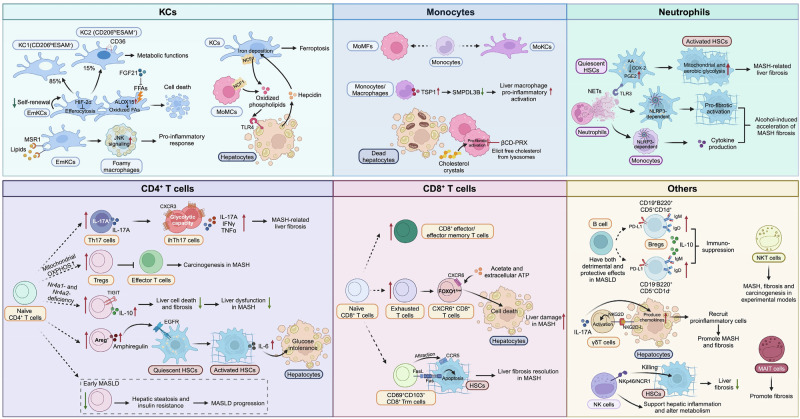
“Neutrophils are the most abundant leukocytes in the blood and form a critical arm of innate immunity, playing a well-recognized role in antibacterial defense by extruding a meshwork of chromatin fi bers to establish neutrophil extracellular traps (NETs) via a process known as NETosis [63, 64].”
A multisociety Delphi consensus statement on new fatty liver disease nomenclature
Rinella, M. E., Lazarus, J. V., Ratziu, V., Francque, S. M., Sanyal, A. J., Kanwal, F., Romero, D., Abdelmalek, M. F., Anstee, Q. M., Arab, J. P., et al. (2023) · Hepatology · doi:10.1097/hep.0000000000000520 · Cited by 3,476 · cited 1×
“These concerns led to a multi-stakeholder effort to develop a consensus on a change in nomenclature and the diagnostic criteria for the condition [7, 8].”
Younossi, Z. M., Golabi, P., Paik, J. M., Henry, A., Van Dongen, C., & Henry, L. (2023) · Hepatology · doi:10.1097/hep.0000000000000004 · Cited by 3,383 · cited 1×
“Metabolic dysfunction-associated steatotic liver disease (MASLD), formerly named nonalcoholic fatty liver disease (NAFLD), affects approximately 30% of the global population, leading to a major health and economic burden [1].”
MAFLD: a consensus-driven proposed nomenclature for metabolic associated fatty liver disease
Eslam, M., Sanyal, A. J., George, J., Sanyal, A., Neuschwander-Tetri, B., Tiribelli, C., Kleiner, D. E., Brunt, E., Bugianesi, E., Yki-Järvinen, H., et al. (2020) · Gastroenterology · doi:10.1053/j.gastro.2019.11.312 · Cited by 3,286
A multisociety Delphi consensus statement on new fatty liver disease nomenclature
Rinella, M. E., Lazarus, J. V., Ratziu, V., Francque, S. M., Sanyal, A. J., Kanwal, F., Romero, D., Abdelmalek, M. F., Anstee, Q. M., Arab, J. P., et al. (2023) · Journal of Hepatology · doi:10.1016/j.jhep.2023.06.003 · Cited by 3,100 · cited 1×
“These concerns led to a multi-stakeholder effort to develop a consensus on a change in nomenclature and the diagnostic criteria for the condition [7, 8].”
Kim, H. J., Park, S. Y., Choi, Y. J., Han, S. J., Lee, K. W., & Kim, D. J. (2013) · Hormone Research in Paediatrics · doi:10.1159/000348848 · Cited by 3,061
Cited by
Sorted by citations
Metabolic dysfunction and alcohol-associated liver disease (MetALD)
Gao, B., Arab, J. P., Liangpunsakul, S., Ding, W., Szabó, G., Mehal, W. Z., Wang, H., He, Y., Stärkel, P., Llorente, C., et al. (2025) · eGastroenterology · doi:10.1136/egastro-2025-100319 · Cited by 43
Self-citationZheng, H., Wu, K., Zheng, H., Chen, G., Lan, Y., Chen, S., Casu, G., Sechi, L. A., Wu, S., Vidili, G., et al. (2025) · JHEP Reports · doi:10.1016/j.jhepr.2025.101602 · Cited by 18
“…Neutrophil extracellular trap formation, M1 macrophages polarization, and T-cell imbalance contribute to hepatic inflammation and fibrosis, 38 while the same mechanisms also drive vascular inflammation, foam cell formation, and plaque instability, linking systemic immune activation to…”
Self-citationThe many pathways driving liver inflammation in MASH
Tilg, H., Adolph, T. E., Romeo, S., & Loomba, R. S. (2026) · Cell Metabolism · doi:10.1016/j.cmet.2026.02.018 · Cited by 17
Youhanna, S., Taebnia, N., Liang, Y., Cheng, N., Wang, Y., Michel, M., & Lauschke, V. M. (2025) · Advanced Biology · doi:10.1002/adbi.202500337 · Cited by 9
Eid, R. A., Hamed, A. M., Elgendy, S. O., Orayj, K. M., Ibrahim, A. S., Hamied, A. M. A., Wahsh, E. A., Youssif, M., Rabea, H., Madney, Y. M., et al. (2025) · Metabolites · doi:10.3390/metabo16010025 · Cited by 9
Nutrigenomics of fat-soluble vitamins and micronutrients in hepatocyte lipotoxicity and MASLD
Migni, A., Bartolini, D., Ceccarini, M. R., Galli, C., Cavaliere, G., Garetto, S., Lucci, J., & Galli, F. (2026) · Progress in Lipid Research · doi:10.1016/j.plipres.2026.101380 · Cited by 10
Xu, Y., Li, Q., & Jiao, X. (2025) · Frontiers in Cell and Developmental Biology · doi:10.3389/fcell.2025.1650677 · Cited by 7
“We will detail the evidence for this cycle and highlight how targeting its key nodes offers novel strategies for breaking the therapeutic bottleneck in MASLD (He et al., 2025).”
Immunometabolic integration in therapeutic strategies for managing MASLD
González-Serrano, S., Onoiu, A.-I., Camps, J., & Joven, J. (2025) · Frontiers in Pharmacology · doi:10.3389/fphar.2025.1693753 · Cited by 8
Liu, J., Wu, H., Zhou, L., Jin, Q., Ji, H., Hu, B., Wu, H., Chen, T., Wang, X., Hong, J., et al. (2026) · Metabolism · doi:10.1016/j.metabol.2025.156453 · Cited by 7
Chen, L.-L., Lu, Y.-J., Deng, A., Cao, J., Jin, F., Huang, H., Pan, F.-F., Liu, Y., Li, Y.-C., Tong, X., et al. (2025) · Cellular & Molecular Biology Letters · doi:10.1186/s11658-025-00794-8 · Cited by 5
“Some patients may eventually develop cirrhosis, and in advanced stages, HCC may arise as a severe complication, significantly impacting patients’ health and quality of life [155].”
Diambra, L. A., Sookoian, S. C., & Pirola, C. J. (2026) · Gut · doi:10.1136/gutjnl-2026-338674 · Cited by 4
Targeting Inflammation and Immune Regulation in Chronic Inflammation Associated Cancers
Rabiu, L., Chinn, M., Chen, F., Yerkinkazhina, Y., Tian, Y., & Zhu, W. (2026) · Cancer Science · doi:10.1111/cas.70360 · Cited by 4
“Chronic hepatic inflammation driven by lipotoxicity and oxidative stress promotes robust MDSC accumulation, leading to suppression of T-cell–mediated antitumor immunity [64].”
Li, Y. C., Hu, Y., He, Y. M., Yang, Y., Xue, D., Xue, E., Jia, J., Zhang, W., & Wang, J. (2026) · Frontiers in Immunology · doi:10.3389/fimmu.2026.1793092 · Cited by 4
“This shift does not imply that metabolites directly determine immune cell fate; rather, they are more likely to constrain metabolic fl exibility required for transitions between distinct functional states by reshaping the relative balance among intracellular metabolic pathways (43).”
Self-citationHu, K., Liu, S., Sheng, S., Zhang, S., Li, B., Sun, B., & Xia, A. (2026) · iLiver · doi:10.1016/j.iliver.2026.100234 · Cited by 5
“100 Recent integrated spatial and single-cell multi-omics investigations in human MASLD livers have identified MITF as a pivotal regulator of the lipid-handling capacity of LAMs, directly controlling their lipid metabolic pathways.”
Wiszniewski, M., Mori, D., Puch, S. I. S., Calejman, C. M., Cymeryng, C. B., & Repetto, E. M. (2025) · Biology · doi:10.3390/biology14081058 · Cited by 5
“Taking this into account, influencing liver inflammation by the inhibition of pro-inflammatory pathways or by stimulating anti-inflammatory pathways seems to be a promising therapeutic approach for MASH treatment [20].”
Herdiana, Y. (2025) · Diabetes Metabolic Syndrome and Obesity · doi:10.2147/dmso.s562536 · Cited by 4
“HCC represents the terminal stage of MASLD progression, with emerging evidence implicating immune regulation in the transition from MASH to HCC. 7 Management of MASLD currently remains a significant clinical challenge.”
The Role of Neutrophils in Non-Alcoholic Fatty Liver Disease: Mechanisms and Clinical Significance
Zhang, F., & Li, W. (2026) · Journal of Inflammation Research · doi:10.2147/jir.s585447 · Cited by 3
Perl, A., & Banki, K. (2026) · Nature Reviews Rheumatology · doi:10.1038/s41584-026-01421-2 · Cited by 3
“Alterations in lipid metabolism associated with MASLD can promote systemic inflammation and immune dysregulation 197 .”
Liu, Z., Zhou, B., Fang, J., Yu, Y., Jiang, X., Chen, X.-Y., Wang, W., Fu, X.-Y., Xia, J.-L., Liu, B.-J., et al. (2025) · Frontiers in Nutrition · doi:10.3389/fnut.2025.1673662 · Cited by 3
“Metabolic dysfunction-associated steatotic liver disease (MASLD), which aected approximately 30% of adults worldwide (13), is a significant metabolic disorder whose prevalence mirrors the increasing prevalence of obesity and T2DM.”
Kumar, G., Shah, Y. R., Shahzad, A., Jameel, K., Guevara-Lazo, D., Khan, N. A., Dahiya, D. S., Gangwani, M. K., Ravichandran, R., Patel, R. J., et al. (2025) · World Journal of Hepatology · doi:10.4254/wjh.v17.i11.111576 · Cited by 2
Lacerda, R. R. E., Barra, N. G., Conn, M. O., Zada, D. K., Robin, N., Fang, H., Marko, D. M., Punde, N. M., Kalyan, S., Gunn, H., et al. (2026) · American Journal of Physiology-Endocrinology and Metabolism · doi:10.1152/ajpendo.00488.2025 · Cited by 2
N 6 ‐Methyladenosine (m 6 A) in Liver Disease: Pathogenic Mechanisms and Therapeutic Potential
Chen, Y., Ya, R., Zhang, Z., Dong, S., Park, S. H., Seo, W., & He, Y. (2026) · iNew Medicine · doi:10.1002/inm3.70036 · Cited by 2
Self-citationComparison and Characteristics of MASLD Mouse Models
Wei, L., 高春辰, & Qin, H. (2026) · Biomedicines · doi:10.3390/biomedicines14040895 · Cited by 3
“…signaling axis in KCs, the NLRP3 inflammasome, and related molecules such as MyD88, iNOS, and HMGB1—drive hepatic inflammation, steatosis, and fibrosis by regulating pro-inflammatory cytokine release; correspondingly, gene knockout can alleviate MASLD-related pathological alterations [102,103].”
Alhawiti, N. M. (2026) · Frontiers in Medicine · doi:10.3389/fmed.2026.1728096 · Cited by 3
“In mouse models of MASH, treatment with aspirin or clopidogrel, a P2Y12 receptor antagonist, resulted in decreased serum ALT levels, reduced hepatic infiltration of immune cells, and lower expression of profibrotic markers such as α -SMA and collagen type I (134).”
Zhao, L., Xu, S., Qiao, S., Wang, Z., 王勝嵐, Liu, C., Zhang, S., Wang, P., Sun, X., Li, S., et al. (2026) · Signal Transduction and Targeted Therapy · doi:10.1038/s41392-026-02704-6 · Cited by 2
“35,36 This process activates Kupffer cells and promotes recruitment of myeloid immune populations; if unresolved, subsequent engagement of adaptive immunity contributes to the development of MASLD.”
Chen, L., Chen, Y., Zhao, M., Yang, X., Sutte, K., Lu, M., Chen, J., Zheng, X., Yang, D., & Liu, J. (2026) · Frontiers in Immunology · doi:10.3389/fimmu.2026.1814879 · Cited by 1
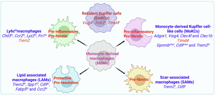
“In addition to competing for survival signals like CSF-1 (9, 33, 34), in fi ltrating monocytes actively replace embryonic KCs (EmKCs) during injury (35 – 37).”
Self-citationDissecting the immune pathogenesis of HBV-ACLF through single-cell multimodal analysis
Tacke, F., & Trebicka, J. (2025) · Gut · doi:10.1136/gutjnl-2025-336545 · Cited by 1
“11 Such interventions need to be tailored to patient characteristics”
Self-citationLiver immunology: new insights from cutting-edge technologies
Guillot, A., & Gao, B. (2025) · Cellular & Molecular Immunology · doi:10.1038/s41423-025-01342-2 · Cited by 1
“1B) [5 – 7].”
Metabolic Liver Disease Beyond the Liver
Gastaldelli, A., Tacke, F., & Holleboom, A. G. (2026) · Clinics in Liver Disease · doi:10.1016/j.cld.2025.12.003 · Cited by 1
Self-citationDaixie recipe ameliorates diet-induced MASH in mice via activating PI3K/AKT and Keap1/Nrf2 signaling
He, X., You, J., Deng, Y., Hu, Y., Qi, S., Li, Q., Yang, Y., Qu, X., Shao, Y., Fu, X., et al. (2026) · Frontiers in Endocrinology · doi:10.3389/fendo.2026.1772033 · Cited by 1
“In fl ammatory responses are a key driver of MASH progression, originating not only from the activation of Kupffer cells and macrophages but also from hepatocyte-derived in fl ammatory mediators (27, 28).”
Self-citationSia, J., Lee, B., & Oon, H. H. B. (2026) · Clinical Reviews in Allergy & Immunology · doi:10.1007/s12016-026-09152-8 · Cited by 1
Xue, L., Wang, X., & Mao, J. (2026) · Frontiers in Cell and Developmental Biology · doi:10.3389/fcell.2026.1808135 · Cited by 1
Gou, Z.-X., Li, N.-W., Yao, J.-Y., & Wang, Y. (2026) · International Journal of Nanomedicine · doi:10.2147/ijn.s597554 · Cited by 1
Guo, Y., Ma, Q., Guo, Z., Chu, J., Wu, N., & Yajuan, Q. I. (2026) · Life Sciences · doi:10.1016/j.lfs.2026.124534 · Cited by 1
Li, X., Jin, K., Fang, J., Ma, X., He, J., Lu, A., Jia, Z., & Shi, A. (2026) · Frontiers in Immunology · doi:10.3389/fimmu.2026.1835197 · Cited by 4
“In murine hepatic tissue, GLP-1R is present on gd T cells, where GLP-1 inhibits their in fl ammatory activity to reduce hepatic in fl ammation (181).”
Shen, K., Zhao, J., Li, K., Xu, L., & Wu, H. (2026) · Frontiers in Endocrinology · doi:10.3389/fendo.2026.1874886 · Cited by 1
“Chronic low-grade in fl ammation is another important feature of MASLD (14).”
Ma, Y., Gao, X., Wang, J., Li, L., Huang, Z., & Li, F. (2026) · Chemical Biology & Drug Design · doi:10.1111/cbdd.70371 · Cited by 1
Meng, Z., Zhou, T., Gou, J., Jiang, C., Li, B., Zhou, H., Liu, J., Feng, Q., & Wen, L. (2025) · Research Square · doi:10.21203/rs.3.rs-7551051/v1 · Cited by 1
Onoiu, A.-I., Murray, L., Gerardin, Y., Singh, G., Egger, R., González-Serrano, S., Cambra-Cortés, V., Jiménez-Franco, A., Guilarte, C., Parada, D., et al. (2026) · Research Square · doi:10.21203/rs.3.rs-8725050/v1 · Cited by 1
Yan, C., Han, S., Chen, Q., He, J., Wang, T., Shen, X., & Gong, X. (2025) · Nutrition Metabolism and Cardiovascular Diseases · doi:10.1016/j.numecd.2025.104208
Huang, J.-W., Man, N., Wang, H., Ai, X., Wei, D.-L., & Dai, Y. (2025) · Hepatic Medicine Evidence and Research · doi:10.2147/hmer.s562237
Guo, S., Li, Z., Han, Z., Ge, Y., Shi, H., Zheng, Y., Tan, W., Cheng, X., Li, Z., Yao, L., et al. (2025) · Journal of Inflammation Research · doi:10.2147/jir.s546860
Frietze, K. K., Brown, A., Das, D., Franks, R. G., Jagadeesan, P., & Nickels, J. T. (2026) · Journal of Biological Chemistry · doi:10.1016/j.jbc.2026.111212
Chen, Q., Liu, L., Feng, J., et al. (2026) · Scientific Reports · doi:10.1038/s41598-026-43311-3
“Accumulating evidence indicates that macrophages and CD8 ⁺ T cells infiltrate extensively during MASLD progression and contribute to the maintenance of chronic inflammation and disease advancement 38 .”
Hemati, H., Blanton, M. B., Rutt, L. N., Keiran, N., Geron, R. D., Lima, F., McCullough, R. L., & Messaoudi, I. (2026) · Frontiers in Endocrinology · doi:10.3389/fendo.2026.1783132 · Cited by 1
The biliary tree: A new frontier in mucosal immunology?
Guo, Y., Chouik, Y., Hao, Y., Kohlhepp, M. S., & Guillot, A. (2026) · Mucosal Immunology · doi:10.1016/j.mucimm.2026.04.006
Kotlyarov, S. N. (2026) · World Journal of Hepatology · doi:10.4254/wjh.v18.i4.116689
ANXA1-FPR1 signaling in myeloid cells drives MASH by elevating S100A4/A11
Yang, S., Qian, S., Yang, L., He, Y., Ge, S., Fu, X., Dong, S., Ya, R., Chen, Y., Ma, N., et al. (2026) · JHEP Reports · doi:10.1016/j.jhepr.2026.101869
“5,11 During the initial phase, the inflammatory response promotes tissue repair and the restoration of hepatic homeostasis, exerting protective effects.”
Self-citationMapping the genomic landscape of MASLD: A framework for molecular subtyping and precision hepatology
Pirola, C. J., & Sookoian, S. C. (2026) · Med · doi:10.1016/j.medj.2026.101131
Adipose tissue inflammation: a main driver of metabolic dysfunction-associated steatohepatitis
Vachliotis, I. D., & Pοlyzos, S. A. (2026) · Immunometabolism · doi:10.1097/in9.0000000000000082
Chen, Z., Yang, Q., Liu, J. S., Zhao, H., Ying, H., Liu, H., Huang, L., Lin, H. Y., Wang, S., Li, M., et al. (2026) · BMC Medicine · doi:10.1186/s12916-026-04888-8
“Our analysis identified a set of downstream protesins enriched in JAK-STAT signaling pathway and cytokine-cytokine receptor interaction, which are critical mediators of the systemic inflammatory response and are well-established drivers of hepatic inflammation and fibrosis progression [82, 83].”
Self-citationLipids and Immunity: From Molecular Mechanisms to Clinical Applications
Wang, Y., Li, X., & (422481), X. T. (2026) · Pharmaceutical science. · doi:10.5772/intechopen.1015581
Integrative Functional Nutrition in Metabolic-Associated Fatty Liver Disease
Güveli, H. (2026) · IntechOpen eBooks · doi:10.5772/intechopen.1016113
Song, L., Hua, S., Zeng, Q., Fu, Y., & Sui, Z. (2026) · Frontiers in Pharmacology · doi:10.3389/fphar.2026.1844294
“The release of damage-associated molecular patterns from injured cells initiates and sustains a potent inflammatory cascade (He et al., 2025).”
Gorke, S., Sharma, S., Malik, A., & Abdullah, I. A. W. A. S. T. (2026) · IntechOpen eBooks · doi:10.5772/intechopen.1015589
LGALS3 signaling and macrophage ferroptosis in steatohepatitis
Liu, W., & Shi, Z. (2026) · World Journal of Hepatology · doi:10.4254/wjh.120591
Yu, H., Chen, X. H., Song, W., Xiong, L., Li, X., Wang, Z., Pan, F., Li, W., Liu, G., Gao, F., et al. (2026) · Journal of Chemical Information and Modeling · doi:10.1021/acs.jcim.6c01470
Dhaou, C. B., Rom, O., & Orr, A. W. (2026) · JACC Basic to Translational Science · doi:10.1016/j.jacbts.2026.101624
Rocca, F. L., Privitera, G., Geraci, C., Morello, V., Geraci, G., Paternò, V., Santoro, C., & Esposito, R. (2026) · Nutrients · doi:10.3390/nu18152527
Yu, H., Liu, S., Zhang, Y., Ji, Y., Min, Y., Yin, D., Wang, T., Ma, X., Chen, J., Yu, W., et al. (2026) · iScience · doi:10.1016/j.isci.2026.117139
Plant-Derived Exosome-like Nanovesicles for Metabolic Dysfunction-Associated Steatotic Liver Disease
Kuang, T., Wang, L., Shi, Y., Cheng, J., & Pan, S. (2026) · Veterinary Sciences · doi:10.3390/vetsci13090851
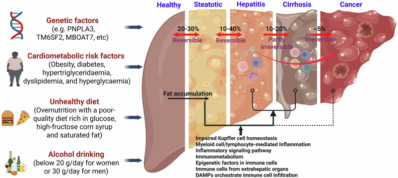
“Hepatic steatosis is a hallmark feature of the disease, resulting from excessive nutrition and a monotonous diet rich in glucose, high-fructose, and saturated fats, which lead to continuous accumulation of triglycerides in the liver through multiple pathways [30,31].”
Ruting, W., Hongxi, W., Liang, J., Huiping, L., Zhenjin, H., Zhang, R., Zhang, R., Hongzhen, T., & Jiang, F. (2026) · Ecotoxicology and Environmental Safety · doi:10.1016/j.ecoenv.2026.120705
Yılmaz, N., Akça, E., Ekinci, Z., Fidan, M. F., Pekel, Z., Kafadar, Z., Yigitbasi, B., Döngelli, H., Özgen, M., & Kehribar, D. Y. (2026) · Current Medical Research and Opinion · doi:10.1080/03007995.2026.2719078
“Consistently, elevated inflammatory markers have been associated with isolated steatosis phenotypes even in the absence of advanced fibrosis 26 .”
Spatial immune dysregulation in MASLD: integration of lobular zoning, metabolism and immune function
Gao, D., Wang, X., Mu, S., Sun, Y., & Wu, Z. (2026) · Frontiers in Immunology · doi:10.3389/fimmu.2026.1922207 · Cited by 1
Kumak, E., Darde, T., & Konu, Ö. (2026) · bioRxiv (Cold Spring Harbor Laboratory) · doi:10.64898/2026.08.26.747338
Onoiu, A.-I., González-Serrano, S., Cambra-Cortés, V., Jiménez-Franco, A., Guilarte, C., Peña, K. B., Riu, F., Camps, J., Joven, J., & Parada, D. (2026) · Frontiers in Immunology · doi:10.3389/fimmu.2026.1816648
Cyclo(Val-Pro) and cyclo(Phe-Phe) alleviate MASLD via distinct CCR2- and ADCY7-associated pathways.
Zhou, J., Jin, H.-J., Zheng, Y.-F., Ding, K.-Y., Long, Y., Yu, C.-J., Zhang, Y.-R., Zhuge, F., Chen, M.-H., Fu, Z.-W., et al. (2026) · Life Science · doi:10.1016/j.lfs.2026.124669
Guo, M., Wu, Q., Deng, C.-H., Jiang, X., Wang, J. H., Zeng, C., Tan, X., Teng, F., Liu, Y., Zeng, Y., et al. (2026) · Metabolism · doi:10.1016/j.metabol.2026.156767
Gaddam, I., Ferreira, M. S., Hastings, C., Calle, M. C., & Andersen, C. J. (2026) · Nutrients · doi:10.3390/nu18182952 · Cited by 1
“Cardiometabolic diseases—such as obesity, T2DM, hypertension, CVD, and MASLD— are among the major causes of morbidity and mortality around the world [51,52].”
Zhong, Q.-Q., Lyu, W., Li, J.-Y., Song, Y.-M., Huang, Y.-X., Wang, C.-B., & Ji, J.-L. (2026) · Frontiers in Immunology · doi:10.3389/fimmu.2026.1963310
“At the same time, cytokines such as IL-6, TGF-b , and IL-10, together with altered glucose, fatty-acid, and amino-acid availability, may in fl uence CD8 + T-cell differentiation, effector function, exhaustion-like features, and tissue adaptation (20 – 22 , 26).”
InfluentialA Neutrophil Defensin–VDAC1 Axis Drives Steatotic Liver DiseasePreprint
Zhang, J., Luo, G., Wang, G., Rao, J., Li, X., & Lu, W. (2026) · bioRxiv (Cold Spring Harbor Laboratory) · doi:10.64898/2026.09.19.752841
Liu, M., Liu, Z., Leng, Y., Zhang, Q., Liu, C., Yao, Y., & Chen, X. (2026) · Biochemical and Biophysical Research Communications · doi:10.1016/j.bbrc.2026.154684
Meng, Z., Chen, Z., Zhou, T., Gou, J., Ma, Z., Yue, A., Jiang, C., Feng, Q., & Wen, L. (2026) · Mediators of Inflammation · doi:10.1155/mi/1367877
Zhang, F., Zhu, C., Li, X., Sun, T., Zhang, Y., Wu, M., Zhang, Q., Dai, Y., Mei, X., & Kong, D. (2026) · Phytotherapy Research · doi:10.1002/ptr.70475
Rao, J., Li, X., Zhang, Y., Guo, J., Feng, B., Lu, Y., & Ma, X. (2026) · Journal of Agricultural and Food Chemistry · doi:10.1021/acs.jafc.6c05636
He, Y., Ma, T., 邢邯英, Cao, X., Song, F., Pan, Y., & Luping, R. (2026) · European Journal of Pharmacology · doi:10.1016/j.ejphar.2026.179418
Self-citationWu, S., li, Y., Zong, Y., Mai, Q., Qiu, R., Song, H., & Chen, H. (2026) · ACS Omega · doi:10.1021/acsomega.5c11799
Genome-wide analyses identify 60 loci associated with liver triglyceride content
Jamialahmadi, O., Mowlaei, S., Gelev, K. Z., Mancina, R. M., Flagiello, V., Kozlitina, J., Pajukanta, P., Valenti, L. V. C., & Romeo, S. (2026) · Nature Genetics · doi:10.1038/s41588-026-02794-6
Xu, L., Hu, L., Zhang, Y., Qi, J., Hou, Y., Cao, W., Duan, C., Liu, S., Ding, R., Lan, T., et al. (2026) · Metabolism · doi:10.1016/j.metabol.2026.156808
Metadata
- Authors
- He, Y., Chen, Y., Qian, S., van Der Merwe, S., Dhar, D., Brenner, D. A., & Tacke, F.
- Affiliations
- Chinese Academy of Sciences (CN)
- Sanford Burnham Prebys Medical Discovery Institute (US)
- Charité – Universitätsmedizin Berlin (DE)
- Corresponding author
- He, Yong
- Tacke, Frank
- Venue
- Cellular & Molecular Immunology
- Published
- 2025-06-10
- Volume
- 22
- Issue
- 10
- Pages
- 1159-1177
- ISSN
- 1672-7681 (ISSN-L)
- 2042-0226
- PMC
- PMC12480724
- OpenAlex
- W4411162672
- Semantic Scholar
- 7f2f36cf67705164236941f4c20e7acd81779cf2
- Cited by
- 81
- References
- 226
- Citations by year
- 2026: 63 · 2025: 18
- FWCI
- 12.44
- Type
- journal article
- Language
- en
- Open access
- hybrid
- doi.org
- Locations
- Cellular and Molecular Immunology (publishedVersion, cc-by)
- Europe PMC (PubMed Central) (publishedVersion, cc-by)
- PubMed (publishedVersion)
- PubMed Central (publishedVersion, cc-by)
- Lirias (KU Leuven) (submittedVersion, cc-by)
- Funders
- U.S. Department of Health and Human Services (R01DK133930, RO1DK137061)
- Deutsche Forschungsgemeinschaft (SFB 1382, DFGTa434/8‐1, Ta434/8‐1, 403224013)
- National Natural Science Foundation of China (82270601, 403224013)
- National Institutes of Health
- National Key Research and Development Program of China (2024ZD0530604, 2023YFA1800804)
- National Institute of Diabetes and Digestive and Kidney Diseases (R01DK137061, R01DK133930)
- Topics
- Health Sciences › Medicine › Hepatology › Liver Disease Diagnosis and Treatment
- Health Sciences › Medicine › Hepatology › Liver physiology and pathology
- Health Sciences › Medicine › Hepatology › Liver Diseases and Immunity
- Keywords
- non-alcoholic fatty liver disease, nonalcoholic steatohepatitis, immunopathogenesis, liver inflammation, immunomodulatory therapy, resmetirom, incretin mimetics, liver fibrosis, genetic susceptibility, overnutrition
- MeSH terms
- Animals, Humans, Liver, Immunomodulation, Non-alcoholic Fatty Liver Disease
About these checks
Registry, retraction, Cites and statistics results are computed, not judged, with one exception: a printed reference that no index lists is read once by a model before it counts as a citation error. Claims are extracted by a model and kept only when their quote appears verbatim in the text; each cited source is then checked, with a model’s help, for whether it says what the claim relies on. Those verdicts can be wrong — read the claim’s quote and the cited source before relying on one.
Statistics and arithmetic are recomputed, not judged.
Confidence bands are shown only for fields calibrated on our annotated test set. None is calibrated yet, so no band is shown.
What counts as a claim ›How we measure confidence ›How we check this ›
Are you an author, or do you see a problem with this page? Request a review
