

The DOI matches its Crossref record; 89 of the 89 references it cites were found in a registry or an index.
Tau exacerbates excitotoxic brain damage in an animal model of stroke
Open accessAbstract
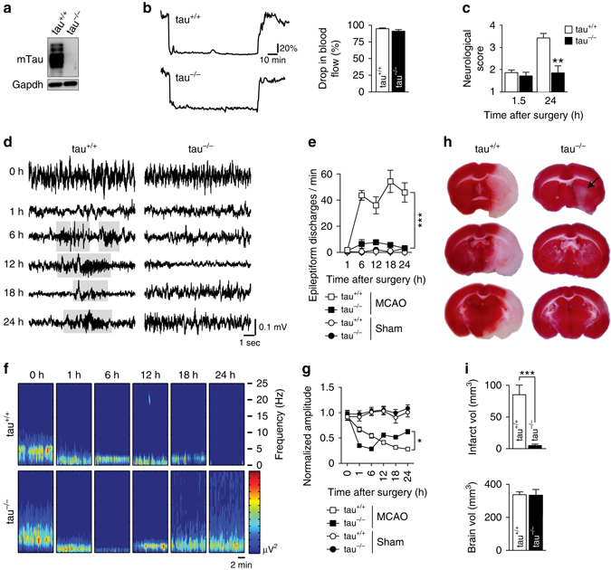
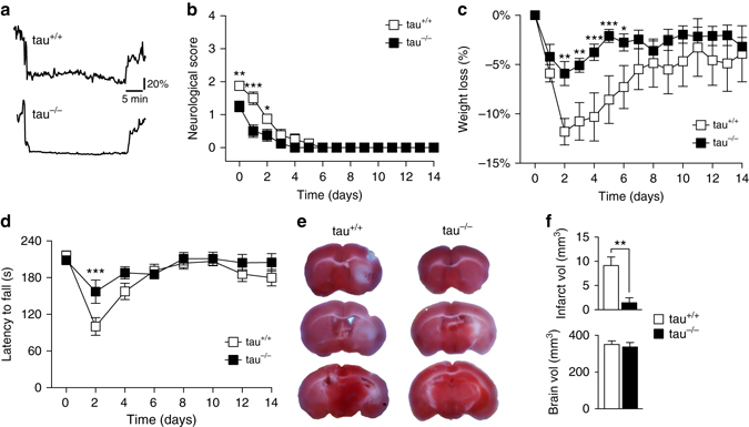
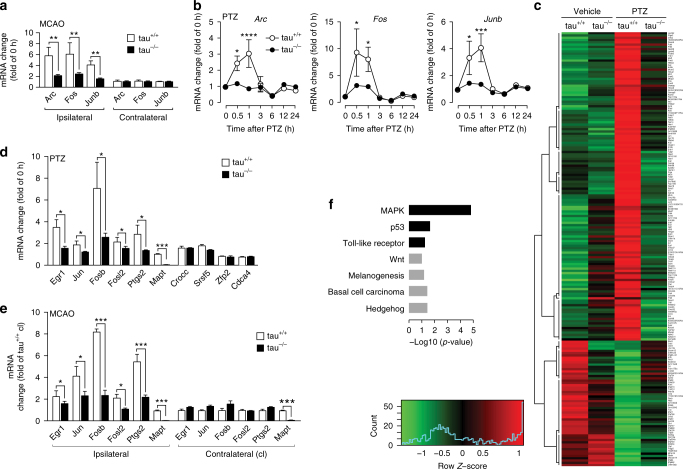
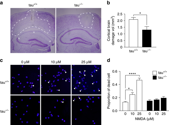
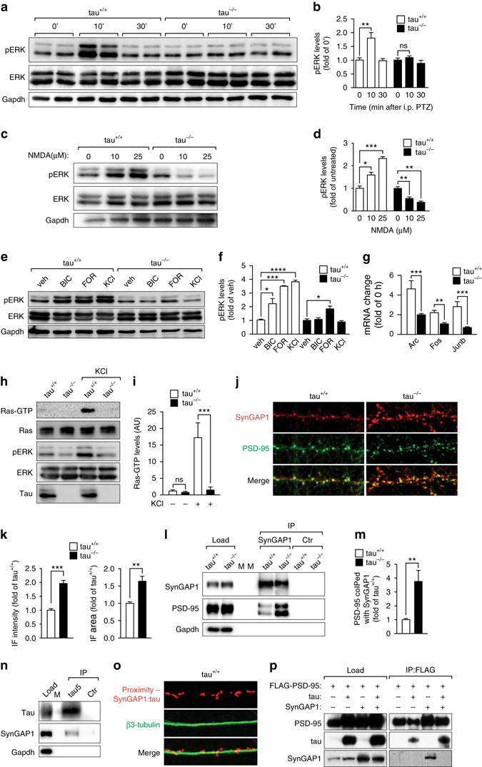
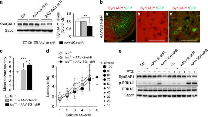
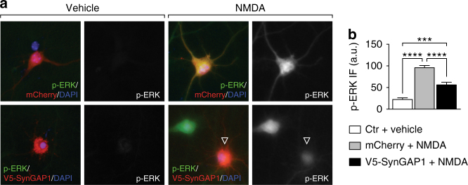
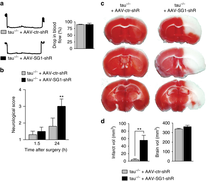
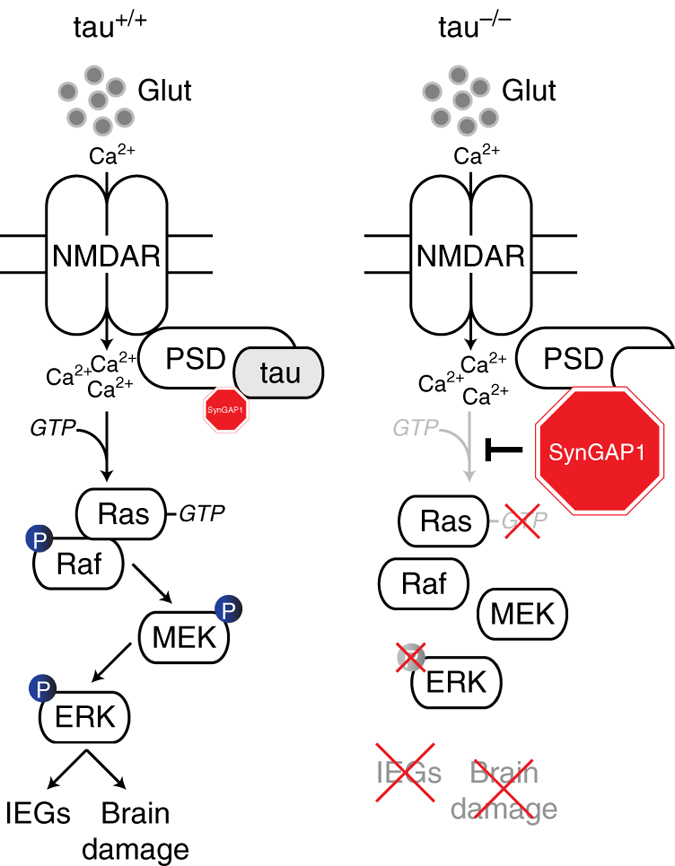
Neuronal excitotoxicity induced by aberrant excitation of glutamatergic receptors contributes to brain damage in stroke. Here we show that tau-deficient (tau −/− ) mice are profoundly protected from excitotoxic brain damage and neurological deficits following experimental stroke, using a middle cerebral artery occlusion with reperfusion model. Mechanistically, we show that this protection is due to site-specific inhibition of glutamate-induced and Ras/ERK-mediated toxicity by accumulation of Ras-inhibiting SynGAP1, which resides in a post-synaptic complex with tau. Accordingly, reducing SynGAP1 levels in tau −/− mice abolished the protection from pharmacologically induced excitotoxicity and middle cerebral artery occlusion-induced brain damage. Conversely, over-expression of SynGAP1 prevented excitotoxic ERK activation in wild-type neurons. Our findings suggest that tau mediates excitotoxic Ras/ERK signaling by controlling post-synaptic compartmentalization of SynGAP1.
Overview
The claims and statistics of this paper haven’t been checked yet.
Run the check — free, no account needed. It reads the open-access text and checks the sources it cites.
Integrity signals
Registry
Cites
Statistics
Figures
Verification
Claims the paper makes
The claims and statistics of this paper haven’t been checked yet.
Run the check — free, no account needed. It reads the open-access text and checks the sources it cites.
Claims other papers make about this one
Each is checked against this paper’s own text: does it say what the citing paper says it does?
The source backs part of the claim, not all of it — or backs it for a different population, condition or comparison.
In the citing paper
“However, these same mechanisms may render neurons vulnerable to excitotoxicity, as tau-dependent Fyn-PSD-95-NMDAR coupling amplifies glutamate receptor activation under pathological conditions [52, 124].”
From Tau Ablation Rewires Brain Cell Programs in Health and Restores Function in Disease (2026) · cites this paper as [124]
Experimental — accuracy not yet measured
Self-check
Percentages, sums and reported differences re-computed from the paper’s own numbers.
The self-check needs the paper’s full text.
Citations
Cites
Fast gapped-read alignment with Bowtie 2
Langmead, B., & Salzberg, S. L. (2012) · Nature Methods · doi:10.1038/nmeth.1923 · Cited by 56,400
Systematic and integrative analysis of large gene lists using DAVID bioinformatics resources
Huang, D. W., Sherman, B. T., & Lempicki, R. A. (2009) · Nature Protocols · doi:10.1038/nprot.2008.211 · Cited by 35,890
Trapnell, C., Roberts, A., Goff, L. A., Pertea, G. M., Kim, D., Kelley, D. R., Pimentel, H., Salzberg, S. L., Rinn, J. L., & Pachter, L. S. (2012) · Nature Protocols · doi:10.1038/nprot.2012.016 · Cited by 12,145
Amyloid oligomers exacerbate tau pathology in a mouse model of tauopathy
Selenica, M.-L. B., Brownlow, M., Jimenez, J. P., Lee, D. C., Pena, G., Dickey, C. A., Gordon, M. N., & Morgan, D. (2013) · Neurodegenerative Diseases · doi:10.1159/000337230 · Cited by 10,359
A Randomized Trial of Intraarterial Treatment for Acute Ischemic Stroke
Berkhemer, O. A., Fransen, P. S. S., Beumer, D., van den Berg, L. A., Lingsma, H. F., Yoo, A. J., Schonewille, W. J., Vos, J. A., Nederkoorn, P. J., Wermer, M. J. H., et al. (2014) · New England Journal of Medicine · doi:10.1056/nejmoa1411587 · Cited by 6,778
Thrombolysis with Alteplase 3 to 4.5 Hours after Acute Ischemic Stroke
Hacke, W., Kaste, M., Bluhmki, E., Brozman, M., Dávalos, A., Guidetti, D., Larrue, V., Lees, K. R., Medeghri, Z., Machnig, T., et al. (2008) · New England Journal of Medicine · doi:10.1056/nejmoa0804656 · Cited by 6,684
Endovascular therapy for ischemic stroke with perfusion-imaging selection.
Campbell, B., Mitchell, P. J., Kleinig, T., Dewey, H., Churilov, L., Yassi, N., Yan, B., Dowling, R., Parsons, M., Oxley, T., et al. (2015) · New England Journal of Medicine · doi:10.1056/nejmoa1414792 · Cited by 5,432
KEGG for integration and interpretation of large-scale molecular data sets
Kanehisa, M., Goto, S., Sato, Y., Furumichi, M., & Tanabe, M. (2012) · Nucleic Acids Research · doi:10.1093/nar/gkr988 · Cited by 4,848
Benardete, E. A., & Nair, A. K. (2015) · Cerebrovascular Diseases Extra · doi:10.1159/000370060 · Cited by 2,559
Treatment Strategies for Acute Ischemic Stroke Caused by Carotid Artery Occlusion
Li, W., Yin, Q., Xu, G., & Liu, X. (2016) · Interventional Neurology · doi:10.1159/000445304 · Cited by 2,319
Cited by
Sorted by citations
Items 101–167 of 167
Pluta, R. M., Bogucka‐Kocka, A., Bogucki, J., Kocki, J., & Czuczwar, S. J. (2024) · Journal of Alzheimer s Disease · doi:10.3233/jad-240401 · Cited by 15
Houben, S., Leroy, K., Ando, K., Yilmaz, Z., Widomski, C., Buée, L., & Brion, J. (2019) · Neurobiology of Disease · doi:10.1016/j.nbd.2019.02.021 · Cited by 16
Pituitary Adenylate Cyclase-Activating Polypeptide: A Potent Therapeutic Agent in Oxidative Stress
Sadanandan, N., Cozene, B., Park, Y. J., Farooq, J., Kingsbury, C., Wang, Z.-J., Moscatello, A., Saft, M., Cho, J., Gonzales‐Portillo, B., et al. (2021) · Antioxidants · doi:10.3390/antiox10030354 · Cited by 16
Treatment of epilepsy using a targeted p38γ kinase gene therapy
Morey, N., Przybyla, M., van der Hoven, J., Ke, Y. D., Delerue, F., van Eersel, J., & Ittner, L. M. (2022) · Science Advances · doi:10.1126/sciadv.add2577 · Cited by 15
Self-citationPluta, R. M., & Czuczwar, S. J. (2024) · International Journal of Molecular Sciences · doi:10.3390/ijms25063091 · Cited by 13
Hong, D. K., Eom, J.-W., Kho, A. R., Lee, S. H., Kang, B. S., Lee, S. H., Koh, J., Kim, Y. H., Choi, B. Y., & Suh, S. W. (2022) · Antioxidants · doi:10.3390/antiox11112192 · Cited by 14
Neves, J. D., Vizuete, A. F. K., Nicola, F., Ré, C. D., Rodrigues, A. F., Schmitz, F., Mestriner, R. G., Aristimunha, D., Wyse, Â. T. D. S., & Netto, C. A. (2018) · Neurochemistry International · doi:10.1016/j.neuint.2018.03.003 · Cited by 13
Decoupling the effects of hydrophilic and hydrophobic moieties at the neuron–nanofibre interface
Martin, A. D., Wojciechowski, J. P., Du, E. Y., Rawal, A., Stefen, H., Au, C. G., Hou, L., Cranfield, C. G., Fath, T., Ittner, L. M., et al. (2019) · Chemical Science · doi:10.1039/c9sc05686f · Cited by 13
Self-citationLiu, J., Tan, S., Wang, Y., Luo, J., Long, Y., Mei, X., & Tang, Y. (2020) · Journal of Molecular Neuroscience · doi:10.1007/s12031-020-01481-3 · Cited by 13
Dioli, C., Patrício, P., Pinto, L., Marie, C., Morais, M., Vyas, S., Bessa, J. M., Pinto, L., & Sotiropoulos, I. (2021) · Cell Proliferation · doi:10.1111/cpr.13027 · Cited by 15
Molecular Insights into Tau Pathology and its Therapeutic Strategies in Alzheimer's Disease
Zhou, F., Zhao, Y., Sun, Y., & Chen, W. (2024) · Journal of Integrative Neuroscience · doi:10.31083/j.jin2311197 · Cited by 13
Tau Acts in Concert With Kinase/Phosphatase Underlying Synaptic Dysfunction
Fan, X., Xia, L., Zhou, Z., Qiu, Y., Zhao, C., Yin, X., & Qian, W. (2022) · Frontiers in Aging Neuroscience · doi:10.3389/fnagi.2022.908881 · Cited by 14
Emerging role of Jumonji domain-containing protein D3 in inflammatory diseases
Li, X., Chen, R.-Y., Shi, J., Li, C., Liu, Y.-J., Gao, C., Gao, M.-R., Zhang, S., Lu, J., Cao, J.-F., et al. (2024) · Journal of Pharmaceutical Analysis · doi:10.1016/j.jpha.2024.100978 · Cited by 11
Epigenetics and Pharmacoepigenetics of Neurodevelopmental and Neuropsychiatric Disorders
Cacabelos, R. (2019) · Elsevier eBooks · doi:10.1016/b978-0-12-813939-4.00022-x · Cited by 11
Deregulated Transcription and Proteostasis in Adult mapt Knockout Mouse
Andrés‐Benito, P., Flores, Á., Busquet-Areny, S., Carmona, M., Ausín, K., Cartas‐Cejudo, P., Lachén‐Montes, M., del Rı́o, J. A., Fernández‐Irigoyen, J., Santamaría, E., et al. (2023) · International Journal of Molecular Sciences · doi:10.3390/ijms24076559 · Cited by 10
Future Perspectives of NMDAR in CNS Disorders
Capó, T., Rebassa, J. B., Raïch, I., Lillo, J., Badia, P., Navarro, G., & Reyes‐Resina, I. (2025) · Molecules · doi:10.3390/molecules30040877 · Cited by 12
Wang, C., Chen, H., Ma, S., Mao, B.-B., Chen, Y., Xu, H., & Yu, H. (2021) · Evidence-based Complementary and Alternative Medicine · doi:10.1155/2021/5582782 · Cited by 10
Acetylated Tau Protein: A New Piece in the Puzzle between Brain Ischemia and Alzheimer’s Disease
Pluta, R. M., Januszewski, S., & Jabłoński, M. (2022) · International Journal of Molecular Sciences · doi:10.3390/ijms23169174 · Cited by 8
Generation and Role of Calpain-Cleaved 17-kDa Tau Fragment in Acute Ischemic Stroke
Chen, Y.-D., Huang, P.-Y., Chiang, C.-S., Huang, Y., & Tang, S. (2021) · Molecular Neurobiology · doi:10.1007/s12035-021-02519-2 · Cited by 9
Zhou, G., & Zha, X. (2021) · Frontiers in Neuroscience · doi:10.3389/fnins.2021.692217 · Cited by 8
Lapin, D., Sharma, A., & Wang, P. (2025) · Journal of Neuroinflammation · doi:10.1186/s12974-025-03340-7 · Cited by 9
Prikas, E., Ahel, H. I., Stefanoska, K., Asih, P. R., Volkerling, A. M., Ittner, L. M., & Ittner, A. (2021) · Biochemistry and Cell Biology · doi:10.1139/bcb-2020-0604 · Cited by 8
Self-citationA model of traumatic brain injury using human iPSC-derived cortical brain organoidsPreprint
Lai, J. D., Berlind, J. E., Fricklas, G., Maria, N. S. S., Jacobs, R. E., Yu, V. L., & Ichida, J. K. (2020) · bioRxiv (Cold Spring Harbor Laboratory) · doi:10.1101/2020.07.05.180299 · Cited by 10
Facal, C. L., Bessone, I. F., Muñiz, J. A., Pereyra, A. E., Pedroncini, O., Páez-Paz, I., Clerici-Delville, R., Arnaiz, C., Urrutia, L., Falasco, G., et al. (2024) · Molecular Therapy · doi:10.1016/j.ymthe.2024.01.033 · Cited by 9
Molecular signatures of regional vulnerability to tauopathy in excitatory cortical neurons
Broekaart, D. W. M., Sharma, A., Ramakrishnan, A., Chongtham, A., Günther, D. M., Subramaniyan, S., Wang, M., Patel, V., Zhang, B., Grinberg, L. T., et al. (2025) · Acta Neuropathologica · doi:10.1007/s00401-025-02879-2 · Cited by 7
Characterizing stroke-related cellular changes in the surviving neurons of mouse ischemic stroke
Ikegami, T., Sasaki, T., Shimbo, T., Kitayama, T., Yamamoto, Y., Ouchi, Y., Yamazaki, S., Sugiyama, S., Nishiyama, K., Gon, Y., et al. (2025) · Neurochemistry International · doi:10.1016/j.neuint.2025.106086 · Cited by 6
Tang, F., Wei, J., Wong, M.-L., & Lei, P. (2025) · Genomic psychiatry : · doi:10.61373/gp025i.0122 · Cited by 7
Are Ischemic Stroke and Alzheimer’s Disease Genetically Consecutive Pathologies?
Filippenkov, I. B., Владимирович, Х. А., Mozgovoy, I. V., Dergunova, L. V., & Limborska, S. A. (2023) · Biomedicines · doi:10.3390/biomedicines11102727 · Cited by 7
cis P-tau Accumulation Triggers Neurodegeneration after Ischemic Stroke
Seddigh, N., Taabodi, D., Dadzadi, M., & Shahpasand, K. (2024) · ACS Omega · doi:10.1021/acsomega.3c07285 · Cited by 7
Czuczwar, S. J., Kocki, J., Miziak, B., Bogucki, J., Bogucka‐Kocka, A., & Pluta, R. M. (2024) · Journal of Alzheimer s Disease · doi:10.3233/jad-231333 · Cited by 7
Common Proteomic and Genomic Contribution to Ischemic Brain Damage and Alzheimer’s Disease
Pluta, R. M., Ułamek-Kozioł, M., Januszewski, S., & Czuczwar, S. J. (2019) · Codon Publications eBooks · doi:10.15586/alzheimersdisease.2019.ch4 · Cited by 6
TDP‐43 pathology and functional deficits in wild‐type and ALS/FTD mutant cyclin F mouse models
van Hummel, A., Sabale, M., Przybyla, M., van der Hoven, J., Chan, G., Feiten, A., Chung, R. S., Ittner, L. M., & Ke, Y. D. (2023) · Neuropathology and Applied Neurobiology · doi:10.1111/nan.12902 · Cited by 5
Self-citationSetti, S. E., & Reed, M. N. (2021) · Metabolic Brain Disease · doi:10.1007/s11011-021-00848-8 · Cited by 7
Tau — a new target in acute brain ischaemia
Sidaway, P. (2017) · Nature Reviews Neurology · doi:10.1038/nrneurol.2017.141 · Cited by 5
Ma, D., An, Q., Zhang, Z., Bian, Q., Li, Y., Yan-li, L., & Zhang, S. (2020) · Therapeutic Hypothermia and Temperature Management · doi:10.1089/ther.2020.0012 · Cited by 5
Song, Y., Zhang, C., Li, N., Fu, Y., Li, H., & Zhang, J. (2020) · Developmental Neuroscience · doi:10.1159/000509108 · Cited by 5
Chen, Y., Jiang, L., Li, M., Shen, Y., Liu, S., & Yang, D. (2024) · Heliyon · doi:10.1016/j.heliyon.2024.e29556 · Cited by 4
HuR deficiency abrogated the enhanced NLRP3 signaling in experimental ischemic stroke
Wang, X., Zhao, M., Lu, X., Du, P., Feng, S., Gong, R., Chen, H., Qi, G., & Yang, F. (2023) · The FASEB Journal · doi:10.1096/fj.202300812r · Cited by 4
Improving Cognition Without Clearing Amyloid: Effects of Tau and Ultrasound Neuromodulation
Leinenga, G., Padmanabhan, P., & Götz, J. (2024) · Journal of Alzheimer s Disease · doi:10.3233/jad-240616 · Cited by 4
Allen, J., Ermine, C. M., Lin, R., CLOUD, G. C., Shultz, S. R., & Casillas-Espinosa, P. M. (2025) · Stroke · doi:10.1161/strokeaha.124.049279 · Cited by 4
Kazmi, S., Farokhi-Sisakht, F., Davoody, S., Bahlakeh, G., Abbaszadeh, F., Rahbarghazi, R., Shekarchi, A. A., & Karimipour, M. (2025) · Molecular Brain · doi:10.1186/s13041-025-01201-1 · Cited by 6
Wu, S.-Y., Tyagi, A., Wu, K., Smith, E. C., Song, Q., Sharma, S., Miller, L. D., Zhang, W., Pan, B., Lin, H., et al. (2026) · Nature Biomedical Engineering · doi:10.1038/s41551-026-01613-x · Cited by 3
Genomic and proteomic conversion of brain ischemia to Alzheimer’s disease
Pluta, R. M., & Ułamek-Kozioł, M. (2026) · Frontiers in Cell and Developmental Biology · doi:10.3389/fcell.2026.1804251 · Cited by 3
Tau-induced mitochondrial reverse electron transport drives neurodegeneration
Li, W., Rimal, S., Bhurtel, S., Yeung, L., Lu, B. G., Grinberg, L. T., Spina, S., Cobos, I., Seeley, W. W., Guo, S., et al. (2026) · Neuron · doi:10.1016/j.neuron.2026.07.012 · Cited by 2
PLP1-Targeting Antisense Oligonucleotides Improve FOXG1 Syndrome Mice
Tan, D. C. S., Jung, S., Deng, Y., Morey, N., Chan, G., Bongers, A., Ke, Y. D., Ittner, L. M., & Delerue, F. (2024) · International Journal of Molecular Sciences · doi:10.3390/ijms251910846 · Cited by 1
Self-citationSadeghzadeh, J., Roqanian, S., Jafarzadeh, J., Zangbar, H. S., Nakhjiri, E., Kalan, A. E., Farhoudi, M., Ahmadian, S., Shahabi, P., & Shahpasand, K. (2025) · International Immunopharmacology · doi:10.1016/j.intimp.2025.114834 · Cited by 1
Pharmacological Effects and Molecular Mechanisms of Lignans in the Treatment of Alzheimer’s Disease
Sun, L., Zhu, X., Fan, C., Wang, Y., Chen, Q., Zhang, L., Zhang, Y., & Liu, Y. (2026) · Molecules · doi:10.3390/molecules31152594 · Cited by 1
Tau Modulates Neurovascular Coupling
Tang, F., Wang, Q., Guo, J., & Lei, P. (2021) · Neuroscience Bulletin · doi:10.1007/s12264-021-00637-4 · Cited by 1
Rayner, S. L., Cheng, F., Yang, S., Grima, N., Ke, Y. D., Au, C. G., Morsch, M., Luca, A. D., Davidson, J. M., Molloy, M. P., et al. (2020) · bioRxiv (Cold Spring Harbor Laboratory) · doi:10.1101/2020.04.30.071365 · Cited by 1
Self-citationWang, P.-F., Zhong, W.-B., Ju, X., Li, Z., & Wang, F.-X. (2020) · doi:10.21203/rs.3.rs-19235/v1 · Cited by 1
Wang, S., Jiang, S., Zheng, G., & Cho, Y. K. (2022) · bioRxiv (Cold Spring Harbor Laboratory) · doi:10.1101/2022.09.01.506229 · Cited by 2
Gil, R. S., Pascovici, D., Venturato, J., Brown-Wright, H., Mehta, P. M., Martin, L. M. S., Wu, J. X., Chui, Y. K., Bademosi, A. T., Swaminathan, S., et al. (2023) · bioRxiv (Cold Spring Harbor Laboratory) · doi:10.1101/2023.06.21.546009 · Cited by 1
Czuczwar, S. J., Kocki, J., Miziak, B., Bogucki, J., Bogucka‐Kocka, A., & Pluta, R. M. (2023) · Preprints.org · doi:10.20944/preprints202309.2168.v1 · Cited by 1
Tau Ablation Rewires Brain Cell Programs in Health and Restores Function in Disease
Chimal-Juárez, E., Patel, H., Jury-Garfe, N., Dabin, L. C., Vidal, R., Kim, J., & Lasagna-Reeves, C. A. (2026) · bioRxiv · doi:10.64898/2026.05.12.724684
TREM2 Limits Progression of Deficits and Spreading of Tau Pathology in MicePreprint
Feiten, A., Au, C. G., van Hummel, A., van der Hoven, J., Deng, Y., Przybyla, M., Bright, F., Butler, T. A., Delerue, F., Toutonji, A., et al. (2021) · Research Square · doi:10.21203/rs.3.rs-741896/v1
Self-citationMa, D., An, Q., Zhang, Z., Bian, Q., Li, Y., Li, Y., & Zhang, S. (2019) · doi:10.21203/rs.2.17383/v1
Parvatikar, P., Bagali, S., Hippargi, S. B., Singh, P. K., Singh, S. B., Biradar, M. S., Patil, A. V., & Das, K. K. (2022) · Letters in Drug Design & Discovery · doi:10.2174/1570180819666220616142153
TREM2 limits progression of deficits and spreading of tau pathology in micePreprint
Feiten, A., Au, C. G., Sabale, M., van Hummel, A., van der Hoven, J., Deng, Y., Przybyla, M., Bright, F., Butler, T., Delerue, F., et al. (2022) · Research Square · doi:10.21203/rs.3.rs-741896/v2
Self-citationThe Role of Phosphorylated-Tau 181 in Enhancing Neuroplasticity After Ischemic Stroke
Arshinta, L., Widyastuti, K., & Widyadharma, I. P. E. (2025) · International Journal Of Scientific Advances · doi:10.51542/ijscia.v6i3.14
Pathoepigenetics of vascular disorders
Cacabelos, R., Naidoo, V., Cacabelos, N., & Martínez-Iglesias, O. A. (2025) · Elsevier eBooks · doi:10.1016/b978-0-443-18722-3.00039-6
He, X.-F., Li, G., Li, L.-L., Li, M.-Y., Liang, F.-Y., Chen, X., & Hu, X. (2020) · Figshare · doi:10.6084/m9.figshare.13064265
Facal, C. L., Páez-Paz, I., Pereyra, A. E., Gaguine, C., Clerici-Delville, R., Foltran, R. B., Soiza‐Reilly, M., & Avale, M. E. (2026) · Molecular Therapy — Nucleic Acids · doi:10.1016/j.omtn.2026.102843
Chen, Y., Man, S., Li, Q., Ji, Y., Peng, B., Ding, Y., & Xu, J. (2026) · Scientific Reports · doi:10.1038/s41598-026-39299-5
Dong, Q., Wu, Z., Li, A., Zhao, X., Qin, X., Xu, W., & Tang, G. (2026) · The FASEB Journal · doi:10.1096/fj.202503776rr
Atypical involvement of Alzheimer’s tau proteins in diseases beyond tauopathies
Meng, J., Deng, Z., Zhang, J., Yu, W., Wu, X., & Lei, P. (2026) · Life Medicine · doi:10.1093/lifemedi/lnag016
m6A Modification Genetic Variants Associated with Autism Spectrum Disorder Risks
Wang, H., Zhao, S., Chen, Y., Hou, F., Zhu, K., Liu, R., Xiang, Z., Chen, Q., Fan, H., Huang, M., et al. (2026) · Molecular Neurobiology · doi:10.1007/s12035-026-05962-1
Metadata
- Authors
- Bi, M., Gladbach, A., van Eersel, J., Ittner, A., Przybyla, M., van Hummel, A., Chua, S. W., van der Hoven, J., Lee, W. S., Müller, J., Parmar, J., Jonquieres, G. V., Stefen, H., Guccione, E., Fath, T., Housley, G. D., Klugmann, M., Ke, Y. D., & Ittner, L. M.
- Affiliations
- UNSW Sydney (AU)
- Agency for Science, Technology and Research (SG)
- Corresponding author
- Ittner, Lars M.
- ORCID
- Venue
- Nature Communications
- Published
- 2017-09-07
- Volume
- 8
- Issue
- 1
- Pages
- 473-473
- ISSN
- 2041-1723 (ISSN-L)
- PMC
- PMC5589746
- OpenAlex
- W2751298713
- Cited by
- 167
- References
- 89
- Citations by year
- 2026: 10 · 2025: 14 · 2024: 20 · 2023: 13 · 2022: 22 · 2021: 25 · 2020: 27 · 2019: 22 · 2018: 13 · 2017: 1
- FWCI
- 8.43
- Type
- journal article
- Language
- en
- Open access
- gold
- doi.org
- Locations
- Nature Communications (publishedVersion, cc-by)
- Europe PMC (PubMed Central) (publishedVersion, cc-by)
- PubMed Central (publishedVersion, cc-by)
- PubMed (publishedVersion)
- National University of Singapore (submittedVersion, cc-by)
- RePEc: Research Papers in Economics (submittedVersion)
- DOAJ (DOAJ: Directory of Open Access Journals) (submittedVersion)
- DOAJ (DOAJ: Directory of Open Access Journals) (submittedVersion, cc-by-sa)
- Funders
- University of New South Wales
- Medical Research Council
- Australian Research Council
- National Health and Medical Research Council
- Topics
- Life Sciences › Neuroscience › Cellular and Molecular Neuroscience › Neuroscience and Neuropharmacology Research
- Life Sciences › Neuroscience › Cellular and Molecular Neuroscience › Alzheimer's disease research and treatments
- Life Sciences › Neuroscience › Neurology › Neuroinflammation and Neurodegeneration Mechanisms
- Keywords
- tau protein, excitotoxicity, stroke, SYNGAP1, Ras/ERK signaling, glutamate receptors, middle cerebral artery occlusion, neuroprotection
- MeSH terms
- Animals, Brain Injuries, Cells, Cultured, Disease Models, Animal, Humans, Male, Mice, Inbred C57BL, Neurons, Synaptosomes, Signal Transduction, tau Proteins, Mice, Knockout, Infarction, Middle Cerebral Artery, Stroke, ras GTPase-Activating Proteins, Gene Expression Profiling
About these checks
Registry, retraction, Cites and statistics results are computed, not judged, with one exception: a printed reference that no index lists is read once by a model before it counts as a citation error. Claims are extracted by a model and kept only when their quote appears verbatim in the text; each cited source is then checked, with a model’s help, for whether it says what the claim relies on. Those verdicts can be wrong — read the claim’s quote and the cited source before relying on one.
Statistics and arithmetic are recomputed, not judged.
Confidence bands are shown only for fields calibrated on our annotated test set. None is calibrated yet, so no band is shown.
What counts as a claim ›How we measure confidence ›How we check this ›
Are you an author, or do you see a problem with this page? Request a review
