

The DOI matches its Crossref record; 59 of the 59 references it cites were found in a registry or an index.
Tumor induces muscle wasting in mice through releasing extracellular Hsp70 and Hsp90
Open accessAbstract
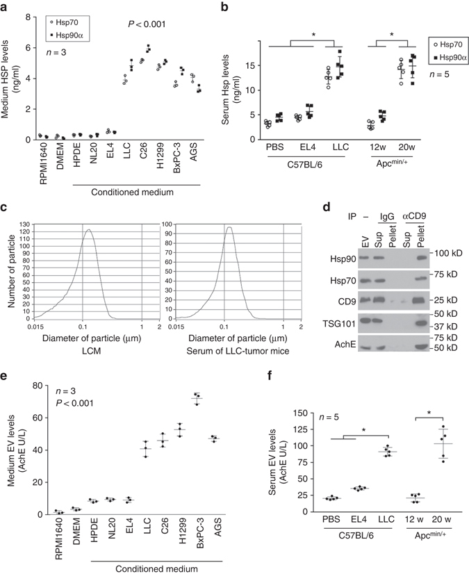
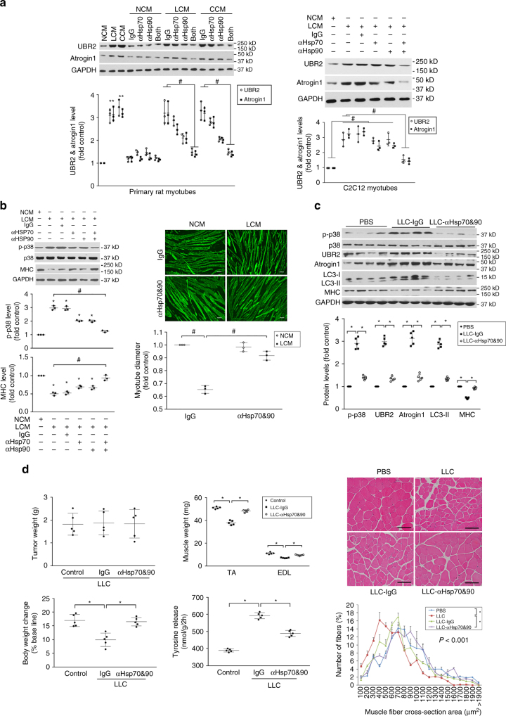
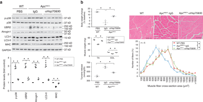
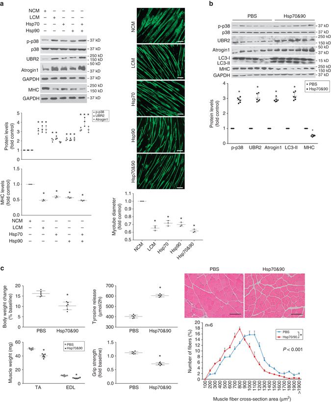
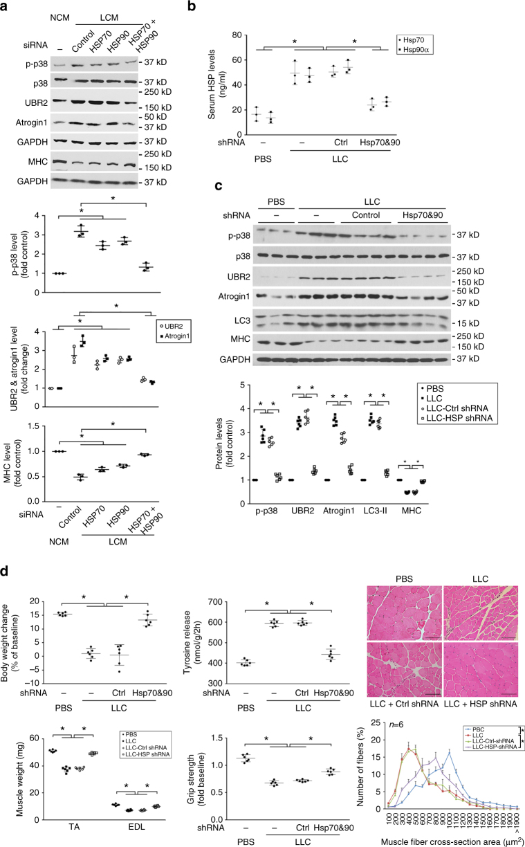
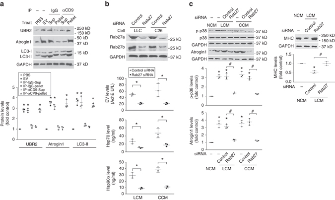
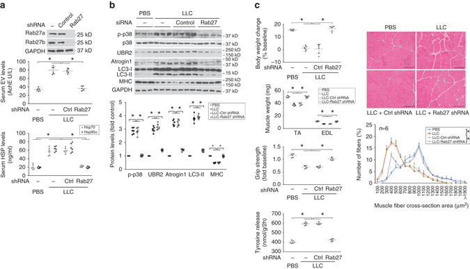
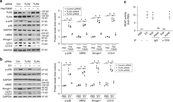
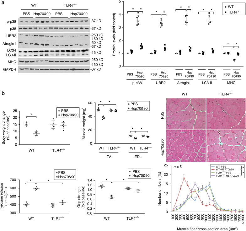
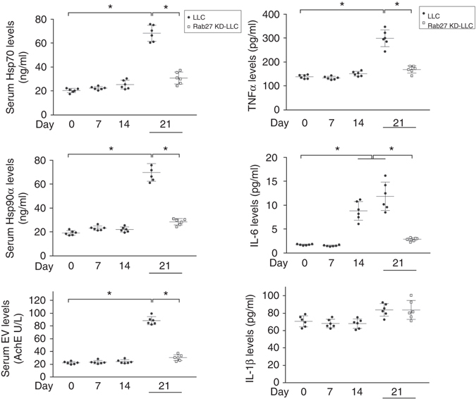
Cachexia, characterized by muscle wasting, is a major contributor to cancer-related mortality. However, the key cachexins that mediate cancer-induced muscle wasting remain elusive. Here, we show that tumor-released extracellular Hsp70 and Hsp90 are responsible for tumor’s capacity to induce muscle wasting. We detected high-level constitutive release of Hsp70 and Hsp90 associated with extracellular vesicles (EVs) from diverse cachexia-inducing tumor cells, resulting in elevated serum levels in mice. Neutralizing extracellular Hsp70/90 or silencing Hsp70/90 expression in tumor cells abrogates tumor-induced muscle catabolism and wasting in cultured myotubes and in mice. Conversely, administration of recombinant Hsp70 and Hsp90 recapitulates the catabolic effects of tumor. In addition, tumor-released Hsp70/90-expressing EVs are necessary and sufficient for tumor-induced muscle wasting. Further, Hsp70 and Hsp90 induce muscle catabolism by activating TLR4, and are responsible for elevation of circulating cytokines. These findings identify tumor-released circulating Hsp70 and Hsp90 as key cachexins causing muscle wasting in mice.
Overview
The claims and statistics of this paper haven’t been checked yet.
Run the check — free, no account needed. It reads the open-access text and checks the sources it cites.
Integrity signals
Registry
Cites
Statistics
Figures
Verification
Claims the paper makes
The claims and statistics of this paper haven’t been checked yet.
Run the check — free, no account needed. It reads the open-access text and checks the sources it cites.
Claims other papers make about this one
Each is checked against this paper’s own text: does it say what the citing paper says it does?
In the citing paper
“Recently, tumor-derived EVs deliver HSP70 and HSP90 to myofibers, where they activate Toll-like receptor 4 (TLR4) and muscle catabolism88 (Fig. 5).”
From Mechanisms of muscle atrophy and hypertrophy: implications in health and disease (2021) · cites this paper as [88]
Self-check
Percentages, sums and reported differences re-computed from the paper’s own numbers.
The self-check needs the paper’s full text.
Citations
Cites
Definition and classification of cancer cachexia: an international consensus
Fearon, K., Strasser, F., Anker, S. D., Bosaeus, I. G., Bruera, E. D., Fainsinger, R. L., Jatoi, A., Loprinzi, C. L., Macdonald, N., Mantovani, G., et al. (2011) · The Lancet Oncology · doi:10.1016/s1470-2045(10)70218-7 · Cited by 6,020
Hoshino, K., Takeuchi, O., Kawai, T., Sanjo, H., Ogawa, T., Takeda, Y., Takeda, K., & Akira, S. (1999) · The Journal of Immunology · doi:10.4049/jimmunol.162.7.3749 · Cited by 3,374
Toll-like receptors in innate immunity
Takeda, K. (2004) · International Immunology · doi:10.1093/intimm/dxh186 · Cited by 3,328
Yang, Y., Li, S., Yang, Q., Shi, Y., Zheng, M., Liu, Y., Chen, F., Song, G., Xu, H., Wan, T., et al. (2014) · Cellular Physiology and Biochemistry · doi:10.1159/000358696 · Cited by 3,263
DAMPs, PAMPs and alarmins: all we need to know about danger
Bianchi, M. E. (2007) · Journal of Leukocyte Biology · doi:10.1189/jlb.0306164 · Cited by 2,794
Rab27a and Rab27b control different steps of the exosome secretion pathway
Ostrowski, M., Carmo, N., Krumeich, S., Fanget, I., Raposo, G., Savina, A., Moita, C. F., Schauer, K., Hume, A. N., Freitas, R. P., et al. (2009) · Nature Cell Biology · doi:10.1038/ncb2000 · Cited by 2,761
Lötvall, J., Hill, A. F., Hochberg, F., Buzás, E. I., Di Vizio, D., Gardiner, C., Gho, Y. S., Kurochkin, I. V., Mathivanan, S., Quesenberry, P., et al. (2014) · Journal of Extracellular Vesicles · doi:10.3402/jev.v3.26913 · Cited by 2,451
Prognostic effect of weight loss prior tochemotherapy in cancer patients
DeWys, W. D., Begg, C. B., Lavin, P. T., Band, P. R., Bennett, J. M., Bertino, J. R., Cohen, M. H., Douglass, H. O., Engstrom, P. F., Ezdinli, E. Z., et al. (1980) · The American Journal of Medicine · doi:10.1016/s0149-2918(05)80001-3 · Cited by 2,350
Exosomal MicroRNA: A Diagnostic Marker for Lung Cancer
Rabinowits, G., Gerçel-Taylor, Ç., Day, J. M., Taylor, D. D., & Kloecker, G. H. (2009) · Clinical Lung Cancer · doi:10.3816/clc.2009.n.006 · Cited by 1,290
Cancer Cachexia: Mediators, Signaling, and Metabolic Pathways
Fearon, K. C. H., Glass, D. J., & Guttridge, D. C. (2012) · Cell Metabolism · doi:10.1016/j.cmet.2012.06.011 · Cited by 1,244
Cited by
Sorted by citations
The biology , function , and biomedical applications of exosomes
Kalluri, R., & LeBleu, V. S. (2020) · Science · doi:10.1126/science.aau6977 · Cited by 10,770
Baracos, V. E., Martin, L., Korc, M., Guttridge, D. C., & Fearon, K. C. H. (2018) · Nature Reviews Disease Primers · doi:10.1038/nrdp.2017.105 · Cited by 1,738
Mechanisms of muscle atrophy and hypertrophy: implications in health and disease
Sartori, R., Romanello, V., & Sandri, M. (2021) · Nature Communications · doi:10.1038/s41467-020-20123-1 · Cited by 940
Release mechanisms of major DAMPs
Murao, A., Aziz, M., Wang, H., Brenner, M., & Wang, P. (2021) · APOPTOSIS · doi:10.1007/s10495-021-01663-3 · Cited by 590
Metal Drugs and the Anticancer Immune Response
Englinger, B., Pirker, C., Heffeter, P., Terenzi, A., Kowol, C. R., Keppler, B. K., & Berger, W. (2019) · Chemical Reviews · doi:10.1021/acs.chemrev.8b00396 · Cited by 355
Tumour extracellular vesicles and particles induce liver metabolic dysfunction
Wang, G., Li, J., Bojmar, L., Chen, H., Li, Z., Tobias, G. C., Hu, M., Homan, E. A., Lucotti, S., Zhao, F., et al. (2023) · Nature · doi:10.1038/s41586-023-06114-4 · Cited by 280
Argilés, J. M. A., López-Soriano, F. J., Stemmler, B., & Busquets, S. (2023) · Nature Reviews Clinical Oncology · doi:10.1038/s41571-023-00734-5 · Cited by 274
Development of Extracellular Vesicle Therapeutics: Challenges, Considerations, and Opportunities
Claridge, B., Lozano, J., Poh, Q. H., & Greening, D. W. (2021) · Frontiers in Cell and Developmental Biology · doi:10.3389/fcell.2021.734720 · Cited by 243
Signaling Pathways That Control Muscle Mass
Vainshtein, A., & Sandri, M. (2020) · International Journal of Molecular Sciences · doi:10.3390/ijms21134759 · Cited by 222
Zhang, H., Zhu, L., Bai, M., Liu, Y., Zhan, Y., Deng, T., Yang, H., Sun, W., Wang, X., Zhu, K., et al. (2018) · International Journal of Cancer · doi:10.1002/ijc.31977 · Cited by 211
RetractedThe crosstalk: exosomes and lipid metabolism
Wang, W., Zhu, N., Yan, T., Shi, Y.-N., Chen, J., Zhang, C.-J., Xie, X.-J., Liao, D.-F., & Qin, L. (2020) · Cell Communication and Signaling · doi:10.1186/s12964-020-00581-2 · Cited by 203
Understanding cachexia in the context of metastatic progression
Biswas, A., & Acharyya, S. (2020) · Nature reviews. Cancer · doi:10.1038/s41568-020-0251-4 · Cited by 199
Extracellular Vesicle-Associated Proteins in Tissue Repair
Roefs, M. T., Sluijter, J. P. G., & Vader, P. (2020) · Trends in Cell Biology · doi:10.1016/j.tcb.2020.09.009 · Cited by 206
Exosomes in Inflammation and Inflammatory Disease
Chan, B., Wong, W., Lee, M. M., Cho, W. C. S., Yee, B. K., Kwan, Y. W., & Tai, W. (2019) · PROTEOMICS · doi:10.1002/pmic.201800149 · Cited by 170
Ono, Y., Maejima, Y., Saito, M., Sakamoto, K., Horita, S., Shimomura, K., Inoue, S., & Kotani, J. (2020) · Scientific Reports · doi:10.1038/s41598-020-57714-3 · Cited by 169
Milk-Derived Extracellular Vesicles in Inter-Organism, Cross-Species Communication and Drug Delivery
Sanwlani, R., Fonseka, P., Chitti, S. V., & Mathivanan, S. (2020) · Proteomes · doi:10.3390/proteomes8020011 · Cited by 170
Yates, A. G., Pink, R. C., Erdbrügger, U., Siljander, P. R. M., Dellar, E. R., Pantazi, P., Akbar, N., Cooke, W. R., Vatish, M., Dias‐Neto, E., et al. (2022) · Journal of Extracellular Vesicles · doi:10.1002/jev2.12190 · Cited by 155
Understanding the common mechanisms of heart and skeletal muscle wasting in cancer cachexia
Rausch, V., Sala, V., Penna, F., Porporato, P. E., & Ghigo, A. (2021) · Oncogenesis · doi:10.1038/s41389-020-00288-6 · Cited by 149
The Role of Epithelial Damage in the Pulmonary Immune Response
Burgoyne, R. A., Fisher, A. J., & Borthwick, L. A. (2021) · Cells · doi:10.3390/cells10102763 · Cited by 153
Yang, J., Zhang, Z., Zhang, Y., Ni, X., Zhang, G., Cui, X., Liu, M., Xu, C., Zhang, Q., Zhu, H., et al. (2018) · Gastroenterology · doi:10.1053/j.gastro.2018.10.026 · Cited by 138
Self-citationExtracellular vesicles and particles impact the systemic landscape of cancer
Lucotti, S., Kenific, C. M., Zhang, H., & Lyden, D. C. (2022) · The EMBO Journal · doi:10.15252/embj.2021109288 · Cited by 145
Fleming, V., Hu, X., Weller, C., Weber, R., Groth, C., Riester, Z., Hüser, L., Sun, Q., Nagibin, V. S., Kirschning, C. J., et al. (2019) · Cancer Research · doi:10.1158/0008-5472.can-19-0053 · Cited by 140
Activation of GPR81 by lactate drives tumour-induced cachexia
Liu, X., Li, S., Cui, Q., Guo, B., Ding, W., Liu, J., Li, Q., Li, X., Xie, P., Jin, L., et al. (2024) · Nature Metabolism · doi:10.1038/s42255-024-01011-0 · Cited by 141
Siddiqui, J. A., Pothuraju, R., Jain, M., Batra, S. K., & Nasser, M. W. (2020) · Biochimica et Biophysica Acta (BBA) - Reviews on Cancer · doi:10.1016/j.bbcan.2020.188359 · Cited by 126
Martin, A., Gallot, Y. S., & Freyssenet, D. G. (2023) · Journal of Cachexia Sarcopenia and Muscle · doi:10.1002/jcsm.13073 · Cited by 127
Wang, W., Li, M., Chen, Z., Xu, L., Chang, M., Wang, K., Deng, C., Gu, Y., Zhou, S., Shen, Y., et al. (2022) · Biochemical Pharmacology · doi:10.1016/j.bcp.2022.114954 · Cited by 110
Cancer cachexia: multilevel metabolic dysfunction
Díaz, M. B., Rohm, M., & Herzig, S. (2024) · Nature Metabolism · doi:10.1038/s42255-024-01167-9 · Cited by 113
Through the back door: Unconventional protein secretion
Cohen, M. J., Chirico, W. J., & Lipke, P. N. (2020) · The Cell Surface · doi:10.1016/j.tcsw.2020.100045 · Cited by 101
Martin, A., & Freyssenet, D. G. (2021) · Journal of Cachexia Sarcopenia and Muscle · doi:10.1002/jcsm.12678 · Cited by 100
Coordination of Tumor Growth and Host Wasting by Tumor-Derived Upd3
Ding, G., Xiang, X., Hu, Y., Xiao, G., Chen, Y., Binari, R. C., Comjean, A., Li, J., Rushworth, E., Fu, Z., et al. (2020) · Cell Reports · doi:10.1016/j.celrep.2021.109553 · Cited by 96
Protein cargo in extracellular vesicles as the key mediator in the progression of cancer
Hánělová, K., Raudenská, M., Masařík, M., & Balvan, J. (2024) · Cell Communication and Signaling · doi:10.1186/s12964-023-01408-6 · Cited by 105
Kandarian, S. C., Nosacka, R. L., Delitto, A. E., Judge, A. R., Judge, S. M., Ganey, J. D., Moreira, J. D., & Jackman, R. W. (2018) · Journal of Cachexia Sarcopenia and Muscle · doi:10.1002/jcsm.12346 · Cited by 94
Crosstalk between exosomes and autophagy: A review of molecular mechanisms and therapies
Xing, H., Tan, J., Miao, Y., Lv, Y., & Zhang, Q. (2021) · Journal of Cellular and Molecular Medicine · doi:10.1111/jcmm.16276 · Cited by 95
Miao, C., Zhang, W., Feng, L., Gu, X., Shen, Q., Lu, S., Fan, M., Li, Y., Guo, X., Ma, Y., et al. (2021) · Molecular Therapy — Nucleic Acids · doi:10.1016/j.omtn.2021.04.015 · Cited by 94
Self-citationHu, W., Ru, Z., Zhou, Y., Xiao, W., Sun, R., Zhang, S., Gao, Y., Li, X., Zhang, X., & Yang, H. (2019) · Biochimica et Biophysica Acta (BBA) - Molecular and Cell Biology of Lipids · doi:10.1016/j.bbalip.2019.04.006 · Cited by 91
Self-citationMarceca, G. P., Londhe, P., & Calore, F. (2020) · Frontiers in Oncology · doi:10.3389/fonc.2020.00298 · Cited by 92
Ferraro, M., D’Annessa, I., Moroni, E., Morra, G., Paladino, A., Rinaldi, S., Compostella, F., & Colombo, G. (2018) · Journal of Medicinal Chemistry · doi:10.1021/acs.jmedchem.8b00825 · Cited by 90
The role of exosomes in immunopathology and potential therapeutic implications
Wang, W., Qiao, S., Kong, X., Zhang, G., & Cai, Z. (2025) · Cellular and Molecular Immunology · doi:10.1038/s41423-025-01323-5 · Cited by 98
Self-citationCancer-cell-secreted miR-122 suppresses O-GlcNAcylation to promote skeletal muscle proteolysis
Yan, W., Cao, M., Ruan, X., Jiang, L., Lee, S. Y., Lemanek, A., Ghassemian, M., Pizzo, D. P., Wan, Y., Qiao, Y., et al. (2022) · Nature Cell Biology · doi:10.1038/s41556-022-00893-0 · Cited by 84
Aguilar‐Cázares, D., Chavez-Dominguez, R., Marroquin-Muciño, M., Perez-Medina, M., Benito-Lopez, J. J., Camarena, Á., Rumbo‐Nava, U., & López-González, J. S. (2022) · Frontiers in Endocrinology · doi:10.3389/fendo.2022.929572 · Cited by 85
Osaid, Z., Haider, M. A., Hamoudi, R., & Harati, R. (2023) · International Journal of Molecular Sciences · doi:10.3390/ijms242115635 · Cited by 89
Contribution of Extracellular Vesicles in Rebuilding Injured Muscles
Bittel, D. C., & Jaiswal, J. K. (2019) · Frontiers in Physiology · doi:10.3389/fphys.2019.00828 · Cited by 87
Pancreatic cancer cachexia: three dimensions of a complex syndrome
Kordes, M., Larsson, L., Engstrand, L., & Löhr, M. J. (2021) · British Journal of Cancer · doi:10.1038/s41416-021-01301-4 · Cited by 83
Mediators of cachexia in cancer patients
Argilés, J. M. A., López-Soriano, F. J., & Busquets, S. (2019) · Nutrition · doi:10.1016/j.nut.2019.03.012 · Cited by 75
80 years of extracellular vesicles: from discovery to clinical translation
Li, Q., Ding, Y., Shi, Y., Qiu, C., Lei, L., Li, S., Zhu, Z., Zheng, J., Qin, C., Wang, K., et al. (2026) · Extracellular Vesicles and Circulating Nucleic Acids · doi:10.20517/evcna.2025.161 · Cited by 81
Self-citationGDF-15 in tumor-derived exosomes promotes muscle atrophy via Bcl-2/caspase-3 pathway
Zhang, W., Sun, W., Gu, X., Miao, C., Feng, L., Shen, Q., Liu, X., & Zhang, X. (2022) · Cell Death Discovery · doi:10.1038/s41420-022-00972-z · Cited by 71
Wu, Q., Sun, S., Li, Z., Yang, Q., Li, B., Zhu, S., Wang, L., Wu, J., Yuan, J., Wang, C., et al. (2018) · Adipocyte · doi:10.1080/21623945.2018.1551688 · Cited by 70
RetractedCancer-Mediated Muscle Cachexia: Etiology and Clinical Management
Siff, T. E., Parajuli, P., Razzaque, M. S., & Atfi, A. (2021) · Trends in Endocrinology and Metabolism · doi:10.1016/j.tem.2021.03.007 · Cited by 73
Extracellular vesicles in cancer therapy
Wang, S. E. (2022) · Seminars in Cancer Biology · doi:10.1016/j.semcancer.2022.06.001 · Cited by 70
The Skeletal Muscle as an Active Player Against Cancer Cachexia
Penna, F., Ballarò, R., Beltrà, M., Lucia, S. D., Garcia‐Castillo, L., & Costelli, P. (2019) · Frontiers in Physiology · doi:10.3389/fphys.2019.00041 · Cited by 73
Hu, Y., Liu, L., Chen, Y., Zhang, X., Zhou, H.-F., Hu, S. S., Xu, L., Li, M., Li, J., Cheng, S., et al. (2023) · Nature Communications · doi:10.1038/s41467-023-40571-9 · Cited by 73
Emerging role of extracellular vesicles in mediating cancer cachexia
Chitti, S. V., Fonseka, P., & Mathivanan, S. (2018) · Biochemical Society Transactions · doi:10.1042/bst20180213 · Cited by 66
Moreira‐Pais, A., Ferreira, R. M., & da Costa, R. M. G. (2018) · Life Sciences · doi:10.1016/j.lfs.2018.07.010 · Cited by 63
Callaway, C. S., Delitto, A. E., D’Lugos, A. C., Patel, R., Nosacka, R. L., Delitto, D., Deyhle, M. R., Trevino, J. G., Judge, S. M., & Judge, A. R. (2019) · Cancers · doi:10.3390/cancers11121863 · Cited by 61
Hu, W., Xiong, H., Ru, Z., Zhao, Y. Q., Zhou, Y., Xie, K., Xiao, W., Xiong, Z., Wang, C., Yuan, C., et al. (2021) · Cell Death and Disease · doi:10.1038/s41419-020-03382-0 · Cited by 64
Self-citationThe systemic hallmarks of cancer
Paul, D. (2020) · Journal of Cancer Metastasis and Treatment · doi:10.20517/2394-4722.2020.63 · Cited by 59
Lima, J. D. C. C., Simoes, E., de Castro, G. S., Morais, M., de Matos‐Neto, E. M., Alves, M. J., Neto, N. I. P., Figuerêdo, R. G., Zorn, T. M. T., Felipe-Silva, A., et al. (2019) · Journal of Cachexia Sarcopenia and Muscle · doi:10.1002/jcsm.12441 · Cited by 59
Chen, Z., Li, S., Li, F., Qin, C., Li, X., Qing, G., Wang, J., Xia, B., Zhang, F., Meng, L., et al. (2023) · Advanced Science · doi:10.1002/advs.202206707 · Cited by 57
ER Stress and Unfolded Protein Response in Cancer Cachexia
Roy, A., & Kumar, A. (2019) · Cancers · doi:10.3390/cancers11121929 · Cited by 55
Metastasis and cachexia: alongside in clinics, but not so in animal models
Tomasin, R., Martin, A. C. B. M., & Cominetti, M. R. (2019) · Journal of Cachexia Sarcopenia and Muscle · doi:10.1002/jcsm.12475 · Cited by 57
Marinho, R., de Alcântara, P. S. M., Otoch, J. P., & Seelaender, M. C. L. (2018) · Frontiers in Nutrition · doi:10.3389/fnut.2017.00069 · Cited by 56
Biomarkers for Cancer Cachexia: A Mini Review
Cao, Z., Zhao, K., Jose, I., Hoogenraad, N. J., & Osellame, L. D. (2021) · International Journal of Molecular Sciences · doi:10.3390/ijms22094501 · Cited by 55
Gao, X., Wang, Y., Lü, F., Chen, X., Yang, D., Cao, Y., Zhang, W., Chen, J., Zheng, L., Wang, G., et al. (2021) · Journal of Extracellular Vesicles · doi:10.1002/jev2.12060 · Cited by 56
Cancer-Associated Muscle Wasting—Candidate Mechanisms and Molecular Pathways
Armstrong, V., Fitzgerald, L. W., & Bathe, O. F. (2020) · International Journal of Molecular Sciences · doi:10.3390/ijms21239268 · Cited by 53
Bohnert, K. R., Goli, P., Roy, A., Sharma, A. K., Xiong, G., Gallot, Y. S., & Kumar, A. (2019) · Molecular and Cellular Biology · doi:10.1128/mcb.00184-19 · Cited by 52
Kim, Y. B., Lee, G. B., & Moon, M. H. (2022) · Analytical Chemistry · doi:10.1021/acs.analchem.2c00806 · Cited by 52
Hu, W., Ru, Z., Xiao, W., Xiong, Z., Wang, C., Yuan, C., Zhang, X., & Yang, H. (2018) · Biochemical and Biophysical Research Communications · doi:10.1016/j.bbrc.2018.09.139 · Cited by 51
Zhou, L., Zhang, T., Shao, W., Lu, R., Wang, L., Liu, H., Jiang, B., Li, S., Zhuo, H., Wang, S.-H., et al. (2021) · Skeletal Muscle · doi:10.1186/s13395-021-00274-5 · Cited by 49
p38β MAPK mediates ULK1-dependent induction of autophagy in skeletal muscle of tumor-bearing mice
Liu, Z., Sin, K. W. T., Ding, H., Doan, H. A., Gao, S., Miao, H., Wei, Y., Wang, Y., Zhang, G., & Li, Y.-P. (2018) · Cell Stress · doi:10.15698/cst2018.11.163 · Cited by 46
Self-citationFan, M., Sun, W., Gu, X., Lu, S., Shen, Q., Liu, X., & Zhang, X. (2022) · Oncogene · doi:10.1038/s41388-021-02151-3 · Cited by 43
Extracellular Heat Shock Protein-90 (eHsp90): Everything You Need to Know
Jay, D. G., Luo, Y., & Li, W. (2022) · Biomolecules · doi:10.3390/biom12070911 · Cited by 46
The Nutritional Challenges of Cancer Cachexia
Gaafer, O., & Zimmers, T. (2021) · JPEN - Journal of Parenteral and Enteral Nutrition · doi:10.1002/jpen.2287 · Cited by 39
Targeting extracellular Hsp90: A unique frontier against cancer
Sager, R., Khan, F. I., Toneatto, L., Votra, S. D., Backe, S. J., Woodford, M. R., Mollapour, M., & Bourboulia, D. (2022) · Frontiers in Molecular Biosciences · doi:10.3389/fmolb.2022.982593 · Cited by 43
Proteomics of Extracellular Vesicles: Recent Updates, Challenges and Limitations
Singh, M., Tiwari, P. K., Kashyap, V. K., & Kumar, S. (2025) · Proteomes · doi:10.3390/proteomes13010012 · Cited by 42
Targeting cancer cachexia: Molecular mechanisms and clinical study
Wang, Y., An, Z.-Y., Lin, D., & Jin, W. (2022) · MedComm · doi:10.1002/mco2.164 · Cited by 45
Zhong, W., Wang, X., Yang, L., Wang, Y., Xiao, Q., Yu, S., Cannon, R. D., Bai, Y., Zhang, C., Chen, D., et al. (2022) · ACS Nano · doi:10.1021/acsnano.2c08146 · Cited by 40
Seclì, L., Avalle, L., Poggio, P., Fragale, G., Cannata, C., Conti, L., Iannucci, A., Carrà, G., Rubinetto, C., Miniscalco, B., et al. (2021) · Cancer Research · doi:10.1158/0008-5472.can-20-3150 · Cited by 39
Ameliorating cancer cachexia by inhibiting cancer cell release of Hsp70 and Hsp90 with omeprazole
Liu, Z., Xiong, J., Gao, S., Zhu, M. X., Sun, K., Li, M., Zhang, G., & Li, Y. (2021) · Journal of Cachexia Sarcopenia and Muscle · doi:10.1002/jcsm.12851 · Cited by 38
Self-citationThakir, T. M., Wang, A. R., Decker-Farrell, A. R., Ferrer, M., Guin, R. N., Kleeman, S. O., Levett, L. M., Zhao, X., & Janowitz, T. (2025) · Journal of Clinical Investigation · doi:10.1172/jci191934 · Cited by 42
Extracellular HSP90 Machineries Build Tumor Microenvironment and Boost Cancer Progression
Poggio, P., Sorge, M., Seclì, L., & Brancaccio, M. (2021) · Frontiers in Cell and Developmental Biology · doi:10.3389/fcell.2021.735529 · Cited by 40
Immunoregulation in cancer-associated cachexia
Wu, Q., Liu, Z., Li, B., Liu, Y., & Wang, P. (2023) · Journal of Advanced Research · doi:10.1016/j.jare.2023.04.018 · Cited by 42
Self-citationZhao, Q., Cheng, X., Hu, S., Zhao, M., Chen, J., Mu, M., Yang, Y., Liu, H., Hu, L., Zhao, B., et al. (2024) · Biosensors and Bioelectronics · doi:10.1016/j.bios.2024.116349 · Cited by 37
Bridging the gap: are animal models consistent with clinical cancer cachexia?
Baracos, V. E. (2018) · Nature Reviews Clinical Oncology · doi:10.1038/nrclinonc.2018.14 · Cited by 37
Exosomal 15-LO2 mediates hypoxia-induced pulmonary artery hypertension in vivo and in vitro
Zhang, M., Xin, W., Ma, C., Zhang, H., Mao, M., Liu, Y., Zheng, X., Zhang, L., Yu, X., Li, H., et al. (2018) · Cell Death and Disease · doi:10.1038/s41419-018-1073-0 · Cited by 35
Extracellular Vesicles as Drug Targets and Delivery Vehicles for Cancer Therapy
Chitti, S. V., Nedeva, C., Manickam, R., Fonseka, P., & Mathivanan, S. (2022) · Pharmaceutics · doi:10.3390/pharmaceutics14122822 · Cited by 35
Olson, B. G., Edwards, J., Stone, L., Jiang, A., Zhu, X., Holland, J. M., Li, R. T., Andersen, P. E., Krasnow, S. M., Marks, D. L., et al. (2020) · JAMA Otolaryngology–Head & Neck Surgery · doi:10.1001/jamaoto.2020.1154 · Cited by 34
Secreted heat shock proteins control airway remodeling: Evidence from bronchial thermoplasty
Fang, L., Li, J., Papakonstantinou, E., Karakioulaki, M., Sun, Q., Schumann, D., Tamm, M., Stolz, D., & Roth, M. (2021) · Journal of Allergy and Clinical Immunology · doi:10.1016/j.jaci.2021.02.022 · Cited by 33
Pin, F., Beltrà, M., Garcia‐Castillo, L., Pardini, B., Birolo, G., Matullo, G., Penna, F., D, D. C. G., & Costelli, P. (2021) · Journal of Cachexia Sarcopenia and Muscle · doi:10.1002/jcsm.12844 · Cited by 33
Extracellular heat shock proteins in cancer: From early diagnosis to new therapeutic approach
Bavisotto, C. C., Gammazza, A. M., Campanella, C., Bucchieri, F., & Cappello, F. (2021) · Seminars in Cancer Biology · doi:10.1016/j.semcancer.2021.09.010 · Cited by 35
Master Regulators of Muscle Atrophy: Role of Costamere Components
Gorza, L., Sorge, M., Seclì, L., & Brancaccio, M. (2021) · Cells · doi:10.3390/cells10010061 · Cited by 31
Gao, S., Zhang, G., Zhang, Z., Zhu, J. Z., Li, L., Zhou, Y., Rodney, G. G., Abo‐Zahrah, R., Anderson, L. J., Garcia, J. M., et al. (2022) · Proceedings of the National Academy of Sciences · doi:10.1073/pnas.2200215119 · Cited by 31
Self-citationThe metabolic basis of cancer-related fatigue
Dantzer, R., Chelette, B. M., Vichaya, E. G., West, A. P., & Grossberg, A. J. (2025) · Neuroscience & Biobehavioral Reviews · doi:10.1016/j.neubiorev.2025.106035 · Cited by 36
Levin, C., Koren, A., Rebibo-Sabbah, A., Koifman, N., Brenner, B., & Aharon, A. (2018) · Frontiers in Physiology · doi:10.3389/fphys.2018.01214 · Cited by 31
Ebner, N., Anker, S. D., & von Haehling, S. (2019) · Journal of Cachexia Sarcopenia and Muscle · doi:10.1002/jcsm.12408 · Cited by 31
Role of Exosomes in Cancer-Related Cognitive Impairment
Koh, Y. Q., Tan, C. J., Toh, Y. L., Sze, S. K., Ho, H. K., Limoli, C. L., & Chan, A. (2020) · International Journal of Molecular Sciences · doi:10.3390/ijms21082755 · Cited by 32
Mediators and clinical treatment for cancer cachexia: a systematic review
Cao, Z., Scott, A. M., Hoogenraad, N. J., & Osellame, L. D. (2021) · JCSM Rapid Communications · doi:10.1002/rco2.30 · Cited by 31
Cancer-Induced Muscle Wasting Requires p38β MAPK Activation of p300
Sin, T. K., Zhang, G., Zhang, Z., Zhu, J. Z., Zuo, Y., Frost, J. A., Li, M., & Li, Y. (2020) · Cancer Research · doi:10.1158/0008-5472.can-19-3219 · Cited by 32
Self-citationCancer cell-derived exosomal miR-425-3p induces white adipocyte atrophy
Liu, A., Pan, W., Zhuang, S., Tang, Y., & Zhang, H. (2022) · Adipocyte · doi:10.1080/21623945.2022.2108558 · Cited by 30
Najaflou, M., Shahgolzari, M., Khosroushahi, A. Y., & Fiering, S. (2022) · Cancers · doi:10.3390/cancers15010082 · Cited by 31
GRP75 triggers white adipose tissue browning to promote cancer-associated cachexia
Chen, X., Wu, Q., Gong, W., Ju, S., Fan, J., Gao, X., Liu, X., Lei, X., Liu, S., Ming, X., et al. (2024) · Signal Transduction and Targeted Therapy · doi:10.1038/s41392-024-01950-w · Cited by 30
Metadata
- Authors
- Zhang, G., Liu, Z., Ding, H., Zhou, Y., Doan, H. A., Sin, K. W. T., Zhu, Z. J., Flores, R., Wen, Y., Gong, X., Liu, Q., & Li, Y.-P.
- Affiliations
- The University of Texas Health Science Center at Houston (US)
- Tongji Hospital (CN)
- Jiangsu University (CN)
- Brown Foundation (US)
- Corresponding author
- Li, Yi-Ping
- Venue
- Nature Communications
- Published
- 2017-09-19
- Volume
- 8
- Issue
- 1
- Pages
- 589-589
- ISSN
- 2041-1723 (ISSN-L)
- PMC
- PMC5605540
- OpenAlex
- W2754168513
- Cited by
- 248
- References
- 59
- Citations by year
- 2026: 17 · 2025: 25 · 2024: 28 · 2023: 28 · 2022: 35 · 2021: 41 · 2020: 28 · 2019: 26 · 2018: 20
- FWCI
- 8.27
- Type
- journal article
- Language
- en
- Open access
- gold
- doi.org
- Locations
- Nature Communications (publishedVersion, cc-by)
- PubMed Central (publishedVersion, cc-by)
- PubMed (publishedVersion)
- RePEc: Research Papers in Economics (submittedVersion)
- DOAJ (DOAJ: Directory of Open Access Journals) (submittedVersion)
- DOAJ (DOAJ: Directory of Open Access Journals) (submittedVersion, cc-by-sa)
- Funders
- University of Texas Health Science Center at Houston
- National Cancer Institute
- National Institute of Arthritis and Musculoskeletal and Skin Diseases (R01AR063786)
- Topics
- Life Sciences › Biochemistry, Genetics and Molecular Biology › Molecular Biology › Muscle Physiology and Disorders
- Health Sciences › Medicine › Physiology (medical) › Nutrition and Health in Aging
- Life Sciences › Biochemistry, Genetics and Molecular Biology › Molecular Biology › Heat shock proteins research
- Keywords
- cancer cachexia, muscle atrophy, extracellular Hsp70, extracellular vesicles, exosomes, TLR4 signaling, serum cytokines, myotubes
- MeSH terms
- Extracellular Vesicles, Animals, Cachexia, Humans, Male, Mice, Inbred C57BL, Neoplasms, Muscle, Skeletal, HSP70 Heat-Shock Proteins, HSP90 Heat-Shock Proteins, Myoblasts, Cell Line, Tumor, Toll-Like Receptor 4, Mice
About these checks
Registry, retraction, Cites and statistics results are computed, not judged, with one exception: a printed reference that no index lists is read once by a model before it counts as a citation error. Claims are extracted by a model and kept only when their quote appears verbatim in the text; each cited source is then checked, with a model’s help, for whether it says what the claim relies on. Those verdicts can be wrong — read the claim’s quote and the cited source before relying on one.
Statistics and arithmetic are recomputed, not judged.
Confidence bands are shown only for fields calibrated on our annotated test set. None is calibrated yet, so no band is shown.
What counts as a claim ›How we measure confidence ›How we check this ›
Are you an author, or do you see a problem with this page? Request a review
