

The DOI matches its Crossref record; 74 of the 75 references it cites were found in a registry or an index.
Homo-PROTACs: bivalent small-molecule dimerizers of the VHL E3 ubiquitin ligase to induce self-degradation
Open accessAbstract
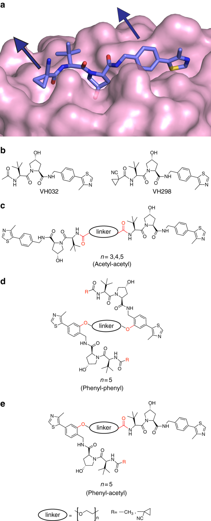
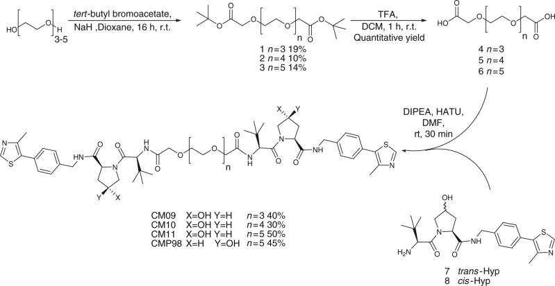
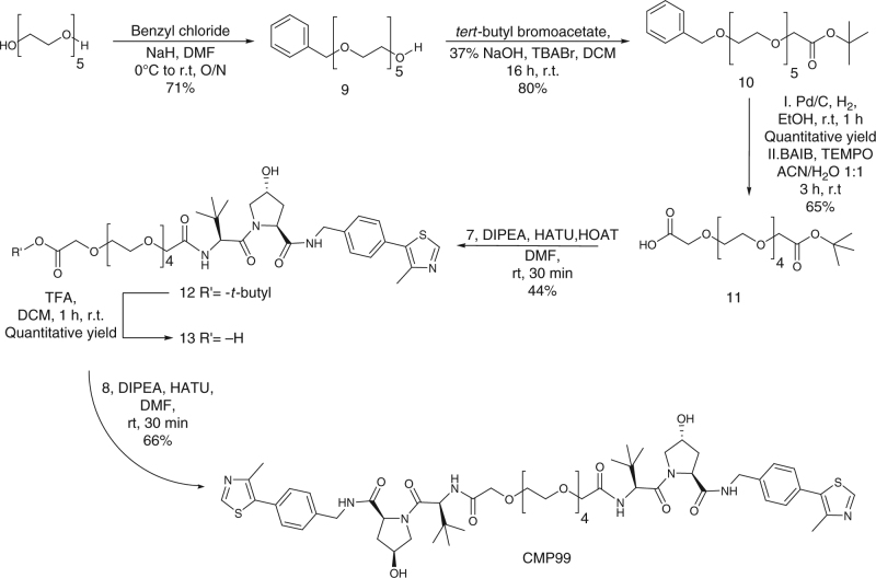
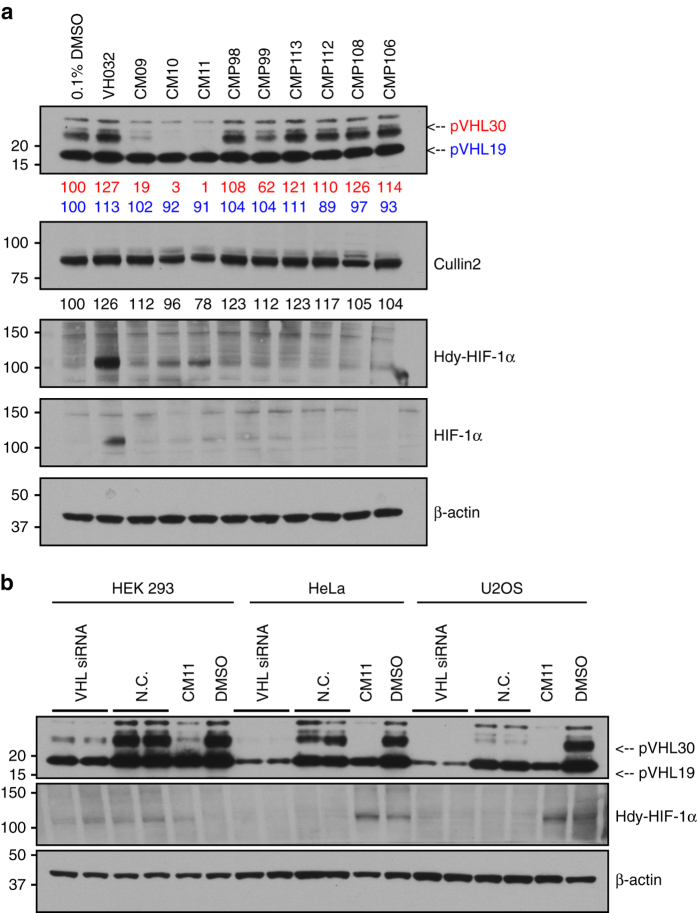
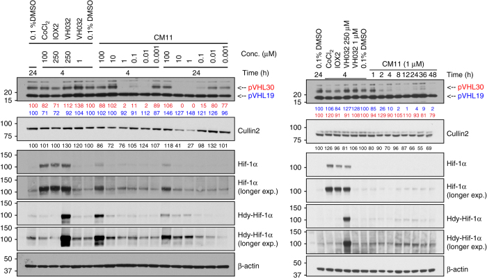
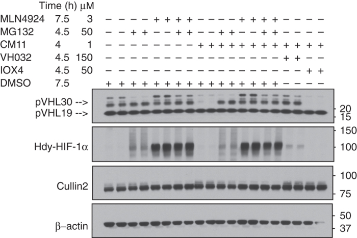
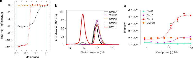
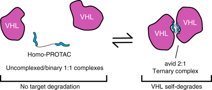
E3 ubiquitin ligases are key enzymes within the ubiquitin proteasome system which catalyze the ubiquitination of proteins, targeting them for proteasomal degradation. E3 ligases are gaining importance as targets to small molecules, both for direct inhibition and to be hijacked to induce the degradation of non-native neo-substrates using bivalent compounds known as PROTACs (for ‘proteolysis-targeting chimeras’). We describe Homo-PROTACs as an approach to dimerize an E3 ligase to trigger its suicide-type chemical knockdown inside cells. We provide proof-of-concept of Homo-PROTACs using diverse molecules composed of two instances of a ligand for the von Hippel-Lindau (VHL) E3 ligase. The most active compound, CM11, dimerizes VHL with high avidity in vitro and induces potent, rapid and proteasome-dependent self-degradation of VHL in different cell lines, in a highly isoform-selective fashion and without triggering a hypoxic response. This approach offers a novel chemical probe for selective VHL knockdown, and demonstrates the potential for a new modality of chemical intervention on E3 ligases.
Overview
The claims and statistics of this paper haven’t been checked yet.
Run the check — free, no account needed. It reads the open-access text and checks the sources it cites.
Integrity signals
Registry
Cites
Statistics
Figures
Verification
Claims the paper makes
The claims and statistics of this paper haven’t been checked yet.
Run the check — free, no account needed. It reads the open-access text and checks the sources it cites.
Claims other papers make about this one
Each is checked against this paper’s own text: does it say what the citing paper says it does?
In the citing paper
“The most active compound, CM11, specifically depleted the long isoform of VHL, VHL30, within 4 h at 10 nM concentration, providing an intriguing tool for research on isoform specific functions of VHL.”
From Advances in targeted degradation of endogenous proteins (2019) · cites this paper as [95]
Self-check
Percentages, sums and reported differences re-computed from the paper’s own numbers.
The self-check needs the paper’s full text.
Citations
Cites
The tumour suppressor protein VHL targets hypoxia-inducible factors for oxygen-dependent proteolysis
Maxwell, P. H., Wiesener, M. S., Chang, G.-W., Clifford, S. C., Vaux, E. C., Cockman, M. E., Wykoff, C. C., Pugh, C. W., Maher, E. R., & Ratcliffe, P. J. (1999) · Nature · doi:10.1038/20459 · Cited by 5,180
In Vivo Activation of the p53 Pathway by Small-Molecule Antagonists of MDM2
Vassilev, L. T., Vu, B. T., Graves, B., Carvajal, D., Podlaski, F., Filipovic, Z., Kong, N., Kammlott, U., Lukacs, C., Klein, C., et al. (2004) · Science · doi:10.1126/science.1092472 · Cited by 4,661
Komander, D., & Rape, M. (2012) · Annual Review of Biochemistry · doi:10.1146/annurev-biochem-060310-170328 · Cited by 3,679
Mechanisms Underlying Ubiquitination
Pickart, C. M. (2001) · Annual Review of Biochemistry · doi:10.1146/annurev.biochem.70.1.503 · Cited by 3,184
Identification of a Primary Target of Thalidomide Teratogenicity
Ito, T., Ando, H., Suzuki, T., Ogura, T., Hotta, K., Imamura, Y., Yamaguchi, Y., & Handa, H. (2010) · Science · doi:10.1126/science.1177319 · Cited by 2,136
An inhibitor of NEDD8-activating enzyme as a new approach to treat cancer
Soucy, T. A., Smith, P. G., Milhollen, M. A., Berger, A. J., Gavin, J. M., Adhikari, S., Brownell, J. E., Burke, K. E., Cardin, D. P., Critchley, S., et al. (2009) · Nature · doi:10.1038/nature07884 · Cited by 1,990
Lenalidomide Causes Selective Degradation of IKZF1 and IKZF3 in Multiple Myeloma Cells
Krönke, J., Udeshi, N. D., Narla, A., Grauman, P., Hurst, S. N., McConkey, M., Svinkina, T., Heckl, D., Comer, E., Li, X., et al. (2014) · Science · doi:10.1126/science.1244851 · Cited by 1,879
Phthalimide conjugation as a strategy for in vivo target protein degradation
Winter, G. E., Buckley, D. L., Paulk, J., Roberts, J. M., Souza, A., Dhe-Paganon, S., & Bradner, J. E. (2015) · Science · doi:10.1126/science.aab1433 · Cited by 1,760
Mechanism of auxin perception by the TIR1 ubiquitin ligase
Tan, X., Calderon-Villalobos, L. I. A., Sharon, M., Zheng, C., Robinson, C. V., Estelle, M., & Zheng, N. (2007) · Nature · doi:10.1038/nature05731 · Cited by 1,688
The Myeloma Drug Lenalidomide Promotes the Cereblon-Dependent Destruction of Ikaros Proteins
Lu, G., Middleton, R. E., Sun, H., Naniong, M., Ott, C. J., Mitsiades, C. S., Wong, K.-K., Bradner, J. E., & Kaelin, W. G. (2014) · Science · doi:10.1126/science.1244917 · Cited by 1,583
Research works printed but not linked: 1
- Uehara, T. et al. Selective degradation of splicing factor CAPERα by anticancer sulfonamides. Nat. Chem. Biol. 13, 675–680 (2017). · DOI · Paper page
Cited by
Sorted by citations
PROteolysis TArgeting Chimeras (PROTACs) — Past, present and future
Pettersson, M., & Crews, C. M. (2019) · Drug Discovery Today Technologies · doi:10.1016/j.ddtec.2019.01.002 · Cited by 830
PROTACs: great opportunities for academia and industry
Sun, X., Gao, H., Yang, Y., He, M., Wu, Y., Song, Y.-G., Tong, Y., & Rao, Y. (2019) · Signal Transduction and Targeted Therapy · doi:10.1038/s41392-019-0101-6 · Cited by 663
Targeted protein degradation: elements of PROTAC design
Paiva, S.-L., & Crews, C. M. (2019) · Current Opinion in Chemical Biology · doi:10.1016/j.cbpa.2019.02.022 · Cited by 598
BAF complex vulnerabilities in cancer demonstrated via structure-based PROTAC design
Farnaby, W., Koegl, M., Roy, M. J., Whitworth, C., Diers, E., Trainor, N., Zollman, D., Steurer, S., Karolyi-Oezguer, J., Riedmueller, C., et al. (2019) · Nature Chemical Biology · doi:10.1038/s41589-019-0294-6 · Cited by 562
Self-citationDifferential PROTAC substrate specificity dictated by orientation of recruited E3 ligase
Smith, B. E., Wang, S. L., Jaime-Figueroa, S., Harbin, A., Wang, J., Hamman, B. D., & Crews, C. M. (2019) · Nature Communications · doi:10.1038/s41467-018-08027-7 · Cited by 517
Metadata
- Authors
- Maniaci, C., Hughes, S. J., Testa, A., Chen, W., Lamont, D. J., Rocha, S., Alessi, D. R., Romeo, R., & Ciulli, A.
- Affiliations
- University of Dundee (GB)
- University of Messina (IT)
- Corresponding author
- Ciulli, Alessio
- ORCID
- Venue
- Nature Communications
- Published
- 2017-10-10
- Volume
- 8
- Issue
- 1
- Pages
- 830-830
- ISSN
- 2041-1723 (ISSN-L)
- PMC
- PMC5635026
- OpenAlex
- W2761240402
- Cited by
- 245
- References
- 75
- Citations by year
- 2026: 16 · 2025: 20 · 2024: 21 · 2023: 36 · 2022: 37 · 2021: 27 · 2020: 37 · 2019: 37 · 2018: 12 · 2017: 1
- FWCI
- 9.94
- Type
- journal article
- Language
- en
- Open access
- gold
- doi.org
- Locations
- Nature Communications (publishedVersion, cc-by)
- Europe PMC (PubMed Central) (publishedVersion, cc-by)
- PubMed Central (publishedVersion, cc-by)
- PubMed (publishedVersion)
- RePEc: Research Papers in Economics (submittedVersion)
- Discovery Research Portal (University of Dundee) (submittedVersion, cc-by)
- DOAJ (DOAJ: Directory of Open Access Journals) (submittedVersion)
- DOAJ (DOAJ: Directory of Open Access Journals) (submittedVersion, cc-by-sa)
- Newcastle University ePrints (Newcastle Univesity) (cc-by)
- Funders
- Wellcome Trust (100476, 100476/Z/12/Z)
- Cancer Research UK (C99667/A12918, 12918)
- Ministero dell’Istruzione, dell’Università e della Ricerca
- Medical Research Council (MC_UU_12016, MC_UU_12016/2)
- FP7 Ideas: European Research Council (311460)
- Topics
- Life Sciences › Biochemistry, Genetics and Molecular Biology › Molecular Biology › Protein Degradation and Inhibitors
- Life Sciences › Biochemistry, Genetics and Molecular Biology › Molecular Biology › Ubiquitin and proteasome pathways
- Physical Sciences › Chemistry › Organic Chemistry › Chemical Synthesis and Analysis
- Keywords
- PROTACs, VHL E3 ubiquitin ligase, ligand-induced dimerization, proteasomal degradation, isoform selectivity, ubiquitin-proteasome system, chemical probes
- MeSH terms
- Humans, Ligands, Dimerization, Ubiquitin, Ubiquitin-Protein Ligases, Proteasome Endopeptidase Complex, Von Hippel-Lindau Tumor Suppressor Protein, Small Molecule Libraries, Proteolysis
About these checks
Registry, retraction, Cites and statistics results are computed, not judged, with one exception: a printed reference that no index lists is read once by a model before it counts as a citation error. Claims are extracted by a model and kept only when their quote appears verbatim in the text; each cited source is then checked, with a model’s help, for whether it says what the claim relies on. Those verdicts can be wrong — read the claim’s quote and the cited source before relying on one.
Statistics and arithmetic are recomputed, not judged.
Confidence bands are shown only for fields calibrated on our annotated test set. None is calibrated yet, so no band is shown.
What counts as a claim ›How we measure confidence ›How we check this ›
Are you an author, or do you see a problem with this page? Request a review
