

The DOI matches its Crossref record; 61 of the 61 references it cites were found in a registry or an index.
Inner lumen proteins stabilize doublet microtubules in cilia and flagella
Open accessPublished version of a preprint
Abstract
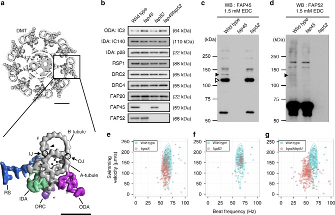
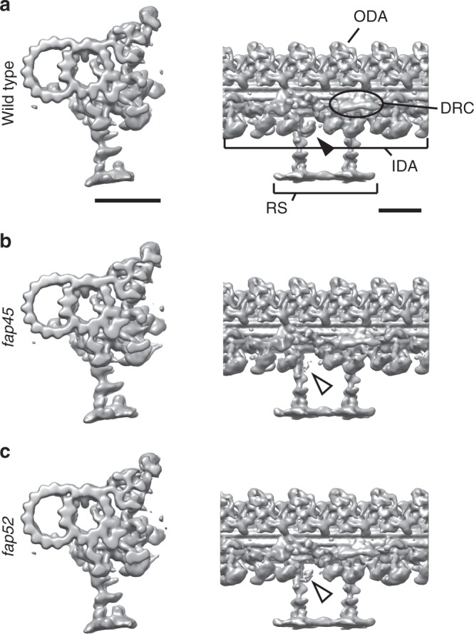
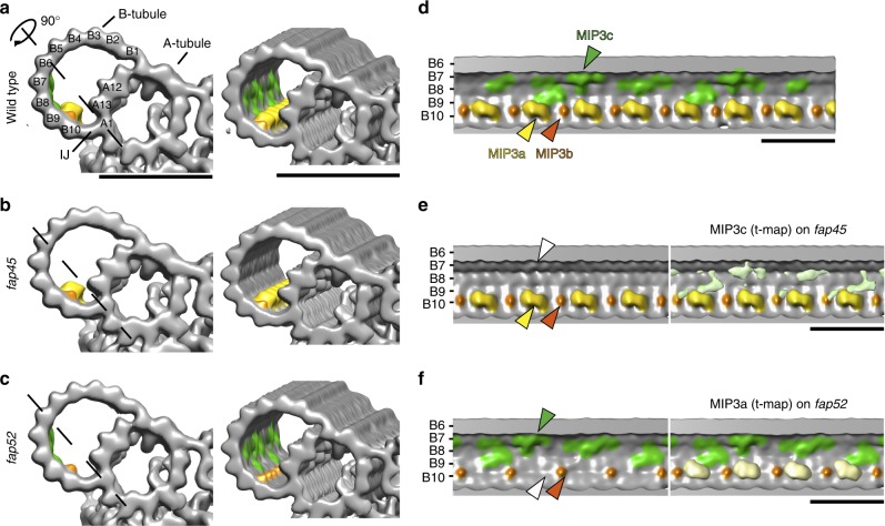
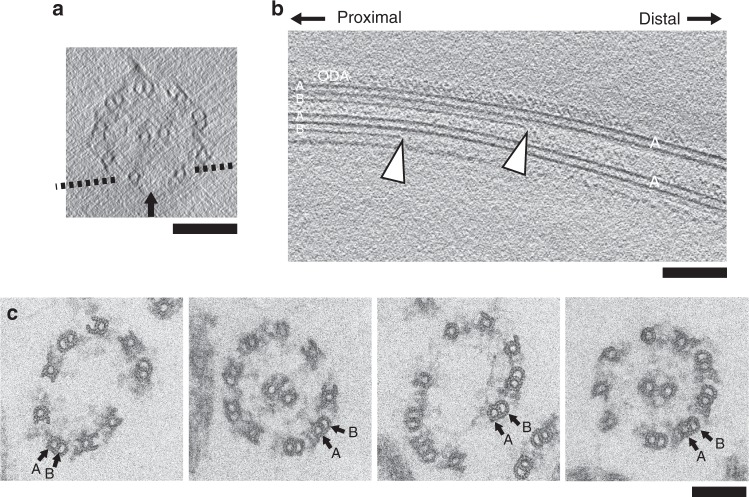
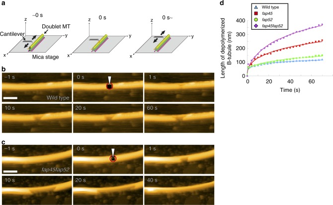
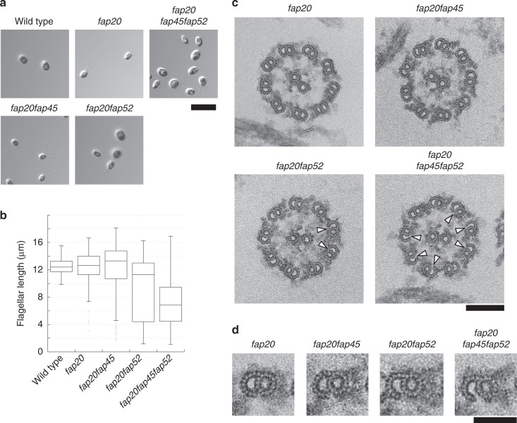
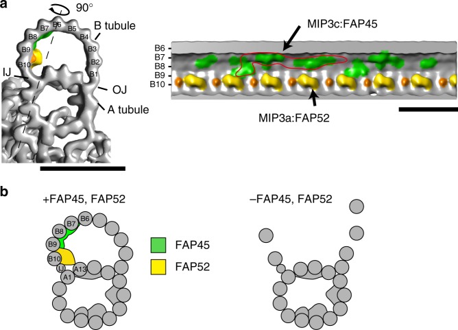
Motile cilia are microtubule-based organelles that play important roles in most eukaryotes. Although axonemal microtubules are sufficiently stable to withstand their beating motion, it remains unknown how they are stabilized while serving as tracks for axonemal dyneins. To address this question, we have identified two uncharacterized proteins, FAP45 and FAP52, as microtubule inner proteins (MIPs) in Chlamydomonas . These proteins are conserved among eukaryotes with motile cilia. Using cryo-electron tomography (cryo-ET) and high-speed atomic force microscopy (HS-AFM), we show that lack of these proteins leads to a loss of inner protrusions in B-tubules and less stable microtubules. These protrusions are located near the inner junctions of doublet microtubules and lack of both FAP52 and a known inner junction protein FAP20 results in detachment of the B-tubule from the A-tubule, as well as flagellar shortening. These results demonstrate that FAP45 and FAP52 bind to the inside of microtubules and stabilize ciliary axonemes.
Overview
The claims and statistics of this paper haven’t been checked yet.
Run the check — free, no account needed. It reads the open-access text and checks the sources it cites.
Integrity signals
Registry
Cites
Statistics
Figures
Verification
Claims the paper makes
The claims and statistics of this paper haven’t been checked yet.
Run the check — free, no account needed. It reads the open-access text and checks the sources it cites.
Claims other papers make about this one
Each is checked against this paper’s own text: does it say what the citing paper says it does?
The source backs part of the claim, not all of it — or backs it for a different population, condition or comparison.
In the citing paper
“Consistent with such a role, in Chlamydomonas or other unicellular eukaryotes, CFAP20, PACRG, CFAP45, CFAP52 and EFHC1/Rib72 are all needed to regulate ciliary beat patterns20,76–78.”
From The inner junction protein CFAP20 functions in motile and non-motile cilia and is critical for vision (2022) · cites this paper as [76]
Experimental — accuracy not yet measured
Self-check
Percentages, sums and reported differences re-computed from the paper’s own numbers.
The self-check needs the paper’s full text.
Citations
Cites
UCSF Chimera—A visualization system for exploratory research and analysis
Pettersen, E. F., Goddard, T. D., Huang, C. C., Couch, G. S., Greenblatt, D. M., Meng, E. C., & Ferrin, T. E. (2004) · Journal of Computational Chemistry · doi:10.1002/jcc.20084 · Cited by 46,047
Computer Visualization of Three-Dimensional Image Data Using IMOD
Kremer, J. R., Mastronarde, D. N., & McIntosh, J. R. (1996) · Journal of Structural Biology · doi:10.1006/jsbi.1996.0013 · Cited by 5,530
Automated electron microscope tomography using robust prediction of specimen movements
Mastronarde, D. N. (2005) · Journal of Structural Biology · doi:10.1016/j.jsb.2005.07.007 · Cited by 5,136
Hildebrandt, F., Benzing, T., & Katsanis, N. (2011) · New England Journal of Medicine · doi:10.1056/nejmra1010172 · Cited by 1,331
A high-speed atomic force microscope for studying biological macromolecules
Ando, T., Kodera, N., Takai, E., Maruyama, D., Saito, K., & Toda, A. (2001) · Proceedings of the National Academy of Sciences · doi:10.1073/pnas.211400898 · Cited by 1,127
Self-citationProteomic analysis of a eukaryotic cilium
Pazour, G. J., Agrin, N. S., Leszyk, J., & Witman, G. B. (2005) · The Journal of Cell Biology · doi:10.1083/jcb.200504008 · Cited by 1,028
The Molecular Architecture of Axonemes Revealed by Cryoelectron Tomography
Nicastro, D., Schwartz, C. L., Pierson, J., Gaudette, R., Porter, M. E., & McIntosh, J. R. (2006) · Science · doi:10.1126/science.1128618 · Cited by 881
Piperno, G., & Fuller, M. T. (1985) · The Journal of Cell Biology · doi:10.1083/jcb.101.6.2085 · Cited by 758
The Primary Cilium as a Complex Signaling Center
Berbari, N. F., O'Connor, A. K., Haycraft, C. J., & Yoder, B. K. (2009) · Current Biology · doi:10.1016/j.cub.2009.05.025 · Cited by 621
High-speed atomic force microscopy for nano-visualization of dynamic biomolecular processes
Ando, T., Uchihashi, T., & Fukuma, T. (2008) · Progress in Surface Science · doi:10.1016/j.progsurf.2008.09.001 · Cited by 561
Self-citationWitman, G. B., Carlson, K., Berliner, J. A., & Rosenbaum, J. L. (1972) · The Journal of Cell Biology · doi:10.1083/jcb.54.3.507 · Cited by 512
Microtubules acquire resistance from mechanical breakage through intralumenal acetylation
Xu, Z., Schaedel, L., Portran, D., Aguilar, A., Gaillard, J., Marinkovich, M. P., Théry, M., & Nachury, M. V. (2017) · Science · doi:10.1126/science.aai8764 · Cited by 505
High-Speed Atomic Force Microscopy Reveals Rotary Catalysis of Rotorless F1-ATPase
Uchihashi, T., Iino, R., Ando, T., & Noji, H. (2011) · Science · doi:10.1126/science.1205510 · Cited by 426
Self-citationWitman, G. B., Plummer, J., & Sander, G. (1978) · The Journal of Cell Biology · doi:10.1083/jcb.76.3.729 · Cited by 396
The dynein regulatory complex is the nexin link and a major regulatory node in cilia and flagella
Heuser, T., Raytchev, M., Krell, J., Porter, M. E., & Nicastro, D. (2009) · The Journal of Cell Biology · doi:10.1083/jcb.200908067 · Cited by 352
Direct observation of microtubule dynamics in living cells
Sammak, P. J., & Borisy, G. G. (1988) · Nature · doi:10.1038/332724a0 · Cited by 325
High-Speed AFM and Applications to Biomolecular Systems
Ando, T., Uchihashi, T., & Kodera, N. (2013) · Annual Review of Biophysics · doi:10.1146/annurev-biophys-083012-130324 · Cited by 303
Self-citationMicrotubules self-repair in response to mechanical stress
Schaedel, L., John, K., Gaillard, J., Nachury, M. V., Blanchoin, L., & Théry, M. (2015) · Nature Materials · doi:10.1038/nmat4396 · Cited by 294
Seeley, E. S., Carrière, C., Goetze, T., Longnecker, D. S., & Korc, M. (2009) · Cancer Research · doi:10.1158/0008-5472.can-08-1290 · Cited by 285
Huang, B.-W., Piperno, G., & Luck, D. (1979) · Journal of Biological Chemistry · doi:10.1016/s0021-9258(17)30186-2 · Cited by 275
Radial spoke proteins ofChlamydomonasflagella
Yang, P., Diener, D. R., Yang, C., Kohno, T., Pazour, G. J., Dienes, J. M., Agrin, N. S., King, S. M., Sale, W. S., Kamiya, R., et al. (2006) · Journal of Cell Science · doi:10.1242/jcs.02811 · Cited by 236
New features of microtubule behaviour observed in vivo
Schulze, E., & Kirschner, M. W. (1988) · Nature · doi:10.1038/334356a0 · Cited by 195
A molecular ruler determines the repeat length in eukaryotic cilia and flagella
Oda, T., Yanagisawa, H., Kamiya, R., & Kikkawa, M. (2014) · Science · doi:10.1126/science.1260214 · Cited by 179
Self-citationChlamydomonas reinhardtii hydin is a central pair protein required for flagellar motility
Lechtreck, K. F., & Witman, G. B. (2007) · The Journal of Cell Biology · doi:10.1083/jcb.200611115 · Cited by 170
Adams, G. M., Huang, B.-W., Piperno, G., & Luck, D. (1981) · The Journal of Cell Biology · doi:10.1083/jcb.91.1.69 · Cited by 164
Microtubule bending and breaking in living fibroblast cells
Odde, D. J., Ma, L., Briggs, A. H., DeMarco, A. L., & Kirschner, M. W. (1999) · Journal of Cell Science · doi:10.1242/jcs.112.19.3283 · Cited by 158
Theory of Q control in atomic force microscopy
Rodríguez, T. R., & Garcia, R. A. (2003) · Applied Physics Letters · doi:10.1063/1.1584790 · Cited by 155
Loss of primary cilia occurs early in breast cancer development
Menzl, I., LeBeau, L. G., Pandey, R., Hassounah, N. B., Li, F. W., Nagle, R. B., Weihs, K. L., & McDermott, K. M. (2014) · Cilia · doi:10.1186/2046-2530-3-7 · Cited by 153
Structural mechanism of the dynein power stroke
Lin, J., Okada, K., Raytchev, M., Smith, M. C., & Nicastro, D. (2014) · Nature Cell Biology · doi:10.1038/ncb2939 · Cited by 151
Yamano, T., Sato, E., Iguchi, H., Fukuda, Y., & Fukuzawa, H. (2015) · Proceedings of the National Academy of Sciences · doi:10.1073/pnas.1501659112 · Cited by 147
Self-citationCryo-electron tomography reveals conserved features of doublet microtubules in flagella
Nicastro, D., Fu, X., Heuser, T., Tso, A., Porter, M. E., & Linck, R. W. (2011) · Proceedings of the National Academy of Sciences · doi:10.1073/pnas.1106178108 · Cited by 147
Ledizet, M., & Piperno, G. (1995) · Molecular Biology of the Cell · doi:10.1091/mbc.6.6.697 · Cited by 140
King, S. M., Wilkerson, C. G., & Witman, G. B. (1991) · Journal of Biological Chemistry · doi:10.1016/s0021-9258(18)92989-3 · Cited by 137
Bower, R., Tritschler, D., Mills, K. V., Perrone, C. A., Mueller, J. M., Fox, L. A., Sale, W. S., & Porter, M. E. (2013) · Molecular Biology of the Cell · doi:10.1091/mbc.e12-11-0801 · Cited by 136
Mechanosignaling between central apparatus and radial spokes controls axonemal dynein activity
Oda, T., Yanagisawa, H., Yagi, T., & Kikkawa, M. (2014) · The Journal of Cell Biology · doi:10.1083/jcb.201312014 · Cited by 130
Self-citationMitchell, D. R., & Sale, W. S. (1999) · The Journal of Cell Biology · doi:10.1083/jcb.144.2.293 · Cited by 128
The Architecture of Outer Dynein Arms in Situ
Ishikawa, T., Sakakibara, H., & Oiwa, K. (2007) · Journal of Molecular Biology · doi:10.1016/j.jmb.2007.02.072 · Cited by 116
Ichikawa, M., Liu, D., Kastritis, P. L., Basu, K., Hsu, T. C., Yang, S.-K., & Bui, K. H. (2017) · Nature Communications · doi:10.1038/ncomms15035 · Cited by 115
Building Blocks of the Nexin-Dynein Regulatory Complex in Chlamydomonas Flagella
Lin, J., Tritschler, D., Song, K., Barber, C. F., Cobb, J. S., Porter, M. E., & Nicastro, D. (2011) · Journal of Biological Chemistry · doi:10.1074/jbc.m111.241760 · Cited by 102
Yanagisawa, H.-A., Mathis, G., Oda, T., Hirono, M., Richey, E. A., Ishikawa, H., Marshall, W. F., Kikkawa, M., & Qin, H. (2014) · Molecular Biology of the Cell · doi:10.1091/mbc.e13-08-0464 · Cited by 89
Self-citationPigino, G., Maheshwari, A., Bui, K. H., Shingyoji, C., Kamimura, S., & Ishikawa, T. (2012) · Journal of Structural Biology · doi:10.1016/j.jsb.2012.02.012 · Cited by 84
Wargo, M. J., Dymek, E. E., & Smith, E. F. (2005) · Journal of Cell Science · doi:10.1242/jcs.02585 · Cited by 81
Insights into the Structure and Function of Ciliary and Flagellar Doublet Microtubules
Linck, R. W., Fu, X., Lin, J., Ouch, C., Schefter, A. M., Steffen, W., Warren, P. G., & Nicastro, D. (2014) · Journal of Biological Chemistry · doi:10.1074/jbc.m114.568949 · Cited by 81
Ikeda, K., Brown, J. A., Yagi, T., Norrander, J. M., Hirono, M., Eccleston, E., Kamiya, R., & Linck, R. W. (2003) · Journal of Biological Chemistry · doi:10.1074/jbc.m210751200 · Cited by 75
A human laterality disorder associated with a homozygous WDR16 deletion
Ta-Shma, A., Perles, Z., Yaacov, B., Werner, M., Frumkin, A., Rein, A. J., & Elpeleg, O. (2015) · European Journal of Human Genetics · doi:10.1038/ejhg.2014.265 · Cited by 70
Detailed structural and biochemical characterization of the nexin-dynein regulatory complex
Oda, T., Yanagisawa, H., & Kikkawa, M. (2014) · Molecular Biology of the Cell · doi:10.1091/mbc.e14-09-1367 · Cited by 69
Self-citationIdentification of the Outer-Inner Dynein Linker as a Hub Controller for Axonemal Dynein Activities
Oda, T., Yagi, T., Yanagisawa, H., & Kikkawa, M. (2013) · Current Biology · doi:10.1016/j.cub.2013.03.028 · Cited by 62
Self-citationNovel structural labeling method using cryo-electron tomography and biotin–streptavidin system
Oda, T., & Kikkawa, M. (2013) · Journal of Structural Biology · doi:10.1016/j.jsb.2013.07.003 · Cited by 61
Self-citationChapter 24 Immunofluorescence Microscopy of Cilia and Flagella
Sanders, M. A., & Salisbury, J. L. (1995) · Methods in cell biology · doi:10.1016/s0091-679x(08)60805-5 · Cited by 59
Norrander, J. M., deCathelineau, A. M., Brown, J. A., Porter, M. E., & Linck, R. W. (2000) · Molecular Biology of the Cell · doi:10.1091/mbc.11.1.201 · Cited by 53
Tetrahymena RIB72A and RIB72B are microtubule inner proteins in the ciliary doublet microtubules
Stoddard, D., Zhao, Y., Bayless, B. A., Gui, L., Louka, P., Dave, D., Suryawanshi, S., Tomasi, R. F.-X., Dupuis-Williams, P., Baroud, C. N., et al. (2018) · Molecular Biology of the Cell · doi:10.1091/mbc.e18-06-0405 · Cited by 53
Thurston, V. C., Pena, P., Pestell, R. G., & Binder, L. I. (1997) · Cell Motility and the Cytoskeleton · doi:10.1002/(sici)1097-0169(1997)38:1 · Cited by 49
Brown, J., DiPetrillo, C. G., Smith, E. F., & Witman, G. B. (2012) · Journal of Cell Science · doi:10.1242/jcs.107151 · Cited by 46
A lumenal interrupted helix in human sperm tail microtubules
Zabeo, D., Heumann, J. M., Schwartz, C. L., Suzuki-Shinjo, A., Morgan, G., Widlund, P. O., & Höög, J. L. (2018) · Scientific Reports · doi:10.1038/s41598-018-21165-8 · Cited by 46
Respiratory and Brain Ependymal Ciliary Function
O’Callaghan, C., Sikand, K. S., & Rutman, A. (1999) · Pediatric Research · doi:10.1203/00006450-199912000-00005 · Cited by 45
Metlagel, Z., Kikkawa, Y. S., & Kikkawa, M. (2006) · Journal of Structural Biology · doi:10.1016/j.jsb.2006.07.015 · Cited by 44
Self-citationHirschner, W., Pogoda, H., Kramer, C., Thiess, U., Hamprecht, B., Wiesmüller, K., Lautner, M., & Verleysdonk, S. (2007) · Journal of Neurochemistry · doi:10.1111/j.1471-4159.2007.04500.x · Cited by 41
Flagellar-associated Protein FAP85 Is a Microtubule Inner Protein That Stabilizes Microtubules
Kirima, J., & Oiwa, K. (2017) · Cell Structure and Function · doi:10.1247/csf.17023 · Cited by 30
Molecular cloning of a novel tissue-specific gene from human nasopharyngeal epithelium
Li, Z., Yao, K., & Cao, Y. (1999) · Gene · doi:10.1016/s0378-1119(99)00234-6 · Cited by 22
High-Throughput Phenotyping of Chlamydomonas Swimming Mutants Based on Nanoscale Video Analysis
Fujita, S., Matsuo, T., Ishiura, M., & Kikkawa, M. (2014) · Biophysical Journal · doi:10.1016/j.bpj.2014.05.033 · Cited by 14
Self-citationChapter 43 Release of the Cytoskeleton and Flagellar Apparatus from Chlamydomonas
Taillon, B. E., & Jarvik, J. W. (1995) · Methods in cell biology · doi:10.1016/s0091-679x(08)60824-9 · Cited by 6
Research works printed but not linked: 1
- Harris, E. in The Chlamydomonas Sourcebook Vol. 2 (Elsevier, San Diego, CA, 2009).
Cited by
Sorted by citations
Khalifa, A. A. Z., Ichikawa, M., Dai, D., Kubo, S., Black, C. S., Peri, K., McAlear, T. S., Veyron, S., Yang, S. K., Vargas, J., et al. (2020) · eLife · doi:10.7554/elife.52760 · Cited by 1,676
Wallmeier, J., Nielsen, K. G., Kuehni, C. E., Lucas, J. S., Leigh, M. W., Zariwala, M. A., & Omran, H. (2020) · Nature Reviews Disease Primers · doi:10.1038/s41572-020-0209-6 · Cited by 403
Structure of the Decorated Ciliary Doublet Microtubule
Ma, M., Stoyanova, M., Rademacher, G., Dutcher, S. K., Brown, A., & Zhang, R. (2019) · Cell · doi:10.1016/j.cell.2019.09.030 · Cited by 290
Cryo-Electron Microscopy Methodology: Current Aspects and Future Directions
Danev, R., Yanagisawa, H., & Kikkawa, M. (2019) · Trends in Biochemical Sciences · doi:10.1016/j.tibs.2019.04.008 · Cited by 231
Self-citationThe molecular structure of mammalian primary cilia revealed by cryo-electron tomography
Kiesel, P. S., Viar, G. A., Tsoy, N., Maraspini, R., Gorilák, P., Varga, V., Honigmann, A., & Pigino, G. (2020) · Nature Structural & Molecular Biology · doi:10.1038/s41594-020-0507-4 · Cited by 183
Metadata
- Authors
- Owa, M., Uchihashi, T., Yanagisawa, H.-A., Yamano, T., Iguchi, H., Fukuzawa, H., Wakabayashi, K.-I., Ando, T., & Kikkawa, M.
- Affiliations
- The University of Tokyo (JP)
- Nagoya University (JP)
- Kyoto University (JP)
- Tokyo Institute of Technology (JP)
- Kanazawa University (JP)
- Corresponding author
- Kikkawa, Masahide
- ORCID
- Venue
- Nature Communications
- Published
- 2019-03-08
- Volume
- 10
- Issue
- 1
- Pages
- 1143-1143
- ISSN
- 2041-1723 (ISSN-L)
- PMC
- PMC6408466
- OpenAlex
- W2885457519
- Cited by
- 140
- References
- 61
- Citations by year
- 2026: 6 · 2025: 17 · 2024: 12 · 2023: 32 · 2022: 20 · 2021: 13 · 2020: 22 · 2019: 17
- FWCI
- 10.81
- Type
- journal article
- Language
- en
- Open access
- gold
- doi.org
- Locations
- Nature Communications (publishedVersion, cc-by)
- bioRxiv (Cold Spring Harbor Laboratory) (acceptedVersion)
- Europe PMC (PubMed Central) (publishedVersion, other-oa)
- PubMed Central (publishedVersion, cc-by)
- PubMed (publishedVersion)
- Institutional Repositories DataBase (IRDB) (submittedVersion, cc-by)
- RePEc: Research Papers in Economics (submittedVersion)
- DOAJ (DOAJ: Directory of Open Access Journals) (submittedVersion)
- DOAJ (DOAJ: Directory of Open Access Journals) (submittedVersion, cc-by-sa)
- Kyoto University Research Information Repository (Kyoto University) (submittedVersion, cc-by)
- Funders
- Japan Society for the Promotion of Science (17H06121, 25120714, #26119003, 18H01837, 16H04805, 15H01206, CREST, 16K07399)
- Japan Science and Technology Agency (CREST, JPMJAL1105)
- Core Research for Evolutional Science and Technology
- Advanced Low Carbon Technology Research and Development Program (JPMJAL1105)
- Topics
- Life Sciences › Biochemistry, Genetics and Molecular Biology › Genetics › Genetic and Kidney Cyst Diseases
- Life Sciences › Biochemistry, Genetics and Molecular Biology › Cell Biology › Microtubule and mitosis dynamics
- Health Sciences › Medicine › Pulmonary and Respiratory Medicine › Cystic Fibrosis Research Advances
- Keywords
- microtubule inner proteins, doublet microtubules, motile cilia, flagella, Chlamydomonas
- MeSH terms
- Cilia, Flagella, Gene Expression, Chlamydomonas reinhardtii, Microscopy, Atomic Force, Cryoelectron Microscopy, Algal Proteins, Axoneme, Electron Microscope Tomography, Axonemal Dyneins
About these checks
Registry, retraction, Cites and statistics results are computed, not judged, with one exception: a printed reference that no index lists is read once by a model before it counts as a citation error. Claims are extracted by a model and kept only when their quote appears verbatim in the text; each cited source is then checked, with a model’s help, for whether it says what the claim relies on. Those verdicts can be wrong — read the claim’s quote and the cited source before relying on one.
Statistics and arithmetic are recomputed, not judged.
Confidence bands are shown only for fields calibrated on our annotated test set. None is calibrated yet, so no band is shown.
What counts as a claim ›How we measure confidence ›How we check this ›
Are you an author, or do you see a problem with this page? Request a review
