

The DOI matches its Crossref record; 23 of the 23 references it cites were found in a registry or an index; none of the 26 sources its claims cite is checked for support yet.
Cerebrospinal fluid tracer efflux to parasagittal dura in humans
Open accessAbstract
The mechanisms behind molecular transport from cerebrospinal fluid to dural lymphatic vessels remain unknown. This study utilized magnetic resonance imaging along with cerebrospinal fluid tracer to visualize clearance pathways to human dural lymphatics in vivo. In 18 subjects with suspicion of various types of cerebrospinal fluid disorders, 3D T2-Fluid Attenuated Inversion Recovery, T1-black-blood, and T1 gradient echo acquisitions were obtained prior to intrathecal administration of the contrast agent gadobutrol (0.5 ml, 1 mmol/ml), serving as a cerebrospinal fluid tracer. Propagation of tracer was followed with T1 sequences at 3, 6, 24 and 48 h after the injection. The tracer escaped from cerebrospinal fluid into parasagittal dura along the superior sagittal sinus at areas nearby entry of cortical cerebral veins. The findings demonstrate that trans-arachnoid molecular passage does occur and suggest that parasagittal dura may serve as a bridging link between human brain and dural lymphatic vessels.
Overview
AI snapshot
Written from the paper’s full text.
TL;DR
In 18 patients with suspected cerebrospinal fluid (CSF) disorders, intrathecal gadobutrol tracer was followed by MRI for up to 48 h and escaped from CSF into parasagittal dura along the superior sagittal sinus. The findings suggest parasagittal dura may bridge molecular exchange between brain/CSF and dural lymphatic vessels in humans.
Key findings
- Parasagittal dura was identified in 18/18 patients; after intrathecal gadobutrol, normalized T1-BB signal increased in parasagittal dura at 6, 24, and 48 h, peaking at 24 h (mean percentage change from pre: 156 ± 148 at 6 h, 194 ± 188 at 24 h, 98 ± 79 at 48 h; Table 2).
- At group level, signal change was highly significant in both parasagittal dura and nearby CSF, was largest within CSF, and signal increases in CSF and parasagittal dura were highly associated (R = 0.92, P < 0.001), with strong positive correlation at all time points.
- Tracer enhancement below skull base outlets was visually detected below the hypoglossal canal in 17/18, the jugular foramen in 17/18, the cribriform plate in 2/18, and the foramen ovale in 0/18.
- Tubular structures resembling dural lymphatic vessels could not be discriminated from other enhancing parasagittal dura tissue, possibly related to the 1 × 1 × 1 mm MRI voxel resolution.
Methods
- Prospective observational study of 18 consecutive individuals referred to neurosurgery for various CSF circulation disorders; no randomization or a priori sample size calculation was used. Exclusion criteria included contrast hypersensitivity, severe allergy, renal dysfunction, age <18 or >80 years, and pregnancy/breastfeeding.
- Scans were obtained on a 3-Tesla MRI with 3D T2-FLAIR, T1-black-blood, and T1-gradient echo acquisitions; T1-weighted scans were repeated at 3, 6, 24, and 48 h after intrathecal injection of 0.5 ml gadobutrol (1 mmol/ml), preceded by iodixanol to confirm needle position.
- Regions of interest were placed in parasagittal dura and adjacent subarachnoid CSF on T1-black-blood images, normalized against the ocular bulb vitreous body; differences used linear mixed models with random intercept, correlations used Pearson coefficient, and significance was set at 0.05.
Limitations
- Stated in the paper: MRI resolution was limited to 1 mm voxel size; lack of T1-mapping techniques restricted measures to semi-quantitative tracer enrichment; the ocular bulb reference may be affected by motion artifacts; and the 48-h multi-phase protocol limited the number of scans per patient, making the peak enhancement time a rough estimate.
- Evident from the text: the cohort was small (18 patients) and clinically heterogeneous, with no healthy controls; the study could not resolve dural lymphatic vessels directly or quantify the relative importance of parasagittal versus basal outflow pathways.
Model-written summary. Check it against the paper before relying on it.
Integrity signals
Registry
Cites
Statistics
Figures
Verification
Claims the paper makes
17 Not checked yet · 12 Own interpretation · 12 Own result · 6 Own method
A result the paper reports from its own data — it cites no other source for it, so there is none to check. The quote shows it is in the paper.
In the paper
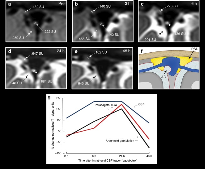
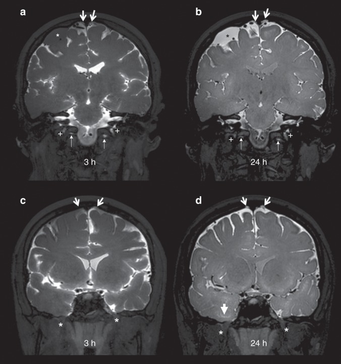
“As expected, CSF tracer also enhanced in macroscopically visible arachnoid granulations, bulging into the sagittal sinus in scattered locations”
§ Cerebrospinal fluid tracer efflux to parasagittal dura in humans > Results > Parasagittal dura
plainSingle case- Asserted: passes — The authors put it forward as true.
- Checkable: passes — It can be checked against evidence.
- Load-bearing: unclear — It is unclear whether the paper’s conclusions depend on it.
- Based: passes — The paper gives a reason why it holds.
A result the paper reports from its own data — it cites no other source for it, so there is none to check. The quote shows it is in the paper.
In the paper
“At late scans (i.e. 24 and 48 h, respectively), the remnants of tracer in CSF typically accumulated at the high cerebral convexities and in regions adjacent to parasagittal dura”
§ Cerebrospinal fluid tracer efflux to parasagittal dura in humans > Results > Parasagittal dura
plain- Asserted: passes — The authors put it forward as true.
- Checkable: passes — It can be checked against evidence.
- Load-bearing: unclear — It is unclear whether the paper’s conclusions depend on it.
- Based: passes — The paper gives a reason why it holds.
A result the paper reports from its own data — it cites no other source for it, so there is none to check. The quote shows it is in the paper.
In the paper
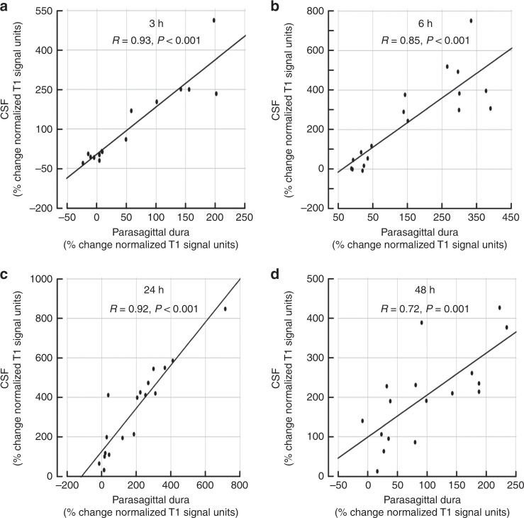
“The tracer enrichment in parasagittal dura was highly dependent on presence of tracer in adjacent CSF, which was shown by their strong positive correlation at all time points (3, 6, 24 and 48 h)”
§ Cerebrospinal fluid tracer efflux to parasagittal dura in humans > Results > Parasagittal dura
boostedcorrelational- Asserted: passes — The authors put it forward as true.
- Checkable: passes — It can be checked against evidence.
- Load-bearing: passes — The paper’s conclusions depend on it.
- Based: passes — The paper gives a reason why it holds.
A result the paper reports from its own data — it cites no other source for it, so there is none to check. The quote shows it is in the paper.
In the paper
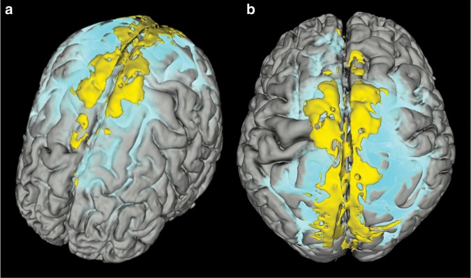
“Tissue within the parasagittal dura having features as specified above (intermediate or high signal at T2-FLAIR (not free CSF) and at T1-BB (not blood)) was visually identified almost exclusively along the middle to posterior segment of the superior sagittal sinus (Fig. 1; Supplementary Movie 2), corresponding well with the levels where cortical veins enter the sinus.”
§ Cerebrospinal fluid tracer efflux to parasagittal dura in humans > Results > Parasagittal dura
plaincorrelational- Asserted: passes — The authors put it forward as true.
- Checkable: passes — It can be checked against evidence.
- Load-bearing: passes — The paper’s conclusions depend on it.
- Based: passes — The paper gives a reason why it holds.
None of the sources cited for this claim have been checked yet.
In the paper
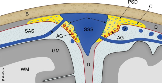
“The region with tracer enhancement, as presented here, stretches laterally to a larger extent than what can be anticipated from the previous anatomical descriptions of intradural human arachnoid granulations14,15”
§ Cerebrospinal fluid tracer efflux to parasagittal dura in humans > Discussion
What the sources say
- Not checked yetcites [14] Han, H., Tao, W. & Zhang, M. The dural entrance of cerebral bridging veins into the superior sagittal sinus: an anatomical comparison between cadavers and digital subtraction angiography. Neuroradiology 49, 169–175 (2007). source
- Not checked yetcites [15] Fox, R. J., Walji, A. H., Mielke, B., Petruk, K. C. & Aronyk, K. E. Anatomic details of intradural channels in the parasagittal dura: a possible pathway for flow of cerebrospinal fluid. Neurosurgery 39, 84–90 (1996). source
plain- Asserted: passes — The authors put it forward as true.
- Checkable: passes — It can be checked against evidence.
- Load-bearing: passes — The paper’s conclusions depend on it.
- Based: passes — The paper gives a reason why it holds.
A result the paper reports from its own data — it cites no other source for it, so there is none to check. The quote shows it is in the paper.
In the paper
“As can be noted, the signal change differed somewhat within the CSF of the subarachnoid space, arachnoid granulations and parasagittal dura, suggesting some level of molecular motion restriction between these structures.”
§ Cerebrospinal fluid tracer efflux to parasagittal dura in humans > Results > Parasagittal dura
hedgedSingle case- Asserted: passes — The authors put it forward as true.
- Checkable: passes — It can be checked against evidence.
- Load-bearing: unclear — It is unclear whether the paper’s conclusions depend on it.
- Based: passes — The paper gives a reason why it holds.
A result the paper reports from its own data — it cites no other source for it, so there is none to check. The quote shows it is in the paper.
In the paper
“The tracer escaped from cerebrospinal fluid into parasagittal dura along the superior sagittal sinus at areas nearby entry of cortical cerebral veins.”
- Also stated in: § Discussion · boosted
“In conclusion, the present study demonstrates in vivo escape of a CSF tracer from the subarachnoid compartment over the arachnoid and into the parasagittal dura along the superior sagittal sinus in humans.”
§ Cerebrospinal fluid tracer efflux to parasagittal dura in humans > Abstract
boosteddirectn = 18- Asserted: passes — The authors put it forward as true.
- Checkable: passes — It can be checked against evidence.
- Load-bearing: passes — The paper’s conclusions depend on it.
- Based: passes — The paper gives a reason why it holds.
- Also stated in: § Discussion · boosted
In the paper
“In our human cohort, tracer was present in CSF at a far longer time span (even after 2 days), and the tail of the remaining tracer bolus at late scans along parasagittal dura (Fig. 1; Supplementary Movie 1) may also indicate a net drift of CSF molecules in this direction, rather than toward the skull base.”
§ Cerebrospinal fluid tracer efflux to parasagittal dura in humans > Discussion
What the sources say
- Not checked yetcites [19] Ma, Q. et al. Rapid lymphatic efflux limits cerebrospinal fluid flow to the brain. Acta Neuropathol. 137, 151–165 (2019). source
hedged- Asserted: passes — The authors put it forward as true.
- Checkable: passes — It can be checked against evidence.
- Load-bearing: passes — The paper’s conclusions depend on it.
- Based: passes — The paper gives a reason why it holds.
A result the paper reports from its own data — it cites no other source for it, so there is none to check. The quote shows it is in the paper.
In the paper
“Tubular structures with any resemblance to dural lymphatic vessels as previously reported could not be discriminated from other enhancing tissue within parasagittal dura, and may be related to the image resolution of our MRI scans being restricted to 1 × 1 × 1 mm voxel size.”
§ Cerebrospinal fluid tracer efflux to parasagittal dura in humans > Results > Parasagittal dura
hedged- Asserted: passes — The authors put it forward as true.
- Checkable: passes — It can be checked against evidence.
- Load-bearing: unclear — It is unclear whether the paper’s conclusions depend on it.
- Based: passes — The paper gives a reason why it holds.
A conclusion the paper draws from its own results — it cites no other source for it, so there is none to check. The quote shows it is in the paper.
In the paper
“The findings demonstrate that trans-arachnoid molecular passage does occur”
- Also stated in: § Discussion · plain
“escape of a CSF tracer from the subarachnoid compartment over the arachnoid and into the parasagittal dura along the superior sagittal sinus in humans”
§ Cerebrospinal fluid tracer efflux to parasagittal dura in humans > Abstract
boosteddirectn = 18- Asserted: passes — The authors put it forward as true.
- Checkable: passes — It can be checked against evidence.
- Load-bearing: passes — The paper’s conclusions depend on it.
- Based: passes — The paper gives a reason why it holds.
- Also stated in: § Discussion · plain
- It may state two separate claims as one. 17
- Probably not a substantial claim. 16
- The quote only partly states this claim. 1
A result the paper reports from its own data — it cites no other source for it, so there is none to check. The quote shows it is in the paper.
In the paper
“Even though there were some variations with regard to prominence, tissue in parasagittal dura with features as here defined was depicted in 18/18 study patients and had the typical appearance of longitudinally interspersed areas of up to several millimeters’ transversal width”
§ Cerebrospinal fluid tracer efflux to parasagittal dura in humans > Results > Parasagittal dura
plainn = 18- Asserted: passes — The authors put it forward as true.
- Checkable: passes — It can be checked against evidence.
- Load-bearing: unclear — It is unclear whether the paper’s conclusions depend on it.
- Based: passes — The paper gives a reason why it holds.
It may state two separate claims as one.
A result the paper reports from its own data — it cites no other source for it, so there is none to check. The quote shows it is in the paper.
In the paper
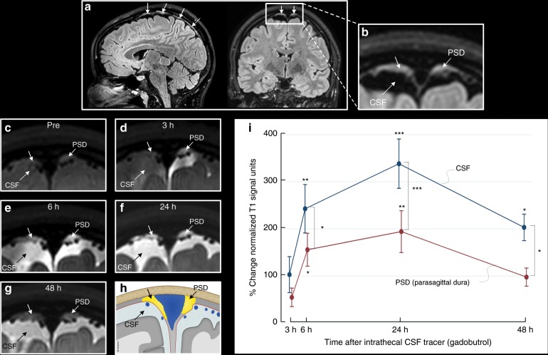
“Also note that signal change peaked at 24 h in both CSF and PSD, and signal increase in these structures was highly associated (R = 0.92, P < 0.001).”
§ Cerebrospinal fluid tracer efflux to parasagittal dura in humans > Results > Parasagittal dura
plaincorrelationaln = 18- Asserted: passes — The authors put it forward as true.
- Checkable: passes — It can be checked against evidence.
- Load-bearing: passes — The paper’s conclusions depend on it.
- Based: passes — The paper gives a reason why it holds.
It may state two separate claims as one.
A result the paper reports from its own data — it cites no other source for it, so there is none to check. The quote shows it is in the paper.
In the paper
“At group level, there was a highly significant increase in normalized T1 signal units within the parasagittal dura, though more pronounced in nearby CSF”
§ Cerebrospinal fluid tracer efflux to parasagittal dura in humans > Results > Parasagittal dura
plaindirect- Asserted: passes — The authors put it forward as true.
- Checkable: passes — It can be checked against evidence.
- Load-bearing: passes — The paper’s conclusions depend on it.
- Based: passes — The paper gives a reason why it holds.
It may state two separate claims as one.
A result the paper reports from its own data — it cites no other source for it, so there is none to check. The quote shows it is in the paper.
In the paper
“After intrathecal injection of CSF tracer, we found enrichment of parasagittal dura at 6, 24 and 48 h and with peak at 24 h”
§ Cerebrospinal fluid tracer efflux to parasagittal dura in humans > Results > Parasagittal dura
plaindirectn = 18- Asserted: passes — The authors put it forward as true.
- Checkable: passes — It can be checked against evidence.
- Load-bearing: passes — The paper’s conclusions depend on it.
- Based: passes — The paper gives a reason why it holds.
It may state two separate claims as one.
A result the paper reports from its own data — it cites no other source for it, so there is none to check. The quote shows it is in the paper.
In the paper
“For the visual assessment of tracer enhancement on T1-BB below selected cranial nerve outlets at the skull base, this was detected below the hypoglossal canal in 17/18, the jugular foramen in 17/18, the cribriform plate in 2/18, and the foramen ovale in 0/18 individuals”
§ Cerebrospinal fluid tracer efflux to parasagittal dura in humans > Results > Parasagittal dura
plainn = 18- Asserted: passes — The authors put it forward as true.
- Checkable: passes — It can be checked against evidence.
- Load-bearing: unclear — It is unclear whether the paper’s conclusions depend on it.
- Based: passes — The paper gives a reason why it holds.
It may state two separate claims as one.
None of the sources cited for this claim have been checked yet.
In the paper
“true lymphatic vessels were shown to reside within the dura mater3,4”
§ Cerebrospinal fluid tracer efflux to parasagittal dura in humans > Introduction
What the sources say
- Not checked yetcites [3] Aspelund, A. et al. A dural lymphatic vascular system that drains brain interstitial fluid and macromolecules. J. Exp. Med. 212, 991–999 (2015). source
- Not checked yetcites [4] Louveau, A. et al. Structural and functional features of central nervous system lymphatic vessels. Nature 523, 337–341 (2015). source
boosted- Asserted: passes — The authors put it forward as true.
- Checkable: passes — It can be checked against evidence.
- Load-bearing: passes — The paper’s conclusions depend on it.
- Based: passes — The paper gives a reason why it holds.
Probably not a substantial claim.
None of the sources cited for this claim have been checked yet.
In the paper
“it seems plausible that molecules within parasagittal dura structures may well reach adjacent lymphatic vessels, and perhaps also vice versa”
§ Cerebrospinal fluid tracer efflux to parasagittal dura in humans > Discussion
What the sources say
- Not checked yetcites [3] Aspelund, A. et al. A dural lymphatic vascular system that drains brain interstitial fluid and macromolecules. J. Exp. Med. 212, 991–999 (2015). source
- Not checked yetcites [4] Louveau, A. et al. Structural and functional features of central nervous system lymphatic vessels. Nature 523, 337–341 (2015). source
- Not checked yetcites [16] Mastorakos, P. & McGavern, D. The anatomy and immunology of vasculature in the central nervous system. Sci. Immunol. 4, eaav0492 (2019). source
hedgedconditional- Asserted: passes — The authors put it forward as true.
- Checkable: passes — It can be checked against evidence.
- Load-bearing: passes — The paper’s conclusions depend on it.
- Based: passes — The paper gives a reason why it holds.
It may state two separate claims as one.
In the paper
“previous findings showing that impairment of meningeal lymphatic function slows paravascular influx of macromolecules into the brain, as well as efflux from the interstitial space5, may suggest that impaired lymphatic clearance function may exert its negative effect directly on the accumulation of molecules within the CSF, and thereby further upstream on molecular exit from brain into CSF”
§ Cerebrospinal fluid tracer efflux to parasagittal dura in humans > Discussion
What the sources say
- Not checked yetcites [5] Da Mesquita, S. et al. Functional aspects of meningeal lymphatics in ageing and Alzheimer’s disease. Nature 560, 185–191 (2018). source
hedgedconditional- Asserted: passes — The authors put it forward as true.
- Checkable: passes — It can be checked against evidence.
- Load-bearing: fails — The paper’s conclusions may not depend on it.
- Based: passes — The paper gives a reason why it holds.
Probably not a substantial claim. The paper’s conclusions may not depend on it. It may state two separate claims as one.
A conclusion the paper draws from its own results — it cites no other source for it, so there is none to check. The quote shows it is in the paper.
In the paper
“which should be highly suggestive for tracer escape to dura outside arachnoid granulations and into the network of intradural channels”
§ Cerebrospinal fluid tracer efflux to parasagittal dura in humans > Discussion
hedgeddirect- Asserted: passes — The authors put it forward as true.
- Checkable: passes — It can be checked against evidence.
- Load-bearing: passes — The paper’s conclusions depend on it.
- Based: passes — The paper gives a reason why it holds.
It may state two separate claims as one.
A conclusion the paper draws from its own results — it cites no other source for it, so there is none to check. The quote shows it is in the paper.
In the paper
“Our findings compare better with previous anatomical reports demonstrating that parasagittal dura along the middle one-third of the superior sagittal sinus harbors a dense carpet of arachnoid granulations medially”
§ Cerebrospinal fluid tracer efflux to parasagittal dura in humans > Discussion
boostedcorrelational- Asserted: passes — The authors put it forward as true.
- Checkable: passes — It can be checked against evidence.
- Load-bearing: passes — The paper’s conclusions depend on it.
- Based: passes — The paper gives a reason why it holds.
It may state two separate claims as one. The quote only partly states this claim.
In the paper
“impairment of these meningeal lymphatic pathways has been shown to slow paravascular efflux of amyloid-β from interstitial brain fluid”
§ Cerebrospinal fluid tracer efflux to parasagittal dura in humans > Introduction
What the sources say
- Not checked yetcites [5] Da Mesquita, S. et al. Functional aspects of meningeal lymphatics in ageing and Alzheimer’s disease. Nature 560, 185–191 (2018). source
plaindirect- Asserted: passes — The authors put it forward as true.
- Checkable: passes — It can be checked against evidence.
- Load-bearing: passes — The paper’s conclusions depend on it.
- Based: passes — The paper gives a reason why it holds.
Probably not a substantial claim.
What the paper did — it cites no source for this, so there is none to check. The quote shows it is in the paper.
In the paper
“Moreover, the long duration of molecular clearance from human CSF, and the high demand on patients and resources associated with multi-phase MR imaging over 48 h, has limited the number of scans we were able to perform in single patients through this time span.”
§ Cerebrospinal fluid tracer efflux to parasagittal dura in humans > Discussion
plaindirect- Asserted: passes — The authors put it forward as true.
- Checkable: passes — It can be checked against evidence.
- Load-bearing: fails — The paper’s conclusions may not depend on it.
- Based: passes — The paper gives a reason why it holds.
Probably not a substantial claim. The paper’s conclusions may not depend on it. It may state two separate claims as one.
What the paper did — it cites no source for this, so there is none to check. The quote shows it is in the paper.
In the paper
“In the lack of T1-mapping techniques with image resolution even close to 1 mm, we did not have the opportunity to go beyond semi-quantitative measures of tracer enrichment.”
§ Cerebrospinal fluid tracer efflux to parasagittal dura in humans > Discussion
plaindirect- Asserted: passes — The authors put it forward as true.
- Checkable: passes — It can be checked against evidence.
- Load-bearing: fails — The paper’s conclusions may not depend on it.
- Based: passes — The paper gives a reason why it holds.
Probably not a substantial claim. The paper’s conclusions may not depend on it.
In the paper
“A recent animal study was, however, unable to reproduce passage of CSF tracer into dural lymphatic vessels7”
§ Cerebrospinal fluid tracer efflux to parasagittal dura in humans > Introduction
What the sources say
- Not checked yetcites [7] Ma, Q., Ineichen, B. V., Detmar, M. & Proulx, S. T. Outflow of cerebrospinal fluid is predominantly through lymphatic vessels and is reduced in aged mice. Nat. Commun. 8, 1434 (2017). source
plain- Asserted: passes — The authors put it forward as true.
- Checkable: passes — It can be checked against evidence.
- Load-bearing: fails — The paper’s conclusions may not depend on it.
- Based: passes — The paper gives a reason why it holds.
Probably not a substantial claim. The paper’s conclusions may not depend on it.
In the paper
“The human subarachnoid space is shown to be continuous with the paravascular compartment of all brain regions1”
§ Cerebrospinal fluid tracer efflux to parasagittal dura in humans > Introduction
What the sources say
- Not checked yetcites [1] Ringstad, G. et al. Brain-wide glymphatic enhancement and clearance in humans assessed with MRI. JCI Insight 3, 121537 (2018). source
boostedcorrelational- Asserted: passes — The authors put it forward as true.
- Checkable: passes — It can be checked against evidence.
- Load-bearing: passes — The paper’s conclusions depend on it.
- Based: passes — The paper gives a reason why it holds.
Probably not a substantial claim.
In the paper
“It has, however, been demonstrated in rats that from the total amount of MRI contrast agent injected into the cisterna magna, merely 19% enriched the brain parenchyma, the remainder escaped via other routes22.”
§ Cerebrospinal fluid tracer efflux to parasagittal dura in humans > Discussion
What the sources say
- Not checked yetcites [22] Lee, H. et al. Quantitative Gd-DOTA uptake from cerebrospinal fluid into rat brain using 3D VFA-SPGR at 9.4T. Magn. Reson. Med. 79, 1568–1578 (2018). source
boosted- Asserted: passes — The authors put it forward as true.
- Checkable: passes — It can be checked against evidence.
- Load-bearing: passes — The paper’s conclusions depend on it.
- Based: passes — The paper gives a reason why it holds.
Probably not a substantial claim.
None of the sources cited for this claim have been checked yet.
In the paper
“previous observations indicating that the arachnoid constitutes an impermeable barrier layer between the CSF and dura mater8,9”
§ Cerebrospinal fluid tracer efflux to parasagittal dura in humans > Introduction
What the sources say
- Not checked yetcites [8] Nabeshima, S., Reese, T. S., Landis, D. M. & Brightman, M. W. Junctions in the meninges and marginal glia. J. Comp. Neurol. 164, 127–169 (1975). source
- Not checked yetcites [9] Coles, J. A., Myburgh, E., Brewer, J. M. & McMenamin, P. G. Where are we? The anatomy of the murine cortical meninges revisited for intravital imaging, immunology, and clearance of waste from the brain. Prog. Neurobiol. 156, 107–148 (2017). source
hedged- Asserted: passes — The authors put it forward as true.
- Checkable: passes — It can be checked against evidence.
- Load-bearing: passes — The paper’s conclusions depend on it.
- Based: passes — The paper gives a reason why it holds.
Probably not a substantial claim.
None of the sources cited for this claim have been checked yet.
In the paper
“Our findings compare better with previous anatomical reports demonstrating that parasagittal dura along the middle one-third of the superior sagittal sinus harbors a dense carpet of arachnoid granulations medially, being in close association with an extensive network of intradural channels from 0.02 to 2.0 mm in diameter, coursing and coalescing into lateral lacunae from laterally toward the super”
§ Cerebrospinal fluid tracer efflux to parasagittal dura in humans > Discussion
What the sources say
- Not checked yetcites [13] Mack, J., Squier, W. & Eastman, J. T. Anatomy and development of the meninges: implications for subdural collections and CSF circulation. Pediatr. Radiol. 39, 200–210 (2009). source
- Not checked yetcites [14] Han, H., Tao, W. & Zhang, M. The dural entrance of cerebral bridging veins into the superior sagittal sinus: an anatomical comparison between cadavers and digital subtraction angiography. Neuroradiology 49, 169–175 (2007). source
boostedcorrelational- Asserted: passes — The authors put it forward as true.
- Checkable: passes — It can be checked against evidence.
- Load-bearing: passes — The paper’s conclusions depend on it.
- Based: passes — The paper gives a reason why it holds.
Probably not a substantial claim. It may state two separate claims as one.
A conclusion the paper draws from its own results — it cites no other source for it, so there is none to check. The quote shows it is in the paper.
In the paper
“The in vivo-based observations from the current MRI study show that parasagittal dura does not drain CSF only, but also molecules, with high level of dependence on their presence in the adjacent subarachnoid compartment.”
§ Cerebrospinal fluid tracer efflux to parasagittal dura in humans > Discussion
boostedcorrelational- Asserted: passes — The authors put it forward as true.
- Checkable: passes — It can be checked against evidence.
- Load-bearing: passes — The paper’s conclusions depend on it.
- Based: passes — The paper gives a reason why it holds.
It may state two separate claims as one.
What the paper did — it cites no source for this, so there is none to check. The quote shows it is in the paper.
In the paper
“An obvious limitation of this study is the MR image resolution of 1 mm voxel size.”
§ Cerebrospinal fluid tracer efflux to parasagittal dura in humans > Discussion
plain- Asserted: passes — The authors put it forward as true.
- Checkable: passes — It can be checked against evidence.
- Load-bearing: fails — The paper’s conclusions may not depend on it.
- Based: passes — The paper gives a reason why it holds.
Probably not a substantial claim. The paper’s conclusions may not depend on it.
A conclusion the paper draws from its own results — it cites no other source for it, so there is none to check. The quote shows it is in the paper.
In the paper
“While our study corroborates findings from animal studies by showing that CSF molecular outflow through skull base foramina occurs also in humans”
§ Cerebrospinal fluid tracer efflux to parasagittal dura in humans > Discussion
plainn = 18- Asserted: passes — The authors put it forward as true.
- Checkable: passes — It can be checked against evidence.
- Load-bearing: passes — The paper’s conclusions depend on it.
- Based: passes — The paper gives a reason why it holds.
It may state two separate claims as one.
None of the sources cited for this claim have been checked yet.
In the paper
“The extent of enhancing tissue within parasagittal dura does not resemble the previously reported appearance of true dural lymphatic vessels, which are tubular-shaped structures of sub-millimeter cross-sectional diameter near the venous sinus wall3,4,11, and also far from the classical MRI appearance of arachnoid granulations bulging into venous sinuses12.”
§ Cerebrospinal fluid tracer efflux to parasagittal dura in humans > Discussion
What the sources say
- Not checked yetcites [3] Aspelund, A. et al. A dural lymphatic vascular system that drains brain interstitial fluid and macromolecules. J. Exp. Med. 212, 991–999 (2015). source
- Not checked yetcites [4] Louveau, A. et al. Structural and functional features of central nervous system lymphatic vessels. Nature 523, 337–341 (2015). source
- Not checked yetcites [11] Absinta, M. et al. Human and nonhuman primate meninges harbor lymphatic vessels that can be visualized noninvasively by MRI. eLife 6, e29738 (2017). source
- Not checked yetcites [12] Trimble, C. R., Harnsberger, H. R., Castillo, M., Brant-Zawadzki, M. & Osborn, A. G. “Giant” arachnoid granulations just like CSF?: NOT!! AJNR Am. J. Neuroradiol. 31, 1724–1728 (2010). source
plain- Asserted: passes — The authors put it forward as true.
- Checkable: passes — It can be checked against evidence.
- Load-bearing: passes — The paper’s conclusions depend on it.
- Based: passes — The paper gives a reason why it holds.
It may state two separate claims as one.
A conclusion the paper draws from its own results — it cites no other source for it, so there is none to check. The quote shows it is in the paper.
In the paper
“may be related to the image resolution of our MRI scans being restricted to 1 × 1 × 1 mm voxel size”
§ Cerebrospinal fluid tracer efflux to parasagittal dura in humans > Results > Parasagittal dura
hedgedconditional- Asserted: passes — The authors put it forward as true.
- Checkable: passes — It can be checked against evidence.
- Load-bearing: fails — The paper’s conclusions may not depend on it.
- Based: passes — The paper gives a reason why it holds.
Probably not a substantial claim. The paper’s conclusions may not depend on it.
What the paper did — it cites no source for this, so there is none to check. The quote shows it is in the paper.
In the paper
“Therefore, the time point for peak tracer enhancement in human parasagittal dura must be considered to represent a rough estimate.”
§ Cerebrospinal fluid tracer efflux to parasagittal dura in humans > Discussion
hedged- Asserted: passes — The authors put it forward as true.
- Checkable: passes — It can be checked against evidence.
- Load-bearing: fails — The paper’s conclusions may not depend on it.
- Based: passes — The paper gives a reason why it holds.
Probably not a substantial claim. The paper’s conclusions may not depend on it.
What the paper did — it cites no source for this, so there is none to check. The quote shows it is in the paper.
In the paper
“Ideally, we could also have used an extra-cranial device as reference for normalization of signal units, as the ocular bulbs may be affected by some motion artifacts during scans of several minutes.”
§ Cerebrospinal fluid tracer efflux to parasagittal dura in humans > Discussion
hedgedconditional- Asserted: passes — The authors put it forward as true.
- Checkable: passes — It can be checked against evidence.
- Load-bearing: fails — The paper’s conclusions may not depend on it.
- Based: passes — The paper gives a reason why it holds.
Probably not a substantial claim. The paper’s conclusions may not depend on it. It may state two separate claims as one.
In the paper
“Gadobutrol is a non-ionic, highly hydrophilic molecule of relatively small size (604 Da), prevented by the intact brain–blood–barrier from leakage into CNS blood vessels when administered intrathecally10.”
§ Cerebrospinal fluid tracer efflux to parasagittal dura in humans > Introduction
What the sources say
- Not checked yetcites [10] Cheng, K. T. Gadobutrol. in Molecular Imaging and Contrast Agent Database (MICAD) (National Center for Biotechnology Information, Bethesda, USA, 2004). https://www.ncbi.nlm.nih.gov/pubmed/20641787.
plaindirect- Asserted: passes — The authors put it forward as true.
- Checkable: passes — It can be checked against evidence.
- Load-bearing: passes — The paper’s conclusions depend on it.
- Based: passes — The paper gives a reason why it holds.
It may state two separate claims as one.
In the paper
“The endothelium of intradural channels was proposed to possibly be the final barrier for the absorption of CSF to venous blood15, but passage of CSF over this cell layer has never since been confirmed in vivo.”
§ Cerebrospinal fluid tracer efflux to parasagittal dura in humans > Discussion
What the sources say
- Not checked yetcites [15] Fox, R. J., Walji, A. H., Mielke, B., Petruk, K. C. & Aronyk, K. E. Anatomic details of intradural channels in the parasagittal dura: a possible pathway for flow of cerebrospinal fluid. Neurosurgery 39, 84–90 (1996). source
hedged- Asserted: passes — The authors put it forward as true.
- Checkable: passes — It can be checked against evidence.
- Load-bearing: fails — The paper’s conclusions may not depend on it.
- Based: passes — The paper gives a reason why it holds.
Probably not a substantial claim. The paper’s conclusions may not depend on it. It may state two separate claims as one.
In the paper
“It has previously been shown that neurotoxic molecules such as amyloid-β and tau are cleared from brain tissue along paravenous pathways (the glymphatic system)20”
§ Cerebrospinal fluid tracer efflux to parasagittal dura in humans > Discussion
What the sources say
- Not checked yetcites [20] Iliff, J. J. et al. A paravascular pathway facilitates CSF flow through the brain parenchyma and the clearance of interstitial solutes, including amyloid beta. Sci. Transl. Med. 4, 147ra111 (2012). source
boosteddirect- Asserted: passes — The authors put it forward as true.
- Checkable: passes — It can be checked against evidence.
- Load-bearing: passes — The paper’s conclusions depend on it.
- Based: passes — The paper gives a reason why it holds.
Probably not a substantial claim.
The paper relies on this without support.
The olfactory system is proportionally much lesser in size in humans than in rodents.
“The olfactory system is indeed of proportionally much lesser size in humans than in rodents”
The paper relies on this without support.
The observed CSF tracer efflux in patients with suspected CSF disorders reflects normal human CSF clearance physiology.
“The study included 18 patients who were under clinical work-up for possible CSF disorders of various etiologies”
“In 18 subjects with suspicion of various types of cerebrospinal fluid disorders”
The paper relies on this without support.
T1 signal change in parasagittal dura reflects gadobutrol tracer concentration or enrichment.
“The CSF tracer enrichment was measured as change in T1-BB signal units, and was normalized against the reference (i.e. the vitreous body of the ocular bulb) to correct for any baseline shift of image grayscale between time points”
Claims other papers make about this one
Each is checked against this paper’s own text: does it say what the citing paper says it does?
In the citing paper
“Gene ontology and KEGG pathway analysis performed with upregulated genes in the absence of neuronal IL-17a signaling revealed several enriched processes involved in γ-aminobutyric acid (GABA)-benzodiazepine activity (Fig. 6f, Extended Data Fig. 6i), the prototypical target system for anxiolytic drugs15.”
From Meningeal γδ T cells regulate anxiety-like behavior via IL-17a signaling in neurons (2020) · cites this paper as [15]
Citations
Cites
Iliff, J. J., Wang, M., Liao, Y., Plogg, B. A., Peng, W., Gundersen, G. A., Benveniste, H., Vates, G. E., Deane, R., Goldman, S. A., et al. (2012) · Science Translational Medicine · doi:10.1126/scitranslmed.3003748 · Cited by 5,824
Structural and functional features of central nervous system lymphatic vessels
Louveau, A., Smirnov, I., Keyes, T. J., Eccles, J. D., Rouhani, S. J., Peske, J. D., Derecki, N. C., Castle, D., Mandell, J. W., Lee, K. S., et al. (2015) · Nature · doi:10.1038/nature14432 · Cited by 4,413
A dural lymphatic vascular system that drains brain interstitial fluid and macromolecules
Aspelund, A., Antila, S., Proulx, S. T., Karlsen, T. V., Karaman, S., Detmar, M., Wiig, H., & Alitalo, K. (2015) · Journal of Experimental Medicine · doi:10.1084/jem.20142290 · Cited by 2,198
Functional aspects of meningeal lymphatics in ageing and Alzheimer’s disease
Da Mesquita, S., Louveau, A., Vaccari, A., Smirnov, I., Cornelison, R. C., Kingsmore, K. M., Contarino, C., Onengut-Gumuscu, S., Farber, E., Raper, D., et al. (2018) · Nature · doi:10.1038/s41586-018-0368-8 · Cited by 1,445
Meningeal lymphatic vessels at the skull base drain cerebrospinal fluid
Ahn, J. H., Cho, H., Kim, J.-H., Kim, S. H., Ham, J.-S., Park, I., Suh, S. H., Hong, S. P., Song, J.-H., Hong, Y.-K., et al. (2019) · Nature · doi:10.1038/s41586-019-1419-5 · Cited by 811
Mandell, D. M., Mossa‐Basha, M., Qiao, Y., Hess, C. P., Hui, F. K., Matouk, C. C., Johnson, M. H., Daemen, M. J. A. P., Vossough, A., Edjlali, M., et al. (2016) · American Journal of Neuroradiology · doi:10.3174/ajnr.a4893 · Cited by 694
Ma, Q., Ineichen, B. V., Detmar, M., & Proulx, S. T. (2017) · Nature Communications · doi:10.1038/s41467-017-01484-6 · Cited by 690
Absinta, M., Ha, S.-K., Nair, G., Sati, P., Luciano, N. J., Palisoc, M., Louveau, A., Zaghloul, K. A., Pittaluga, S., Kipnis, J., et al. (2017) · eLife · doi:10.7554/elife.29738 · Cited by 643
Johnston, M., Zakharov, A., Papaiconomou, C., Salmasi, G., & Armstrong, D. (2004) · Cerebrospinal Fluid Research · doi:10.1186/1743-8454-1-2 · Cited by 572
Brain-wide glymphatic enhancement and clearance in humans assessed with MRI.
Ringstad, G., Valnes, L. M., Dale, A. M., Pripp, A. H., Vatnehol, S.-A. S., Emblem, K. E., Mardal, K.-A., & Eide, P. K. (2018) · JCI Insight · doi:10.1172/jci.insight.121537 · Cited by 533
Self-citationThe Meningeal Lymphatic System: A New Player in Neurophysiology
Da Mesquita, S., Fu, Z., & Kipnis, J. (2018) · Neuron · doi:10.1016/j.neuron.2018.09.022 · Cited by 479
Junctions in the meninges and marginal glia.
Nabeshima, S., Reese, T. S., Landis, D. M. D., & Brightman, M. W. (1975) · Journal of Comparative Neurology · doi:10.1002/cne.901640202 · Cited by 444
The function and structure of the cerebrospinal fluid outflow system
Pollay, M. (2010) · Cerebrospinal Fluid Research · doi:10.1186/1743-8454-7-9 · Cited by 371
The anatomy and immunology of vasculature in the central nervous system
Mastorakos, P., & McGavern, D. (2019) · Science Immunology · doi:10.1126/sciimmunol.aav0492 · Cited by 333
Eide, P. K., Vatnehol, S. A. S., Emblem, K. E., & Ringstad, G. (2018) · Scientific Reports · doi:10.1038/s41598-018-25666-4 · Cited by 299
Self-citationAnatomy and development of the meninges: implications for subdural collections and CSF circulation
Mack, J., Squier, W., & Eastman, J. T. (2009) · Pediatric Radiology · doi:10.1007/s00247-008-1084-6 · Cited by 233
Bakshi, R., Ariyaratana, S., Benedict, R. H. B., & Jacobs, L. R. (2001) · Archives of Neurology · doi:10.1001/archneur.58.5.742 · Cited by 220
Rapid lymphatic efflux limits cerebrospinal fluid flow to the brain
Ma, Q., Ries, M., Decker, Y., Müller, A., Riner, C., Bücker, A., Fassbender, K., Detmar, M., & Proulx, S. T. (2019) · Acta Neuropathologica · doi:10.1007/s00401-018-1916-x · Cited by 210
Coles, J. A., Myburgh, E., Brewer, J. M., & McMenamin, P. G. (2017) · Progress in Neurobiology · doi:10.1016/j.pneurobio.2017.05.002 · Cited by 123
Quantitative Gd‐DOTA uptake from cerebrospinal fluid into rat brain using 3D VFA‐SPGR at 9.4T
Lee, H., Mortensen, K., Sanggaard, S., Koch, P., Brunner, H., Quistorff, B., Nedergaard, M., & Benveniste, H. (2018) · Magnetic Resonance in Medicine · doi:10.1002/mrm.26779 · Cited by 110
Fox, R. J., Walji, A. H., Mielke, B., Petruk, K. C., & Aronyk, K. E. (1996) · Neurosurgery · doi:10.1097/00006123-199607000-00017 · Cited by 105
Han, H., Tao, W., & Zhang, M. (2007) · Neuroradiology · doi:10.1007/s00234-006-0175-z · Cited by 76
“Giant” Arachnoid Granulations Just Like CSF?: NOT!!
Trimble, C. R., Harnsberger, H. R., Castillo, M., Brant-Zawadzki, M., & Osborn, A. G. (2010) · American Journal of Neuroradiology · doi:10.3174/ajnr.a2157 · Cited by 63
Research works printed but not linked: 1
- Cheng, K. T. Gadobutrol. in Molecular Imaging and Contrast Agent Database (MICAD) (National Center for Biotechnology Information, Bethesda, USA, 2004). https://www.ncbi.nlm.nih.gov/pubmed/20641787.
Cited by
Sorted by citations
Functional characterization of the dural sinuses as a neuroimmune interface
Rustenhoven, J., Drieu, A., Mamuladze, T., de Lima, K. A., Dykstra, T., Wall, M., Papadopoulos, Z., Kanamori, M., Salvador, A. F., Baker, W., et al. (2021) · Cell · doi:10.1016/j.cell.2020.12.040 · Cited by 685
Rasmussen, M. K., Mestre, H., & Nedergaard, M. (2021) · Physiological Reviews · doi:10.1152/physrev.00031.2020 · Cited by 642
Neuroinflammation in Alzheimer disease
Heneka, M. T., van der Flier, W. M., Jessen, F., Hoozemanns, J., Thal, D. R., Boche, D., Brosseron, F., Teunissen, C., Zetterberg, H., Jacobs, A. H., et al. (2025) · Nature Reviews Immunology · doi:10.1038/s41577-024-01104-7 · Cited by 638
Meningeal γδ T cells regulate anxiety-like behavior via IL-17a signaling in neurons
Alves de Lima, K., Rustenhoven, J., Da Mesquita, S., Wall, M., Salvador, A. F., Smirnov, I., Martelossi Cebinelli, G., Mamuladze, T., Baker, W., Papadopoulos, Z., et al. (2020) · Nature Immunology · doi:10.1038/s41590-020-0776-4 · Cited by 416
Proulx, S. T. (2021) · Cellular and Molecular Life Sciences · doi:10.1007/s00018-020-03706-5 · Cited by 412
Metadata
- Authors
- Ringstad, G., & Eide, P. K.
- Affiliations
- Oslo University Hospital (NO)
- Corresponding author
- Eide, Per Kristian
- Venue
- Nature Communications
- Published
- 2020-01-17
- Volume
- 11
- Issue
- 1
- Pages
- 354-354
- ISSN
- 2041-1723 (ISSN-L)
- PMC
- PMC6969040
- OpenAlex
- W3000117238
- Semantic Scholar
- 4ebc1d939b7f614713ef61edcc2b62fdfbbad002
- Cited by
- 292
- References
- 23
- Citations by year
- 2026: 42 · 2025: 50 · 2024: 53 · 2023: 54 · 2022: 45 · 2021: 33 · 2020: 15
- FWCI
- 16.85
- Type
- journal article
- Language
- en
- Open access
- gold
- doi.org
- Locations
- Nature Communications (publishedVersion, cc-by)
- PubMed Central (publishedVersion, cc-by)
- PubMed (publishedVersion)
- RePEc: Research Papers in Economics (submittedVersion)
- DOAJ (DOAJ: Directory of Open Access Journals) (submittedVersion)
- DOAJ (DOAJ: Directory of Open Access Journals) (submittedVersion, cc-by-sa)
- NORA - Norwegian Open Research Archives (submittedVersion, cc-by)
- Funders
- Universitetet i Oslo
- Topics
- Health Sciences › Medicine › Clinical Neurology › Cerebrospinal fluid and hydrocephalus
- Health Sciences › Medicine › Clinical Neurology › Cerebral Venous Sinus Thrombosis
- Health Sciences › Medicine › Clinical Neurology › Neurosurgical Procedures and Complications
- Keywords
- cerebrospinal fluid, superior sagittal sinus, gadobutrol, cerebrospinal fluid clearance, cerebral veins
- MeSH terms
- Adult, Aged, Arachnoid, Biological Transport, Brain, Cerebrospinal Fluid, Contrast Media, Dura Mater, Humans, Lymphatic System, Magnetic Resonance Imaging, Middle Aged, Organometallic Compounds, Lymphatic Vessels, Young Adult
About these checks
Registry, retraction, Cites and statistics results are computed, not judged, with one exception: a printed reference that no index lists is read once by a model before it counts as a citation error. Claims are extracted by a model and kept only when their quote appears verbatim in the text; each cited source is then checked, with a model’s help, for whether it says what the claim relies on. Those verdicts can be wrong — read the claim’s quote and the cited source before relying on one.
A language model drew the claims from the paper on 2026-10-09, under version 3 of the claim definition (model deepseek-v4.1-flash, prompt extract-v3).
A second model labelled each claim on 2026-10-09 (model jev-1.13-20260917, definition revision gold-guideline-v0.2).
Statistics and arithmetic are recomputed, not judged.
Confidence bands are shown only for fields calibrated on our annotated test set. None is calibrated yet, so no band is shown.
What counts as a claim ›How we measure confidence ›How we check this ›
Are you an author, or do you see a problem with this page? Request a review
