

The DOI matches its Crossref record; 72 of the 72 references it cites were found in a registry or an index.
Naturally-occurring cholesterol analogues in lipid nanoparticles induce polymorphic shape and enhance intracellular delivery of mRNA
Open accessAbstract
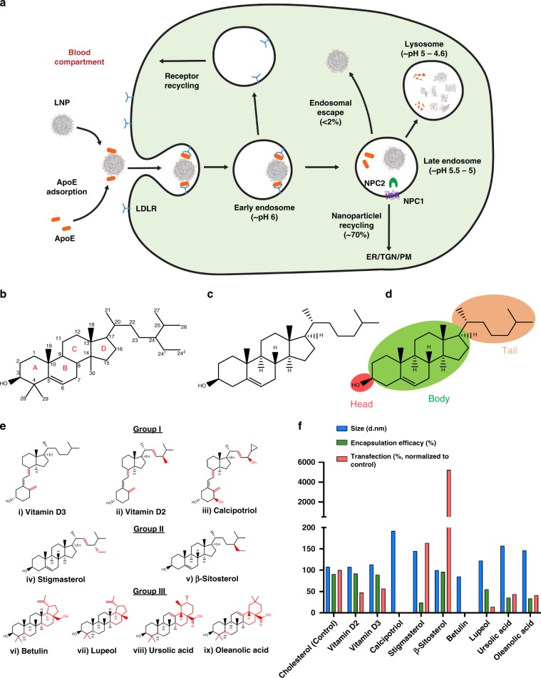
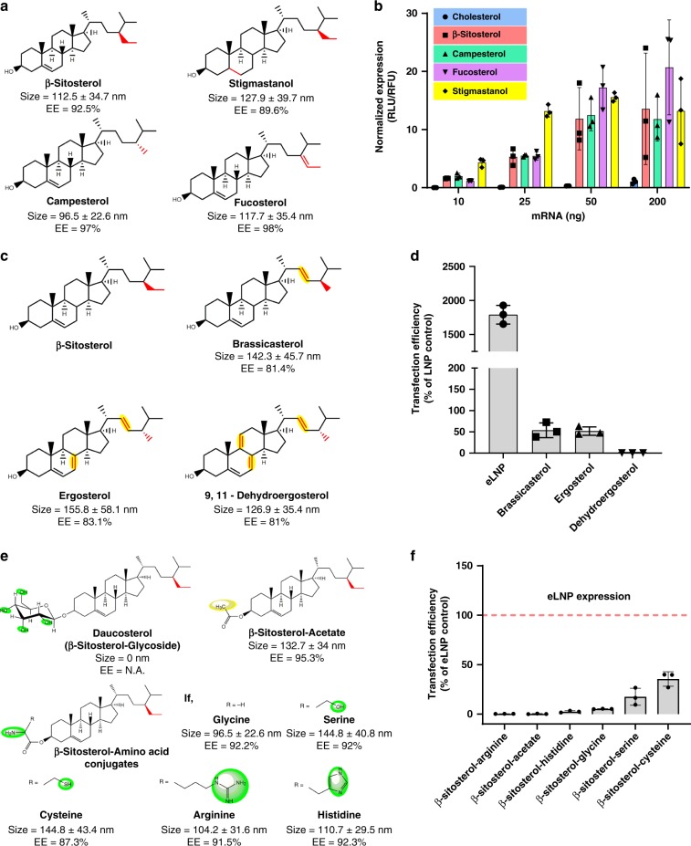
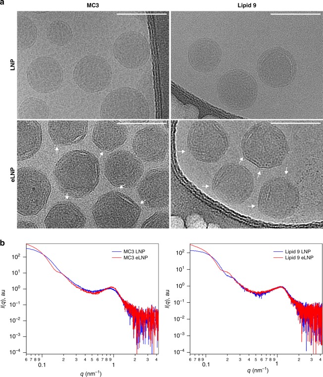
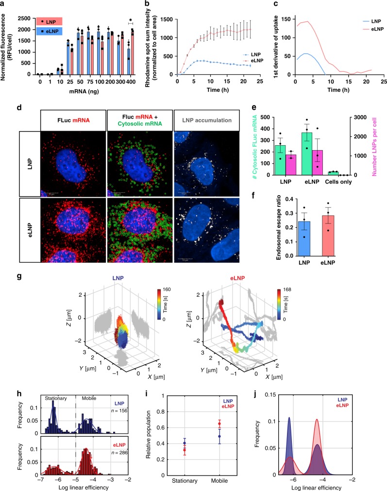
Endosomal sequestration of lipid-based nanoparticles (LNPs) remains a formidable barrier to delivery. Herein, structure-activity analysis of cholesterol analogues reveals that incorporation of C-24 alkyl phytosterols into LNPs (eLNPs) enhances gene transfection and the length of alkyl tail, flexibility of sterol ring and polarity due to -OH group is required to maintain high transfection. Cryo-TEM displays a polyhedral shape for eLNPs compared to spherical LNPs, while x-ray scattering shows little disparity in internal structure. eLNPs exhibit higher cellular uptake and retention, potentially leading to a steady release from the endosomes over time. 3D single-particle tracking shows enhanced intracellular diffusivity of eLNPs relative to LNPs, suggesting eLNP traffic to productive pathways for escape. Our findings show the importance of cholesterol in subcellular transport of LNPs carrying mRNA and emphasize the need for greater insights into surface composition and structural properties of nanoparticles, and their subcellular interactions which enable designs to improve endosomal escape.
Overview
The claims and statistics of this paper haven’t been checked yet.
Run the check — free, no account needed. It reads the open-access text and checks the sources it cites.
Integrity signals
Registry
Cites
Statistics
Figures
Verification
Claims the paper makes
The claims and statistics of this paper haven’t been checked yet.
Run the check — free, no account needed. It reads the open-access text and checks the sources it cites.
Claims other papers make about this one
Each is checked against this paper’s own text: does it say what the citing paper says it does?
The source backs part of the claim, not all of it — or backs it for a different population, condition or comparison.
In the citing paper
“Other researchers propose that the outer shell of mRNA-LNPs consists of one or multiple bilayers, based on cryo-TEM analysis (Fig. 2) (Patel et al., 2020, Tanaka et al., 2020) or SANS (Sebastiani et al., 2021).”
From mRNA-lipid nanoparticle COVID-19 vaccines: structure and stability (2021) · cites this paper as Patel et al., 2020
Experimental — accuracy not yet measuredIn the citing paper
“In mRNA-LNP formulations, such as those used in mRNA vaccines, the fraction of accessible mRNA is very low (Yanez Arteta et al., 2018, Patel et al., 2020) and thus, encapsulation efficiencies, derived from RiboGreen assays, are typically > 90%.”
From mRNA-lipid nanoparticle COVID-19 vaccines: structure and stability (2021) · cites this paper as Patel et al., 2020
Self-check
Percentages, sums and reported differences re-computed from the paper’s own numbers.
The self-check needs the paper’s full text.
Citations
Cites
MotionCor2: anisotropic correction of beam-induced motion for improved cryo-electron microscopy
Zheng, S. Q., Palovcak, E. J., Armache, J., Verba, K. A., Cheng, Y., & Agard, D. A. (2017) · Nature Methods · doi:10.1038/nmeth.4193 · Cited by 7,496
mRNA-based therapeutics--developing a new class of drugs
Sahin, U., Karikó, K., & Türeci, Ö. (2014) · Nature Reviews Drug Discovery · doi:10.1038/nrd4278 · Cited by 2,474
Gilleron, J., Querbes, W., Zeigerer, A., Borodovsky, A., Marsico, G., Schubert, U., Manygoats, K., Seifert, S., Andree, C., Stöter, M., et al. (2013) · Nature Biotechnology · doi:10.1038/nbt.2612 · Cited by 1,681
New developments in liposomal drug delivery
Pattni, B. S., Chupin, V. V., & Torchilin, V. P. (2015) · Chemical Reviews · doi:10.1021/acs.chemrev.5b00046 · Cited by 1,600
An Inverted Hexagonal Phase of Cationic Liposome-DNA Complexes Related to DNA Release and Delivery
Koltover, I., Salditt, T., Rädler, J. O., & Safinya, C. R. (1998) · Science · doi:10.1126/science.281.5373.78 · Cited by 1,336
Ebola virus entry requires the cholesterol transporter Niemann–Pick C1
Carette, J. E., Raaben, M., Wong, A., Herbert, A. S., Obernosterer, G., Mulherkar, N., Kuehne, A. I., Kranzusch, P. J., Griffin, A. M., Ruthel, G. T., et al. (2011) · Nature · doi:10.1038/nature10348 · Cited by 1,327
Maximizing the potency of siRNA lipid nanoparticles for hepatic gene silencing in vivo
Jayaraman, M., Ansell, S. M., Mui, B. L., Tam, Y. K., Chen, J., Du, X., Butler, D., Eltepu, L., Matsuda, S., Narayanannair, J. K., et al. (2012) · Angewandte Chemie International Edition · doi:10.1002/anie.201203263 · Cited by 1,267
Targeted delivery of RNAi therapeutics with endogenous and exogenous ligand-based mechanisms
Akinc, A., Querbes, W., De, S., Qin, J., Frank-Kamenetsky, M., Jayaprakash, K. N., Jayaraman, M., Rajeev, K. G., Cantley, W. L., Dorkin, J. R., et al. (2010) · Molecular Therapy · doi:10.1038/mt.2010.85 · Cited by 1,261
Moreau, R. A., Whitaker, B. D., & Hicks, K. B. (2002) · Progress in Lipid Research · doi:10.1016/s0163-7827(02)00006-1 · Cited by 1,162
Efficiency of siRNA delivery by lipid nanoparticles is limited by endocytic recycling
Sahay, G., Querbes, W., Alabi, C., Eltoukhy, A., Sarkar, S., Zurenko, C., Karagiannis, E., Love, K., Chen, D., Zoncu, R., et al. (2013) · Nature Biotechnology · doi:10.1038/nbt.2614 · Cited by 948
Sabnis, S., Kumarasinghe, E. S., Salerno, T., Mihai, C., Ketova, T., Senn, J. J., Lynn, A., Bulychev, A., McFadyen, I., Chan, J., et al. (2018) · Molecular Therapy · doi:10.1016/j.ymthe.2018.03.010 · Cited by 820
Tools for translation: non-viral materials for therapeutic mRNA delivery
Hajj, K. A., & Whitehead, K. A. (2017) · Nature Reviews Materials · doi:10.1038/natrevmats.2017.56 · Cited by 799
Kauffman, K. J., Dorkin, J. R., Yang, J. H., Heartlein, M. W., DeRosa, F., Mir, F. F., Fenton, O. S., & Anderson, D. G. (2015) · Nano Letters · doi:10.1021/acs.nanolett.5b02497 · Cited by 796
Kulkarni, J. A., Darjuan, M. M., Mercer, J. E., Chen, S., van der Meel, R., Thewalt, J. L., Tam, Y. Y. C., & Cullis, P. R. (2018) · ACS Nano · doi:10.1021/acsnano.8b01516 · Cited by 663
Biosynthesis of Cholesterol and Other Sterols
Nes, W. D. (2011) · Chemical Reviews · doi:10.1021/cr200021m · Cited by 659
P HYTOSTEROLS IN H UMAN N UTRITION
Ostlund, R. E. (2002) · Annual Review of Nutrition · doi:10.1146/annurev.nutr.22.020702.075220 · Cited by 645
Lysosomal cholesterol activates mTORC1 via an SLC38A9–Niemann-Pick C1 signaling complex
Castellano, B. M., Thelen, A. M., Moldavski, O., Feltes, M., van der Welle, R. E. N., Mydock-McGrane, L., Jiang, X., van Eijkeren, R. J., Davis, O. B., Louie, S. M., et al. (2017) · Science · doi:10.1126/science.aag1417 · Cited by 562
Yanez Arteta, M., Kjellman, T., Bartesaghi, S., Wallin, S., Wu, X., Kvist, A. J., Dabkowska, A., Székely, N., Radulescu, A., Bergenholtz, J., et al. (2018) · Proceedings of the National Academy of Sciences · doi:10.1073/pnas.1720542115 · Cited by 534
Dufourc, É. J. (2008) · Journal of Chemical Biology · doi:10.1007/s12154-008-0010-6 · Cited by 485
Bidirectional cargo transport: moving beyond tug of war
Hancock, W. O. (2014) · Nature Reviews Molecular Cell Biology · doi:10.1038/nrm3853 · Cited by 447
Brief update on endocytosis of nanomedicines
Patel, S., Kim, J., Herrera‐Barrera, M., Mukherjee, A., Kabanov, A. V., & Sahay, G. (2019) · Advanced Drug Delivery Reviews · doi:10.1016/j.addr.2019.08.004 · Cited by 409
Nucleic Acid Delivery: The Missing Pieces of the Puzzle?
Nguyen, J., & Szoka, F. C. (2012) · Accounts of Chemical Research · doi:10.1021/ar3000162 · Cited by 356
Leung, A. K. K., Hafez, I. M., Baoukina, S., Belliveau, N. M., Zhigaltsev, I. V., Afshinmanesh, E., Tieleman, D. P., Hansen, C., Hope, M. J., & Cullis, P. (2012) · The journal of physical chemistry. C, Nanomaterials and interfaces · doi:10.1021/jp303267y · Cited by 339
Gong, X., Qian, H., Zhou, X., Wu, J., Wan, T., Cao, P., Huang, W., Zhao, X., Wang, X., Wang, P., et al. (2016) · Cell · doi:10.1016/j.cell.2016.05.022 · Cited by 314
The Structural Role of Cholesterol in Cell Membranes: From Condensed Bilayers to Lipid Rafts
Krause, M. R., & Regen, S. L. (2014) · Accounts of Chemical Research · doi:10.1021/ar500260t · Cited by 288
An Orthogonal Array Optimization of Lipid-like Nanoparticles for mRNA Delivery in Vivo
Li, B., Luo, X., Deng, B., Wang, J., McComb, D. W., Shi, Y., Gaensler, K. M. L., Tan, X., Dunn, A. L., Kerlin, B. A., et al. (2015) · Nano Letters · doi:10.1021/acs.nanolett.5b03528 · Cited by 261
Regulation of cholesterol homeostasis
Goedeke, L., & Fernandez-Hernando, C. F. (2011) · Cellular and Molecular Life Sciences · doi:10.1007/s00018-011-0857-5 · Cited by 232
Xu, S., Benoff, B., Liou, H.-L., Lobel, P., & Stock, A. (2007) · Journal of Biological Chemistry · doi:10.1074/jbc.m703848200 · Cited by 221
Cheng, Q., Wei, T., Jia, Y., Farbiak, L., Zhou, K., Zhang, S., Wei, Y., Zhu, H., & Siegwart, D. J. (2018) · Advanced Materials · doi:10.1002/adma.201805308 · Cited by 221
Plant sterols and cardiovascular disease: a systematic review and meta-analysis†
Genser, B., Silbernagel, G., De Backer, G., Bruckert, E., Carmena, R., Chapman, M. J., Deanfield, J., Descamps, O. S., Rietzschel, E. R., Dias, K. C., et al. (2012) · European Heart Journal · doi:10.1093/eurheartj/ehr441 · Cited by 209
Paunovska, K., Da Silva Sanchez, A. J., Sago, C. D., Gan, Z., Lokugamage, M. P., Islam, F. Z., Kalathoor, S., Krupczak, B. R., & Dahlman, J. E. (2019) · Advanced Materials · doi:10.1002/adma.201807748 · Cited by 205
Viger-Gravel, J., Schantz, A., Pinon, A. C., Rossini, A. J., Schantz, S., & Emsley, L. (2018) · The Journal of Physical Chemistry B · doi:10.1021/acs.jpcb.7b10795 · Cited by 202
High-speed varifocal imaging with a tunable acoustic gradient index of refraction lens
Mermillod‐Blondin, A., McLeod, E. J., & Arnold, C. B. (2008) · Optics Letters · doi:10.1364/ol.33.002146 · Cited by 193
Membrane composition and dynamics: A target of bioactive virgin olive oil constituents
López, S., Bermúdez, B., la Paz, S. M., Jaramillo, S., Varela, L. M., Ortega‐Gómez, A., Abia, R., & Muriana, F. J. G. (2014) · Biochimica et Biophysica Acta (BBA) - Biomembranes · doi:10.1016/j.bbamem.2014.01.007 · Cited by 175
Li, X., Saha, P., Li, J., Blobel, G., & Pfeffer, S. R. (2016) · Proceedings of the National Academy of Sciences · doi:10.1073/pnas.1611956113 · Cited by 170
Emerging roles of the intestine in control of cholesterol metabolism
Kruit, J. K. (2006) · World Journal of Gastroenterology · doi:10.3748/wjg.v12.i40.6429 · Cited by 165
Paunovska, K., Gil, C. J., Lokugamage, M. P., Sago, C. D., Sato, M., Lando, G. N., Gamboa Castro, M., Bryksin, A. V., & Dahlman, J. E. (2018) · ACS Nano · doi:10.1021/acsnano.8b03640 · Cited by 160
Differential Modulation of Membrane Structure and Fluctuations by Plant Sterols and Cholesterol
Hodžić, A., Rappolt, M., Amenitsch, H. W., Laggner, P., & Pabst, G. (2008) · Biophysical Journal · doi:10.1529/biophysj.107.123224 · Cited by 151
Halling, K. K., & Slotte, J. P. (2004) · Biochimica et Biophysica Acta (BBA) - Biomembranes · doi:10.1016/j.bbamem.2004.05.006 · Cited by 150
FDA approves patisiran to treat hereditary transthyretin amyloidosis
Wood, H. (2018) · Nature Reviews Neurology · doi:10.1038/s41582-018-0065-0 · Cited by 145
Transfection efficiency boost of cholesterol-containing lipoplexes
Pozzi, D., Marchini, C., Cardarelli, F., Amenitsch, H. W., Garulli, C., Bifone, A., & Caracciolo, G. (2012) · Biochimica et Biophysica Acta (BBA) - Biomembranes · doi:10.1016/j.bbamem.2012.05.017 · Cited by 141
Wagner, T., Kröll, A., Haramagatti, C. R., Lipinski, H., & Wiemann, M. (2017) · PLoS ONE · doi:10.1371/journal.pone.0170165 · Cited by 136
Bernsdorff, C., & Winter, R. (2003) · The Journal of Physical Chemistry B · doi:10.1021/jp034922a · Cited by 132
Cubic Phases in Phosphatidylcholine-Cholesterol Mixtures: Cholesterol as Membrane “Fusogen”
Tenchov, B., MacDonald, R. C., & Siegel, D. P. (2006) · Biophysical Journal · doi:10.1529/biophysj.106.083766 · Cited by 130
Mochizuki, S., Kanegae, N., Nishina, K., Kamikawa, Y., Koiwai, K., Masunaga, H., & Sakurai, K. (2013) · Biochimica et Biophysica Acta · doi:10.1016/j.bbamem.2012.10.017 · Cited by 129
Andersson, M. S., Hammarstroem, L., & Edwards, K. (1995) · The Journal of Physical Chemistry · doi:10.1021/j100039a047 · Cited by 108
Disc formation in cholesterol-free liposomes during phase transition
Ickenstein, L. M., Arfvidsson, M. C., Needham, D., Mayer, L. D., & Edwards, K. (2003) · Biochimica et Biophysica Acta (BBA) - Biomembranes · doi:10.1016/s0005-2736(03)00196-2 · Cited by 102
NPC1/NPC2 function as a tag team duo to mobilize cholesterol
Subramanian, K., & Balch, W. E. (2008) · Proceedings of the National Academy of Sciences · doi:10.1073/pnas.0808256105 · Cited by 97
Cholesterol Is Required for Endocytosis and Endosomal Escape of Adenovirus Type 2
Imelli, N., Meier, O. C., Boucke, K., Hemmi, S., & Greber, U. F. (2004) · Journal of Virology · doi:10.1128/jvi.78.6.3089-3098.2004 · Cited by 96
Biosynthesis of marine sterol side chains
Giner, J. L. (1993) · Chemical Reviews · doi:10.1021/cr00021a004 · Cited by 80
Polymorphism of DNA–anionic liposome complexes reveals hierarchy of ion-mediated interactions
Liang, H., Harries, D., & Wong, G. C. L. (2005) · Proceedings of the National Academy of Sciences · doi:10.1073/pnas.0502416102 · Cited by 79
LDL–cholesterol transport to the endoplasmic reticulum
Pfisterer, S. G., Peränen, J., & Ikonen, E. (2016) · Current Opinion in Lipidology · doi:10.1097/mol.0000000000000292 · Cited by 77
Cationic phospholipids: structure–transfection activity relationships
Koynova, R. D., & Tenchov, B. G. (2009) · Soft Matter · doi:10.1039/b902027f · Cited by 74
Gindy, M. E., DiFelice, K., Kumar, V., Prud’homme, R. K., Celano, R., Haas, R. M., Smith, J. S., & Boardman, D. (2014) · Langmuir · doi:10.1021/la500630h · Cited by 70
Su, Y., Li, Q., Chen, L., & Yu, Z. (2006) · Colloids and Surfaces A Physicochemical and Engineering Aspects · doi:10.1016/j.colsurfa.2006.07.016 · Cited by 67
Lai, S. K., Hida, K., Chen, C., & Hanes, J. S. (2007) · Journal of Controlled Release · doi:10.1016/j.jconrel.2007.10.015 · Cited by 67
Plant pentacyclic triterpenic acids as modulators of lipid membrane physical properties
Prades, J., Vögler, O., Alemany, R., Gómez‐Florit, M., Funari, S. S., Ruíz-Gutiérrez, V., & Barceló, F. (2010) · Biochimica et Biophysica Acta (BBA) - Biomembranes · doi:10.1016/j.bbamem.2010.12.007 · Cited by 65
Investigation of charge ratio variation in mRNA – DEAE-dextran polyplex delivery systems
Siewert, C., Haas, H., Nawroth, T., Ziller, A., Nogueira, S. S., Schroer, M. A., Blanchet, C. E., Svergun, D. I., Rădulescu, A., Bates, F., et al. (2018) · Biomaterials · doi:10.1016/j.biomaterials.2018.10.020 · Cited by 65
Modulation of signal transduction in cancer cells by phytosterols
Bradford, P. G., & Awad, A. B. (2010) · BioFactors · doi:10.1002/biof.97 · Cited by 62
Chen, C., & Tripp, C. P. (2012) · Biochimica et Biophysica Acta (BBA) - Biomembranes · doi:10.1016/j.bbamem.2012.03.009 · Cited by 57
Zidovska, A., Evans, H. M., Ahmad, A. K., Ewert, K. K., & Safinya, C. R. (2009) · The Journal of Physical Chemistry B · doi:10.1021/jp809000e · Cited by 54
Robust real-time 3D single-particle tracking using a dynamically moving laser spot
Hou, S., Lang, X., & Welsher, K. (2017) · Optics Letters · doi:10.1364/ol.42.002390 · Cited by 52
Benesch, M. G. K., & McElhaney, R. N. (2014) · Biochimica et Biophysica Acta (BBA) - Biomembranes · doi:10.1016/j.bbamem.2014.03.019 · Cited by 37
Ashwanikumar, N., Kumar, N. A., Nair, S. A., & Kumar, G. S. V. (2014) · Acta Biomaterialia · doi:10.1016/j.actbio.2014.07.032 · Cited by 32
Imam, Z. I., Kenyon, L. E., Ashby, G., Nagib, F., Mendicino, M., Zhao, C., Gadok, A. K., & Stachowiak, J. C. (2017) · Cellular and Molecular Bioengineering · doi:10.1007/s12195-017-0489-4 · Cited by 31
Koynova, R. D., Wang, L., & MacDonald, R. C. (2007) · The Journal of Physical Chemistry B · doi:10.1021/jp071286y · Cited by 27
Side-chain structure is critical for the transport of sterols from lysosomes to cytoplasm
Sato, Y., Nishikawa, K., Aikawa, K., Mimura, K., Murakami‐Murofushi, K., Arai, H., & Inoue, K. (1995) · Biochimica et Biophysica Acta (BBA) - Lipids and Lipid Metabolism · doi:10.1016/0005-2760(95)00053-f · Cited by 25
Imam, Z. I., Kenyon, L. E., Ashby, G., Nagib, F., Mendicino, M., Zhao, C., Gadok, A. K., & Stachowiak, J. C. (2017) · Cellular and Molecular Bioengineering · doi:10.1007/s12195-017-0491-x · Cited by 18
Bangham, A. D. (1994) · Biophysical Journal · doi:10.1016/s0006-3495(94)80607-1 · Cited by 14
A Protocol for Real-time 3D Single Particle Tracking
Hou, S., & Welsher, K. (2018) · Journal of Visualized Experiments · doi:10.3791/56711 · Cited by 12
Popov, A. M. (2003) · Journal of Evolutionary Biochemistry and Physiology · doi:10.1023/a:1026147925642 · Cited by 9
Popov, A. M. (2004) · PubMed · Cited by 8
Cited by
Sorted by citations
Lipid nanoparticles for mRNA delivery
Hou, X., Zaks, T., Langer, R., & Dong, Y. (2021) · Nature Reviews Materials · doi:10.1038/s41578-021-00358-0 · Cited by 4,076
mRNA-lipid nanoparticle COVID-19 vaccines: structure and stability
Schoenmaker, L., Witzigmann, D., Kulkarni, J. A., Verbeke, R., Kersten, G., Jiskoot, W., & Crommelin, D. J. A. (2021) · International Journal of Pharmaceutics · doi:10.1016/j.ijpharm.2021.120586 · Cited by 1,566
mRNA vaccines for infectious diseases: principles, delivery and clinical translation
Chaudhary, N., Weissman, D., & Whitehead, K. A. (2021) · Nature Reviews Drug Discovery · doi:10.1038/s41573-021-00283-5 · Cited by 1,555
Chemistry of Lipid Nanoparticles for RNA Delivery
Eygeris, Y., Gupta, M., Kim, J., & Sahay, G. (2021) · Accounts of Chemical Research · doi:10.1021/acs.accounts.1c00544 · Cited by 917
Dilliard, S. A., & Siegwart, D. J. (2023) · Nature Reviews Materials · doi:10.1038/s41578-022-00529-7 · Cited by 744
Metadata
- Authors
- Patel, S., Ashwanikumar, N., Robinson, E., Xia, Y., Mihai, C., Griffith, J. P., Hou, S., Esposito, A. A., Ketova, T., Welsher, K., Joyal, J. L., Almarsson, Ö., & Sahay, G.
- Affiliations
- Oregon State University (US)
- Moderna Therapeutics (United States) (US)
- Duke University (US)
- Corresponding author
- Sahay, Gaurav
- ORCID
- Venue
- Nature Communications
- Published
- 2020-02-20
- Volume
- 11
- Issue
- 1
- Pages
- 983-983
- ISSN
- 2041-1723 (ISSN-L)
- PMC
- PMC7033178
- OpenAlex
- W3006764150
- Cited by
- 601
- References
- 72
- Citations by year
- 2026: 108 · 2025: 156 · 2024: 127 · 2023: 72 · 2022: 64 · 2021: 50 · 2020: 23
- FWCI
- 16.94
- Type
- journal article
- Language
- en
- Update notices
- correction (2020-07-06)
- Open access
- gold
- doi.org
- Locations
- Nature Communications (publishedVersion, cc-by)
- Europe PMC (PubMed Central) (publishedVersion, cc-by)
- PubMed Central (publishedVersion, cc-by)
- PubMed (publishedVersion)
- RePEc: Research Papers in Economics (submittedVersion)
- DOAJ (DOAJ: Directory of Open Access Journals) (submittedVersion, cc-by-sa)
- Funders
- U.S. Department of Health & Human Services | NIH | National Institute of Biomedical Imaging and Bioengineering (1R15EB021581)
- U.S. Department of Health & Human Services | NIH | National Heart, Lung, and Blood Institute (1R01HL146736-01)
- Cystic Fibrosis Foundation (SAHAY18G0)
- Topics
- Life Sciences › Biochemistry, Genetics and Molecular Biology › Molecular Biology › RNA Interference and Gene Delivery
- Physical Sciences › Materials Science › Biomaterials › Nanoparticle-Based Drug Delivery
- Life Sciences › Pharmacology, Toxicology and Pharmaceutics › Pharmaceutical Science › Advancements in Transdermal Drug Delivery
- Keywords
- cholesterol analogues, lipid nanoparticles, mRNA delivery, endosomal escape, nanoparticle morphology, transfection, cellular uptake, intracellular diffusion, structure-activity relationship
- MeSH terms
- RAW 264.7 Cells, Animals, Biological Transport, Active, Cell Line, Cholesterol, HeLa Cells, Humans, Lipids, Endosomes, RNA, Messenger, Sitosterols, Transfection, X-Ray Diffraction, Cryoelectron Microscopy, Mice, Nanoparticles, HEK293 Cells
About these checks
Registry, retraction, Cites and statistics results are computed, not judged, with one exception: a printed reference that no index lists is read once by a model before it counts as a citation error. Claims are extracted by a model and kept only when their quote appears verbatim in the text; each cited source is then checked, with a model’s help, for whether it says what the claim relies on. Those verdicts can be wrong — read the claim’s quote and the cited source before relying on one.
Statistics and arithmetic are recomputed, not judged.
Confidence bands are shown only for fields calibrated on our annotated test set. None is calibrated yet, so no band is shown.
What counts as a claim ›How we measure confidence ›How we check this ›
Are you an author, or do you see a problem with this page? Request a review
