

The DOI matches its Crossref record; 92 of the 92 references it cites were found in a registry or an index.
Acetylated tau inhibits chaperone-mediated autophagy and promotes tau pathology propagation in mice
Open accessAbstract
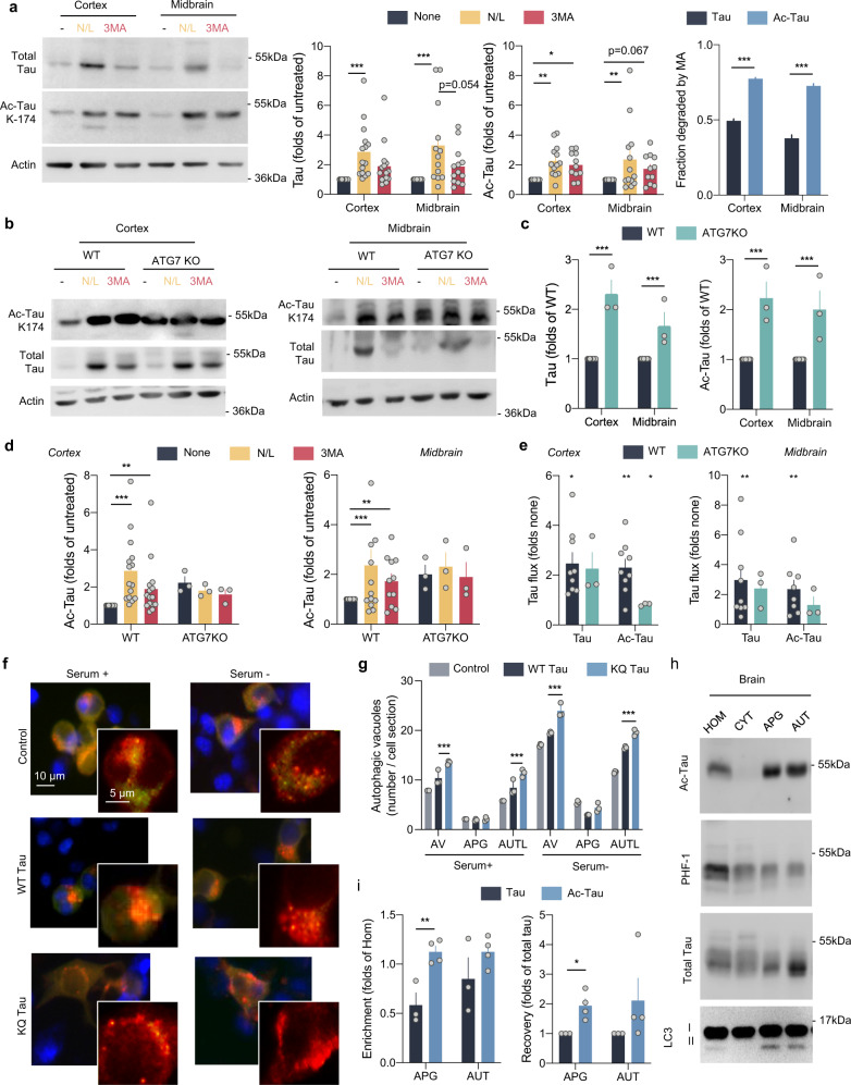
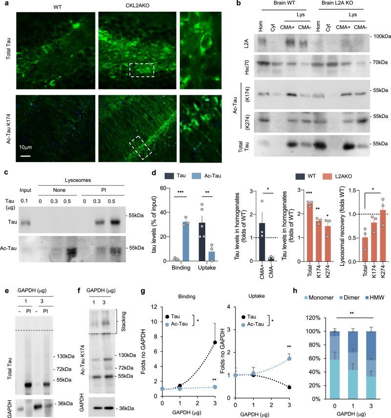
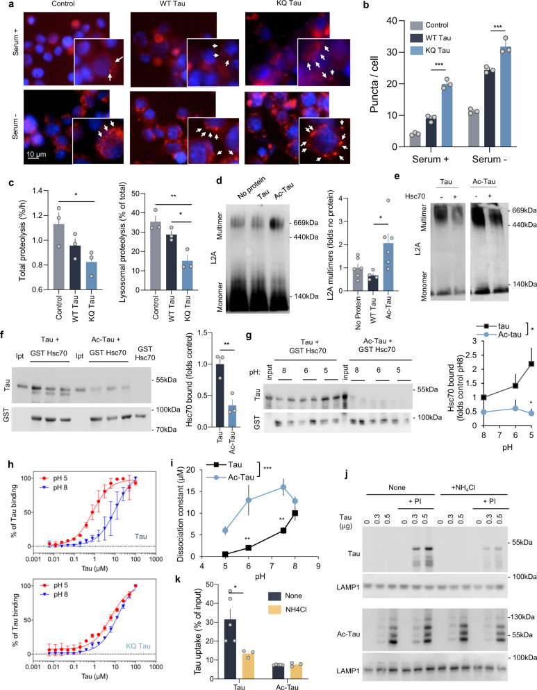
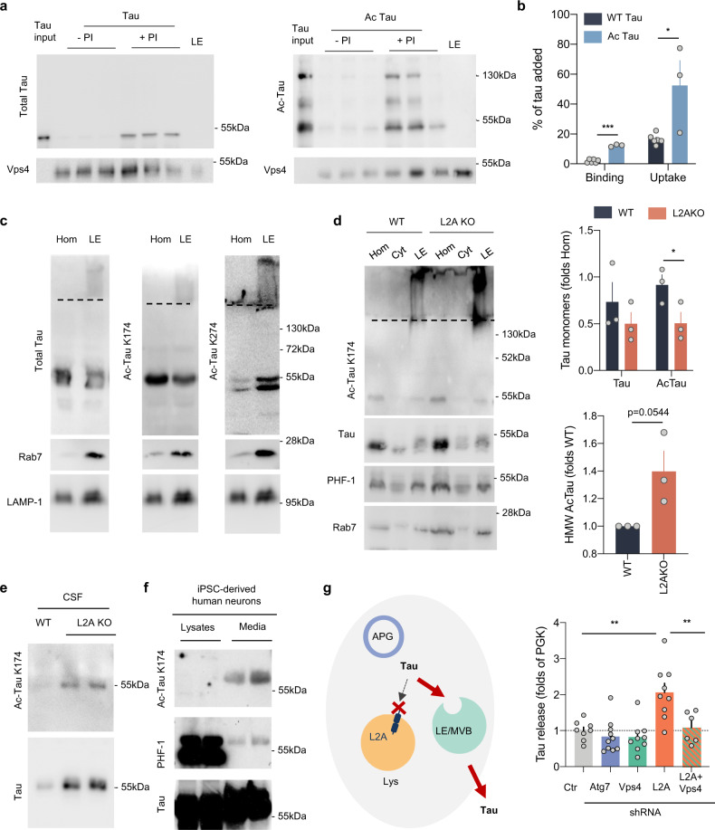
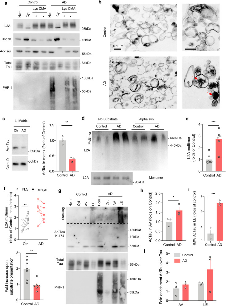
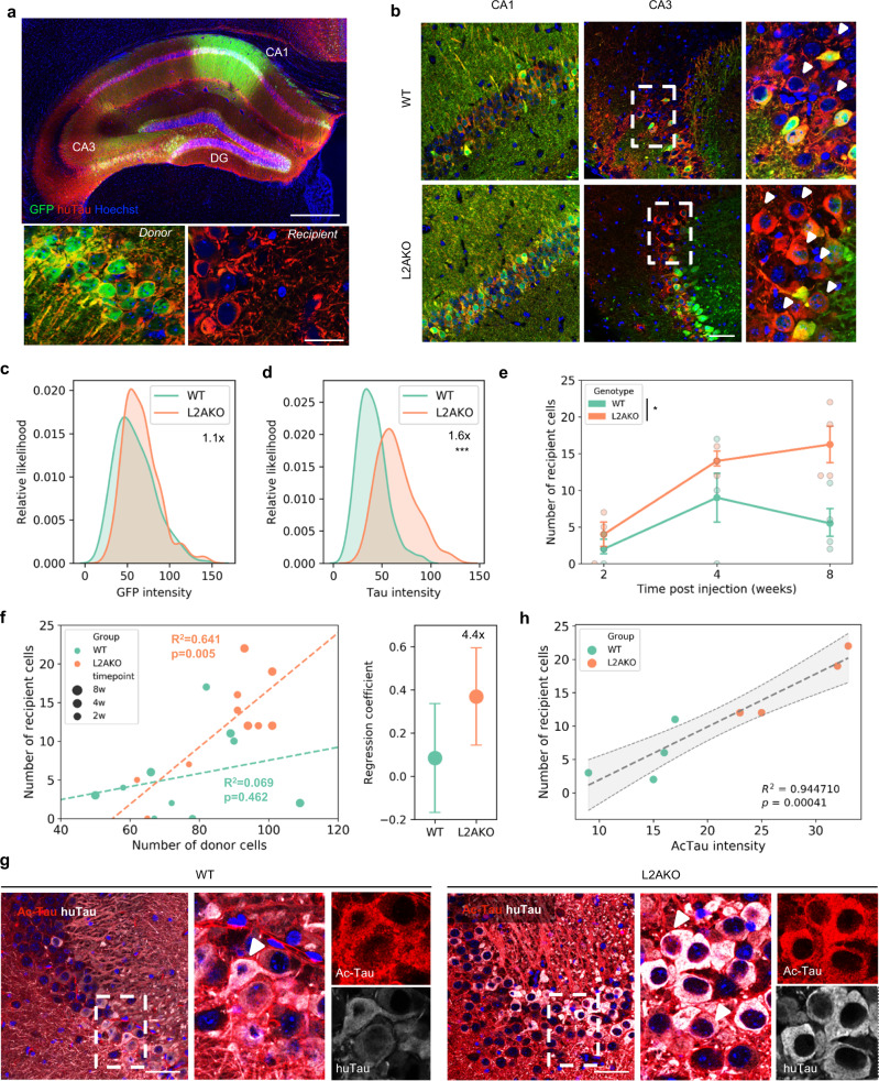
Disrupted homeostasis of the microtubule binding protein tau is a shared feature of a set of neurodegenerative disorders known as tauopathies. Acetylation of soluble tau is an early pathological event in neurodegeneration. In this work, we find that a large fraction of neuronal tau is degraded by chaperone-mediated autophagy (CMA) whereas, upon acetylation, tau is preferentially degraded by macroautophagy and endosomal microautophagy. Rerouting of acetylated tau to these other autophagic pathways originates, in part, from the inhibitory effect that acetylated tau exerts on CMA and results in its extracellular release. In fact, experimental blockage of CMA enhances cell-to-cell propagation of pathogenic tau in a mouse model of tauopathy. Furthermore, analysis of lysosomes isolated from brains of patients with tauopathies demonstrates similar molecular mechanisms leading to CMA dysfunction. This study reveals that CMA failure in tauopathy brains alters tau homeostasis and could contribute to aggravate disease progression.
Overview
The claims and statistics of this paper haven’t been checked yet.
Run the check — free, no account needed. It reads the open-access text and checks the sources it cites.
Integrity signals
Registry
Cites
Statistics
Figures
Verification
Claims the paper makes
The claims and statistics of this paper haven’t been checked yet.
Run the check — free, no account needed. It reads the open-access text and checks the sources it cites.
Claims other papers make about this one
Each is checked against this paper’s own text: does it say what the citing paper says it does?
The check wasn’t confident either way, so it gives no verdict — never disputed.
In the citing paper
“The balance between cell proliferation and programmed cell death is altered in LSDs as well as neurodegenerative disorders.26,27”
From The lysosome and proteostatic stress at the intersection of pediatric neurological disorders and adult neurodegenerative diseases (2025) · cites this paper as [27]
Experimental — accuracy not yet measuredIn the citing paper
“In addition, the acetylation of tau is an early pathological feature of neurodegeneration. Acetylated tau inhibits its degradation, promotes pathological aggregation and propagation, and contributes to tauopathy [46–51].”
From Tau and neuroinflammation in Alzheimer’s disease: interplay mechanisms and clinical translation (2023) · cites this paper as [50]
In the citing paper
“Blockage of CMA leads to rerouting of some of the pathogenic forms of MAPT/tau toward endosomal microautophagy, as both pathways share the same chaperone, HSPA8, and this promotes fusion of late endosomes with the plasma membrane and subsequent extraneuronal release of the MAPT/tau variants, thus contributing to MAPT/tau propagation (Caballero et al, 2021).”
From Autophagy in major human diseases (2021) · cites this paper as Caballero et al, 2021
In the citing paper
“However, mutations and posttranslational modifications of this protein, such as acetylation, not only prevent MAPT/tau degradation by CMA but also inhibit normal CMA functioning (Caballero et al, 2018; Caballero et al, 2021).”
From Autophagy in major human diseases (2021) · cites this paper as Caballero et al, 2021
In the citing paper
“Chaperone‐mediated autophagy also contributes to degradation of a large fraction of neuronal MAPT/tau under physiological conditions (Caballero et al, 2018; Caballero et al, 2021).”
From Autophagy in major human diseases (2021) · cites this paper as Caballero et al, 2021
In the citing paper
“Reduction in neuronal CMA activity has been recently shown in AD patient's brains (Bourdenx et al, 2021; Caballero et al, 2021)”
From Autophagy in major human diseases (2021) · cites this paper as Caballero et al, 2021
Self-check
Percentages, sums and reported differences re-computed from the paper’s own numbers.
The self-check needs the paper’s full text.
Citations
Cites
PROTEIN MEASUREMENT WITH THE FOLIN PHENOL REAGENT
Lowry, O. H., Rosebrough, N., Farr, A. L., & Randall, R. (1951) · Journal of Biological Chemistry · doi:10.1016/s0021-9258(19)52451-6 · Cited by 316,394
Analysis of Relative Gene Expression Data Using Real-Time Quantitative PCR and the 2−ΔΔCT Method
Livak, K. J., & Schmittgen, T. D. (2001) · Methods · doi:10.1006/meth.2001.1262 · Cited by 180,860
Towbin, H., Staehelin, T., & Gordon, J. (1979) · Proceedings of the National Academy of Sciences · doi:10.1073/pnas.76.9.4350 · Cited by 53,218
Raza, H., John, A., & Nemmar, A. (2013) · Cellular Physiology and Biochemistry · doi:10.1159/000350087 · Cited by 47,311
Repertoires of Autophagy in the Pathogenesis of Ocular Diseases
Li, Y., Jiang, Q. L., Cao, G.-F., Yao, J., & Yan, B. (2015) · Cellular Physiology and Biochemistry · doi:10.1159/000373980 · Cited by 16,351
Neuropathological stageing of Alzheimer-related changes.
Braak, H., & Braak, E. (1991) · Acta Neuropathologica · doi:10.1007/bf00308809 · Cited by 16,123
Amyloid oligomers exacerbate tau pathology in a mouse model of tauopathy
Selenica, M.-L. B., Brownlow, M., Jimenez, J. P., Lee, D. C., Pena, G., Dickey, C. A., Gordon, M. N., & Morgan, D. (2013) · Neurodegenerative Diseases · doi:10.1159/000337230 · Cited by 10,359
Autophagy: Renovation of Cells and Tissues
Mizushima, N., & Komatsu, M. (2011) · Cell · doi:10.1016/j.cell.2011.10.026 · Cited by 6,598
Cui, C., Cui, J., Jin, F., Cui, Y., Li, R., Jiang, X., Tian, Y., Wang, R. C., Jiang, P., & Gao, J. (2017) · Cellular Physiology and Biochemistry · doi:10.1159/000479571 · Cited by 5,214
Mutation of the mouse klotho gene leads to a syndrome resembling ageing
Kuro-o, M., Matsumura, Y., Aizawa, H., Kawaguchi, H., Suga, T., Utsugi, T., Ohyama, Y., Kurabayashi, M., Kaname, T., Kume, E., et al. (1997) · Nature · doi:10.1038/36285 · Cited by 3,837
Research works printed but not linked: 1
- Morris, M., Maeda, S., Vossel, K. & Mucke, L. The many faces of Tau. Neuron 70, 410–426 (2011). · DOI · Paper page
Cited by
Sorted by citations
Items 101–200 of 223
Exploring the landscape of post-translational modification in drug discovery
Cao, Y., Yu, T., Zhu, Z., Zhang, Y., Sun, S., Li, N., Gu, C., & Yang, Y. (2024) · Pharmacology & Therapeutics · doi:10.1016/j.pharmthera.2024.108749 · Cited by 17
Cannabidiol-Induced Autophagy Ameliorates Tau Protein Clearance
Vrechi, T. A. D. M., Guarache, G. C., Oliveira, R. B., Guedes, É. D. C., Erustes, A. G., Leão, A. H. F. F., Abílio, V. C., Zuardi, A. W., Hallak, J. E. C., Crippa, J. A. S., et al. (2025) · Neurotoxicity Research · doi:10.1007/s12640-025-00729-3 · Cited by 18
Khawaja, R. R., Griego, E., Lindenau, K. L., Salek, A. B., Gambardella, J., Scrivo, A., Monday, H. R., Bourdenx, M., Madero‐Pérez, J., Khan, Z. N., et al. (2025) · Nature Cell Biology · doi:10.1038/s41556-025-01771-1 · Cited by 17
Self-citationThe connection between autophagy and Alzheimer’s disease
Nechushtai, L., Chen, D., Dan, F., & Pinkas‐Kramarski, R. (2025) · Inflammation Research · doi:10.1007/s00011-025-02118-0 · Cited by 19
Autophagy and Glycative Stress: A Bittersweet Relationship in Neurodegeneration
Gómez, O., Perini-Villanueva, G., Yuste, A., Rodríguez‐Navarro, J. A., Poch, E., & Bejarano, E. (2021) · Frontiers in Cell and Developmental Biology · doi:10.3389/fcell.2021.790479 · Cited by 14
Davis, S. E., Cook, A. K., Hall, J. A., Voskobiynyk, Y., Carullo, N. V. N., Boyle, N. R., Hakim, A. R., Anderson, K. M., Hobdy, K. P., Pugh, D. A., et al. (2023) · Acta Neuropathologica Communications · doi:10.1186/s40478-023-01571-4 · Cited by 12
García-Cruz, V. M., & Arias, C. (2024) · Molecular Neurobiology · doi:10.1007/s12035-023-03886-8 · Cited by 17
SON-dependent nuclear speckle rehabilitation alleviates proteinopathies
Dion, W. A., Tao, Y., Chambers, M., Zhao, S., Arbuckle, R. K., Sun, M., Kubra, S., Schaich, M. A., Nie, Y., Ye, M., et al. (2025) · Nature Communications · doi:10.1038/s41467-025-62242-7 · Cited by 17
Self-citationDisrupting the Balance of Protein Quality Control Protein UBQLN2 Accelerates Tau Proteinopathy
Gerson, J. E., Sandoval-Pistorius, S. S., Welday, J. P., Rodriguez, A. L., Gregory, J. D., Liggans, N., Schache, K. J., Li, X., Trzeciakiewicz, H., Barmada, S. J., et al. (2022) · Journal of Neuroscience · doi:10.1523/jneurosci.1116-21.2021 · Cited by 14
Chaperone-Mediated Autophagy: A Potential Target for Metabolic Diseases
Yang, M., Luo, S., Chen, W., Zhao, L., & Wang, X. (2022) · Current Medicinal Chemistry · doi:10.2174/0929867329666220811141955 · Cited by 15
Benyair, R., Giridharan, S. S. P., Rivero-Ríos, P., Hasegawa, J., Bristow, E., Eskelinen, E., Shmueli, M. D., Fishbain-Yoskovitz, V., Merbl, Y., Sharkey, L. M., et al. (2023) · Autophagy Reports · doi:10.1080/27694127.2023.2166722 · Cited by 13
Emerging roles of p300/CBP in autophagy and autophagy-related human disorders
Xu, Y., & Wan, W. (2023) · Journal of Cell Science · doi:10.1242/jcs.261028 · Cited by 13
Small molecules targeting molecular chaperones for tau regulation: Achievements and challenges
He, C., Gu, J., Wang, D., Wang, K., Wang, Y., You, Q., & Wang, L. (2023) · European Journal of Medicinal Chemistry · doi:10.1016/j.ejmech.2023.115859 · Cited by 12
Kim, S., Hyun, D. G., Nam, Y., Shin, S. J., Im, D., Kim, H. S., Leem, S. H., Park, H. H., Kim, B., Park, Y., et al. (2023) · Biomedicine & Pharmacotherapy · doi:10.1016/j.biopha.2023.115770 · Cited by 13
Zaki, M. O., El-Desouky, S., Elsherbiny, D. A., Salama, M., & Azab, S. S. (2022) · Inflammopharmacology · doi:10.1007/s10787-022-01023-w · Cited by 12
RetractedLee, J., Xu, Y., & Ye, Y. (2022) · Frontiers in Cell and Developmental Biology · doi:10.3389/fcell.2022.906453 · Cited by 11
Targeting autophagy in astrocytes: a potential for neurodegenerative disease intervention
Potokar, M., & Jorgačevski, J. (2025) · Frontiers in Cellular Neuroscience · doi:10.3389/fncel.2025.1584767 · Cited by 14
“Dirty Dancing” of Calcium and Autophagy in Alzheimer’s Disease
Zhang, H., & Борисович, Б. И. (2023) · Life · doi:10.3390/life13051187 · Cited by 11
Min, J.-H., Sarlus, H., & Harris, R. A. (2024) · Frontiers in Neuroscience · doi:10.3389/fnins.2024.1467333 · Cited by 11
Sun, K. T., & Mok, S. (2025) · Neurotherapeutics · doi:10.1016/j.neurot.2024.e00512 · Cited by 12
ESCRT-I and PTPN23 mediate microautophagy of ubiquitylated tau aggregates
Men, Y., Hirayama, S., Ao, S., Sakurai, Y., Shibata, Y., Lo, M., Sato, Y., & Murata, S. (2025) · The Journal of Cell Biology · doi:10.1083/jcb.202406120 · Cited by 10
Yao, J., He, Z., You, G., Liu, Q., & Li, N. (2023) · Current Issues in Molecular Biology · doi:10.3390/cimb45080402 · Cited by 9
Re-Arranging the Puzzle between the Amyloid-Beta and Tau Pathology: An APP-Centric Approach
Haut, F., Argyrousi, E. K., & Arancio, O. (2023) · International Journal of Molecular Sciences · doi:10.3390/ijms25010259 · Cited by 11
Lipoprotein Metabolism, Protein Aggregation, and Alzheimer’s Disease: A Literature Review
Grao‐Cruces, E., Claro, C., la Paz, S. M., & Nóbrega, C. (2023) · International Journal of Molecular Sciences · doi:10.3390/ijms24032944 · Cited by 15
Dysregulation of neuroproteasomes by ApoE isoforms drives endogenous Tau aggregationPreprint
Paradise, V., Sabu, M., Bafia, J., Sharif, N., Nguyen, C., Konrad-Vicario, K., Mukim, R. D., Wang, X., Corjuc, B., Fu, J. M., et al. (2022) · bioRxiv (Cold Spring Harbor Laboratory) · doi:10.1101/2022.11.29.518293 · Cited by 10
Self-citationMurakami, R., Watanabe, H., Hashimoto, H., Kashiwagi-Hakozaki, M., Hashimoto, T., Karch, C. M., the Dominantly Inherited Alzheimer Network (DIAN), Iwatsubo, T., & Okano, H. (2024) · The Journal of Neuroscience · doi:10.1523/jneurosci.1709-23.2024 · Cited by 9
Self-citationDaly, T. P., Henry, V., & Bourdenx, M. (2023) · Journal of Alzheimer s Disease · doi:10.3233/jad-221004 · Cited by 11
Self-citationSelective autophagy: a therapeutic target for healthy aging?
Manastireanu, D. M., Salazar, N. A., Bejarano, E., & Nieto-Torres, J. L. (2024) · Aging advances. · doi:10.4103/agingadv.agingadv-d-24-00002 · Cited by 10
Targeting tau in Alzheimer’s Disease: rationale, approach and challenges
Aslanyan, A., Foiani, M. S., Giovannucci, T. A., McDade, E., Duff, K., Mummery, C. J., Schöll, M., Wildsmith, K. R., & Paterson, R. W. (2026) · Molecular Neurodegeneration · doi:10.1186/s13024-026-00925-5 · Cited by 10
Mirfakhar, F. S., Marsh, J. A., Sato, C., Schache, K. J., Minaya, M., Dolle, R. E., Pak, S. C., Silverman, G. A., Perlmutter, D. H., Macauley, S. L., et al. (2026) · Nature Communications · doi:10.1038/s41467-026-70473-5 · Cited by 9
Self-citationHan, M., Saxton, A. D., Currey, H. N., Waldherr, S. M., Liachko, N. F., & Kraemer, B. C. (2024) · Disease Models & Mechanisms · doi:10.1242/dmm.050473 · Cited by 9
Symmetric and Asymmetric Synapses Driving Neurodegenerative Disorders
Romaus‐Sanjurjo, D., Custodia, A., Aramburu-Núñez, M., Posado-Fernández, A., Vázquez‐Vázquez, L., Camino-Castiñeiras, J., Leira, Y., Peleteiro, J. M. P., Aldrey, J. M., Ouro, A., et al. (2021) · Symmetry · doi:10.3390/sym13122333 · Cited by 8
Diverse influences on tau aggregation and implications for disease progressionPreprint
Alstyne, M. V., Pratt, J., & Parker, R. (2025) · Genes & Development · doi:10.1101/gad.352551.124 · Cited by 11
Oh, Y. R., & Shin, M. (2025) · BMB Reports · doi:10.5483/bmbrep.2025-0077 · Cited by 9
Bryan, M. R., Tian, X., Tseng, J., Evangelista, B., Ragusa, J. V., Bryan, A. F., Trotman, W., Irwin, D. J., & Cohen, T. J. (2024) · Acta Neuropathologica Communications · doi:10.1186/s40478-024-01865-1 · Cited by 10
Chaperone-mediated autophagy regulates the metastatic state of mesenchymal tumors
Zhou, X., Berenger, E., Shi, Y., Shirokova, V., Kochetkova, E. Y., Becirovic, T., Zhang, B., Kaminskyy, V. O., Esmaeilian, Y., Hosaka, K., et al. (2025) · EMBO Molecular Medicine · doi:10.1038/s44321-025-00210-w · Cited by 9
Role of saturated fatty acid metabolism in posttranslational modifications of the Tau protein
García-Cruz, V. M., Coria, R., & Arias, C. (2025) · Molecular and Cellular Biochemistry · doi:10.1007/s11010-025-05275-2 · Cited by 7
Tittelmeier, J., & Nussbaum-Krammer, C. I. (2025) · Cells · doi:10.3390/cells14110845 · Cited by 11
Fan, J., Zhu, T., Tian, X., Wang, M., Li, Z., & Li, W. (2025) · Phytotherapy Research · doi:10.1002/ptr.70027 · Cited by 9
Autophagy: mechanisms, roles in human diseases, and therapeutic perspectives
He, J., & Qi, T. (2026) · Frontiers in Cell and Developmental Biology · doi:10.3389/fcell.2026.1776289 · Cited by 7
Inhibition of tau aggregation by the CCT3 and CCT7 apical domains
Ben‐Maimon, M., Elad, N., Naveh‐Tassa, S., Levy, Y., & Horovitz, A. (2025) · Protein Science · doi:10.1002/pro.70162 · Cited by 7
Anisimov, S., Takahashi, M., Kakihana, T., Katsuragi, Y., Sango, J., Abe, T., & Fujii, M. (2025) · Journal of Biological Chemistry · doi:10.1016/j.jbc.2025.110292 · Cited by 6
Yu, L., Yang, X., Lei, H.-P., Hu, Y., Wu, L., Zhou, S., & Jin, F. (2025) · Molecular Neurobiology · doi:10.1007/s12035-025-05162-3 · Cited by 7
Lin, A., Dai, X., Chen, J., Han, T., Du, Q., Wu, M., Cheng, J., Chen, W., Ye, Q.-Y., Chen, X., et al. (2025) · Alzheimer s & Dementia · doi:10.1002/alz.70919 · Cited by 6
Self-citationWu, J., Geng, C., Liu, L., & Tang, Y. (2025) · Medicine Plus · doi:10.1016/j.medp.2025.100112 · Cited by 8
Rsp5/NEDD4 and ESCRT regulate TDP-43 toxicity and turnover via an endolysosomal clearance mechanism
Byrd, A. T., Marmorale, L. J., Marcinowski, S., Dykstra, M. M., Addison, V., Barmada, S. J., & Buchan, J. R. (2025) · The Journal of Cell Biology · doi:10.1083/jcb.202212064 · Cited by 7
Hamano, T., & Endo, Y. (2022) · Elsevier eBooks · doi:10.1016/b978-0-323-89906-2.00006-x · Cited by 7
Rosenzweig-Lipson, S. J. (2023) · The Journal of Prevention of Alzheimer s Disease · doi:10.14283/jpad.2023.106 · Cited by 6
Luo, T.-Y., Li, C., Zhou, L., Sun, H., & Yang, M. M. (2025) · Investigative Ophthalmology & Visual Science · doi:10.1167/iovs.66.5.30 · Cited by 5
Zheng, N., Wang, Z., Cao, J., Li, K., Xu, H., Wang, J., Zhang, L., Meng, J., Tang, Z., Luo, H., et al. (2025) · Alzheimer s & Dementia · doi:10.1002/alz.70369 · Cited by 5
Waqar, Z., Sethi, P., Jain, D., Singh, K., Alsaidan, O. A., Alzarea, S. I., Gupta, J. K., Saxena, S., & Sharma, M. C. (2025) · Current Genomics · doi:10.2174/0113892029372437251010114053 · Cited by 6
Honey, M. I. J., Hok‐A‐Hin, Y. S., Thijssen, E. H., Cousins, K. A. Q., van der Weijden, B., Veld, L. I. ‘., Stoops, E., de Boer, S. C. M., Duits, F. H., Houwer, J. F. H. D., et al. (2026) · Nature Medicine · doi:10.1038/s41591-026-04341-6 · Cited by 8
Self-citationFOXO family and neurodegenerative diseases: Mechanisms of action and therapeutic potential
Lv, Z.-X., Liu, X., Zhou, Z., Huang, Q., Liu, H.-J., Xu, Z.-C., & Xu, P. (2026) · Redox Biology · doi:10.1016/j.redox.2026.104218 · Cited by 7
Rate of tau propagation is a heritable disease trait in genetically diverse mouse strains
Welikovitch, L. A., Dujardin, S., Dunn, A. R., Fernandes, A. R., Khasnavis, A., Chibnik, L. B., Kaczorowski, C. C., & Hyman, B. T. (2023) · iScience · doi:10.1016/j.isci.2023.105983 · Cited by 6
Self-citationMechanistic insights of molecular chaperone Hsp70: Pathogenesis of neurodegenerative diseases
Choudhary, A., Kumar, P., Jagtap, Y. A., Kinger, S., Jha, H. C., Gutti, R. K., Sharma, V., Prajapati, V. K., & Mishra, A. (2025) · Brain Network Disorders · doi:10.1016/j.bnd.2025.05.001 · Cited by 5
GFAP degradation in TBI: linking novel modified products to astrocyte pathology and patient outcome
Wanner, I. B., Halford, J., Lopez, J., Shen, S., Chen, Y., Zhao, H., Salas, C., Loo, R. R. O., Robicsek, S. A., Ellingson, B. M., et al. (2026) · Acta Neuropathologica Communications · doi:10.1186/s40478-026-02240-y · Cited by 5
Wu, Z., Jiang, Z., Hong, C., Yang, S., Jin, S., Wu, C., Guo, K., Liu, J., Xu, S., Wang, C., et al. (2025) · Advanced Science · doi:10.1002/advs.202516015 · Cited by 4
Self-citationAdvances and emerging challenges in detecting Tau proteoforms for Alzheimer's disease diagnosis
Cai, Q., Miao, M., & Li, Z. (2026) · TrAC Trends in Analytical Chemistry · doi:10.1016/j.trac.2026.118897 · Cited by 3
Mohl, G. A., Dixon, G., Marzette, E., McKetney, J., Samelson, A. J., Serras, C. P., Jin, J., Ariqat, N., Keys, A., Chavira, C., et al. (2026) · npj Dementia · doi:10.1038/s44400-026-00076-w · Cited by 4
Zhang, Y., Wu, H., Zhang, Q., Cong, P., Li, Z., Wu, Q., Huang, X., Li, X., Feng, B., Liu, Q., et al. (2025) · Cell Reports · doi:10.1016/j.celrep.2025.115489 · Cited by 4
Fu, H., Chen, S., & Acosta, D. (2022) · Neural Regeneration Research · doi:10.4103/1673-5374.361540 · Cited by 4
Lane-Donovan, C., Paredes, M., & Kao, A. W. (2025) · Progress in Neurobiology · doi:10.1016/j.pneurobio.2025.102854 · Cited by 3
The role of Aha1 in cancer and neurodegeneration
Blagg, B. S. J., & Catalfano, K. C. (2024) · Frontiers in Molecular Neuroscience · doi:10.3389/fnmol.2024.1509280 · Cited by 3
A novel lncRNA FAM151B-DT regulates degradation of aggregation prone proteins
Renganathan, A., Minaya, M. A., Broder, M., Alfradique-Dunham, I., Miller, R. L., Dhavale, D. D., Moritz, M., Bhagat, R., Marsh, J. A., Verbeck, A. C., et al. (2025) · Molecular Psychiatry · doi:10.1038/s41380-025-03277-6 · Cited by 4
Self-citationMontgomery, K. M., Samelson, A. J., & Gestwicki, J. E. (2023) · Israel Journal of Chemistry · doi:10.1002/ijch.202300104 · Cited by 4
Self-citationEndosomal microautophagy is activated by specific cellular stresses in trout hepatocytes
Vélez, E. J., Véron, V., Gouis, J., Reji, S., Dias, K., Herpin, A., Beaumatin, F., & Seiliez, I. (2025) · Scientific Reports · doi:10.1038/s41598-025-23022-x · Cited by 4
Xiong, R., Shao, D., Do, S., & Chan, W. K. (2023) · International Journal of Molecular Sciences · doi:10.3390/ijms242015116 · Cited by 4
Zou, J., Liu, L., Chai, F., Shan, Q., & Wang, Y. (2025) · Molecular Neurobiology · doi:10.1007/s12035-025-05084-0 · Cited by 3
Chaperone‐Mediated Autophagy: A Critical Regulator of Neuroinflammation and Neurodegeneration
Han, Y., Huang, X., Su, Y., Ma, J., & Wu, J. (2025) · Advanced Biology · doi:10.1002/adbi.202500191 · Cited by 3
Shen, M., Yu, X., Wu, S., Qiu, J., Wang, R., Li, K., & Zhang, Q. (2025) · Trends in Food Science & Technology · doi:10.1016/j.tifs.2025.105182 · Cited by 3
Rogers, E. A., Diorio, T. C., Beauclair, T., Martinez, J., Mufti, S. J., Kim, D., Krishnan, N., Rayz, V. L., & Shi, R. (2025) · Lab on a Chip · doi:10.1039/d5lc00067j · Cited by 2
Bridging structure and selectivity in chaperone‐mediated autophagy: towards targeted therapeutics
Sahu, D., Patel, I., Lakshman, K., Mapa, K., & Malhotra, N. (2025) · FEBS Journal · doi:10.1111/febs.70262 · Cited by 2
Takahashi, M., Kitaura, H., Nakahara, A., Kakita, A., Watanabe, K., Kakihana, T., Hara, T., Katsuragi, Y., Yoshita‐Takahashi, M., Anisimov, S., et al. (2025) · Molecular and Cellular Biology · doi:10.1080/10985549.2025.2575950 · Cited by 2
Goniotaki, D., Hausherr, M., Lynham, S., Ale, A., Chennell, G., Marcotti, S., Marcus, K., Noble, W. J., Hanger, D. P., Fraser, G. M., et al. (2025) · Alzheimer s & Dementia · doi:10.1002/alz.70977 · Cited by 2
Presenilin-dependent regulation of neuronal tau pathology via the autophagy and proteasome pathways
del Ser‐Badia, A., Soto-Faguás, C. M., Vecino, R., Vendrell, C., Molina‐Porcel, L., Sánchez‐Valle, R., Rodrı́guez-Álvarez, J., Vicario‐Abejón, C., & Saura, C. A. (2026) · Acta Neuropathologica Communications · doi:10.1186/s40478-026-02270-6 · Cited by 3
Salinas, M. D., Martínez, I. M., Naranjo, E., Molina, M. L., Cuervo, A. M., Muller, S., & Valdor, R. (2026) · Autophagy · doi:10.1080/15548627.2026.2672700 · Cited by 3
Self-citationBioenergetic impairment-induced Tau acetylation: Converging pathways in Alzheimer’s disease
C, B. K., & G., P. P. C. (2026) · International review of neurobiology · doi:10.1016/bs.irn.2026.05.020 · Cited by 2
Molecular and environmental drivers of tau post-translational modifications and tau pathology
Price, T. M., Tucker, A. E., & Funk, K. E. (2026) · Ageing Research Reviews · doi:10.1016/j.arr.2026.103223 · Cited by 2
Curbing Rhes Actions: Mechanism-based Molecular Target for Huntington’s Disease and Tauopathies
Subramaniam, S., & Boregowda, S. V. (2023) · CNS & Neurological Disorders - Drug Targets · doi:10.2174/1871527322666230320103518 · Cited by 3
Chloroquine Sensitizes Esophageal Carcinoma EC109 Cells to Paclitaxel by Inhibiting Autophagy
Yuan, Z., Cai, J., Du, Q., Ma, Q., Xu, L., Cai, Y., Zhong, X., & Guo, X. (2023) · Critical Reviews in Eukaryotic Gene Expression · doi:10.1615/critreveukaryotgeneexpr.2023046722 · Cited by 2
Mitochondrial bioenergetics stimulates autophagy for pathological tau clearance in tauopathy neuronsPreprint
Jia, N., Ganesan, D., Guan, H., Jeong, Y. Y., Han, S., Nissenbaum, M., Kusnecov, A. W., & Cai, Q. (2024) · bioRxiv (Cold Spring Harbor Laboratory) · doi:10.1101/2024.02.12.579959 · Cited by 2
Sphingolipid imbalance aggravates tau pathology by endomembrane rigidification and rupturePreprint
Tittelmeier, J., Sandhof, C. A., Martin, N., El-Kabarity, D., Ngonza-Nito, S.-B., Melki, R., & Nussbaum-Krammer, C. I. (2025) · eLife · doi:10.7554/elife.106865.1 · Cited by 2
Deng, L., Wu, L., Chen, D., & Cao, Y. (2024) · Journal of Cellular Physiology · doi:10.1002/jcp.31283 · Cited by 1
Liang, Q., Xiang, S., Wang, P., Chen, L., Cao, H., Cheng, T., Jiang, T., Zuo, H., Tian, Z., & Bhattacharyya, S. (2025) · Advanced Therapeutics · doi:10.1002/adtp.202400462 · Cited by 1
Synaptotoxic effects of extracellular tau are mediated by its microtubule-binding region
Ondrejčák, T., Hu, N., Coode, E., Campbell, T., Corbett, G. T., Doykov, I., Mills, K. A., Walsh, D. M., Livesey, F. J., Rowan, M. J., et al. (2025) · Acta Neuropathologica · doi:10.1007/s00401-025-02897-0 · Cited by 1
Impact of Lysine Acetylation Mutations on the Structure of Full-Length Tau Fibrils
Dregni, A. J., & Hong, M. (2025) · ACS Chemical Neuroscience · doi:10.1021/acschemneuro.5c00149 · Cited by 1
Chaperone-Mediated Autophagy in Health and Disease
Kaushik, S. (2025) · Learning materials in biosciences · doi:10.1007/978-3-031-88121-3_11 · Cited by 1
Understanding the impact of cytoskeleton disruption on synaptic plasticity in Alzheimer’s disease
Basavaraju, K. C., & Priyadarshini, P. (2025) · Advances in protein chemistry and structural biology · doi:10.1016/bs.apcsb.2025.10.020 · Cited by 1
Alzheimer’s Tau seeds-induced pathology enhances hippocampal extracellular diffusion
Estaún‐Panzano, J., Lovisotto, A., Mazzocco, C., Piva, C., Darricau, M., Calaresu, I., Gresil, Q., Planche, V., Dehay, B., Groc, L., et al. (2025) · Communications Biology · doi:10.1038/s42003-025-09054-z · Cited by 2
Garrahy, J. P. (2025) · Journal of Alzheimer s Disease · doi:10.1177/13872877251404970 · Cited by 1
Autophagy in emerging and highly concerned severe zoonotic infectious diseases
Qi, Y., Wang, L., Wu, S., Meng, C., Chu, Y., Yin, Y., & Jiao, H. (2026) · Frontiers in Immunology · doi:10.3389/fimmu.2026.1761571 · Cited by 1
Ge, C., Wang, K., Tang, H., Ke, Y., Wang, H., Fu, Q., Xiu, Y., Guo, Y., Jia, Y., Long, Z., et al. (2026) · Translational Neurodegeneration · doi:10.1186/s40035-026-00549-1 · Cited by 1
Spatiotemporal characterization of Alzheimer disease pathology in living human brain tissue
Aslanyan, A., Mukherjee, S., Moncur, E. M., Szadziewska, A., He, Y., Kac, P. R., Coyle, R. A., Camporesi, E., Sato, C., Ghoshal, N., et al. (2026) · Nature Communications · doi:10.1038/s41467-026-76494-4 · Cited by 1
Autophagic plasmid expressing SIRT3 and SIRT4 to improve Alzheimer’s diseasePreprint
Li, S., Deng, J., Mo, X., Sun, H., Wang, J., Dai, Z., Chen, Y., Zheng, W., Li, Z., Zhong, Z., et al. (2022) · Research Square · doi:10.21203/rs.3.rs-1717742/v1 · Cited by 1
Autophagic enhancer rescues Tau accumulation in a stem cell model of frontotemporal dementiaPreprint
Mirfakhar, F. S., Marsh, J. A., Minaya, M., Pak, S. C., Silverman, G. A., Perlmutter, D. H., Macauley, S. L., & Karch, C. M. M. (2024) · bioRxiv (Cold Spring Harbor Laboratory) · doi:10.1101/2024.09.30.615921 · Cited by 1
Self-citation(2022) · The Neuroscientist · doi:10.1177/10738584221078777
Tau acetylation reduces its autophagic degradation and is a targetable pathway for human tauopathies
Johnson, N. R. (2022) · BioEssays · doi:10.1002/bies.202200062
Rodriguez-Rodriguez, P., Arroyo-García, L., Li, L.-C., Tsagkogianni, C., Wang, W., Salas-Allende, I., Plautz, Z. R., Cedazo-Mínguez, A., Sinha, S. C., Troyanskaya, O., et al. (2022) · bioRxiv · doi:10.1101/2022.05.14.491965
Zhou, X., Shi, Y., Shirokova, V., Kochetkova, E. Y., Becirovic, T., Zhang, B., Kaminskyy, V. O., Lindskog, C., Hydbring, P., Ekman, S., et al. (2022) · bioRxiv (Cold Spring Harbor Laboratory) · doi:10.1101/2022.07.07.499098
Uddin, M. N., Shimonaka, S., & Motoi, Y. (2022) · Elsevier eBooks · doi:10.1016/b978-0-323-89906-2.00014-9
Metadata
- Authors
- Caballero, B., Bourdenx, M., Luengo, E., Diaz, A., Sohn, P. D., Chen, X., Wang, C., Juste, Y. R., Wegmann, S., Patel, B., Young, Z. T., Kuo, S. Y., Rodriguez-Navarro, J. A., Shao, H., Lopez, M. G., Karch, C. M., Goate, A. M., Gestwicki, J. E., Hyman, B. T., Gan, L., & Cuervo, A. M.
- Affiliations
- Albert Einstein College of Medicine (US)
- Centre National de la Recherche Scientifique (FR)
- Weill Cornell Medicine (US)
- Harvard University (US)
- University of California, San Francisco (US)
- Washington University in St. Louis (US)
- Icahn School of Medicine at Mount Sinai (US)
- ORCID
- Venue
- Nature Communications
- Published
- 2021-04-14
- Volume
- 12
- Issue
- 1
- Pages
- 2238-2238
- ISSN
- 2041-1723 (ISSN-L)
- PMC
- PMC8047017
- OpenAlex
- W3154991192
- Cited by
- 223
- References
- 92
- Citations by year
- 2026: 29 · 2025: 61 · 2024: 30 · 2023: 47 · 2022: 42 · 2021: 14
- FWCI
- 20.89
- Type
- journal article
- Language
- en
- Open access
- gold
- doi.org
- Locations
- Nature Communications (publishedVersion, cc-by)
- PubMed Central (publishedVersion, cc-by)
- PubMed (publishedVersion)
- Digital Commons@Becker (Washington University School of Medicine) (submittedVersion, cc-by)
- RePEc: Research Papers in Economics (submittedVersion)
- DOAJ (DOAJ: Directory of Open Access Journals) (submittedVersion, cc-by-sa)
- eScholarship (California Digital Library) (submittedVersion)
- eScholarship (California Digital Library) (submittedVersion, cc-by)
- DZNE Pub (submittedVersion, other-oa)
- Funders
- Fundación Tatiana Pérez de Guzmán el Bueno (Fellowship)
- U.S. Department of Health & Human Services | National Institutes of Health (T32 GM007491)
- Charles H. Revson Foundation (CP13-00234)
- Rainwaters Foundation
- U.S. Department of Health & Human Services | NIH | National Institute on Aging (AG05681, AG058674, AG036884, AG021904, AG054108, AG031782, AG038072)
- Rainwaters Foundation JPB Foundation
- U.S. Department of Health & Human Services | NIH | National Institute of Neurological Disorders and Stroke (NS100717)
- Rainwaters Foundation Backus Foundation JPB Foundation
- Topics
- Life Sciences › Neuroscience › Cellular and Molecular Neuroscience › Alzheimer's disease research and treatments
- Life Sciences › Biochemistry, Genetics and Molecular Biology › Cell Biology › Autophagy in Disease and Therapy
- Life Sciences › Biochemistry, Genetics and Molecular Biology › Molecular Biology › Heat shock proteins research
- Keywords
- tau acetylation, chaperone-mediated autophagy, tauopathy, tau degradation, macroautophagy, microautophagy, lysosomes, cell-to-cell transmission, neurodegeneration
- MeSH terms
- Chaperone-Mediated Autophagy, Acetylation, Animals, Brain, Female, Humans, Male, Mice, Inbred C57BL, Neurons, tau Proteins, Tauopathies, Mice
About these checks
Registry, retraction, Cites and statistics results are computed, not judged, with one exception: a printed reference that no index lists is read once by a model before it counts as a citation error. Claims are extracted by a model and kept only when their quote appears verbatim in the text; each cited source is then checked, with a model’s help, for whether it says what the claim relies on. Those verdicts can be wrong — read the claim’s quote and the cited source before relying on one.
Statistics and arithmetic are recomputed, not judged.
Confidence bands are shown only for fields calibrated on our annotated test set. None is calibrated yet, so no band is shown.
What counts as a claim ›How we measure confidence ›How we check this ›
Are you an author, or do you see a problem with this page? Request a review
