

The DOI matches its Crossref record; 92 of the 92 references it cites were found in a registry or an index.
Acetylated tau inhibits chaperone-mediated autophagy and promotes tau pathology propagation in mice
Open accessAbstract
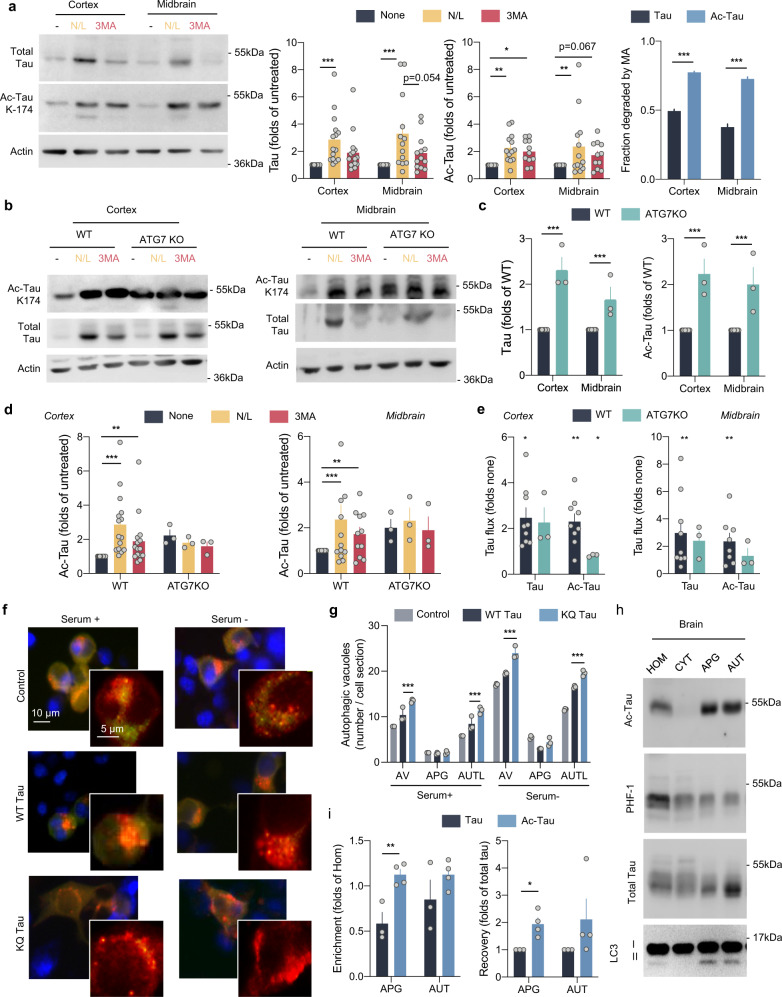
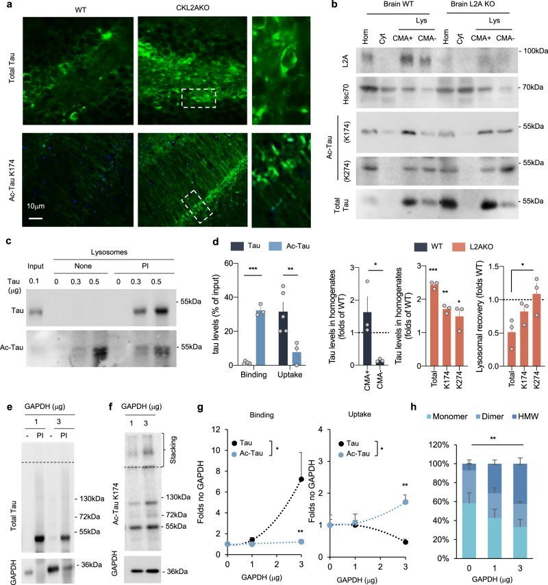
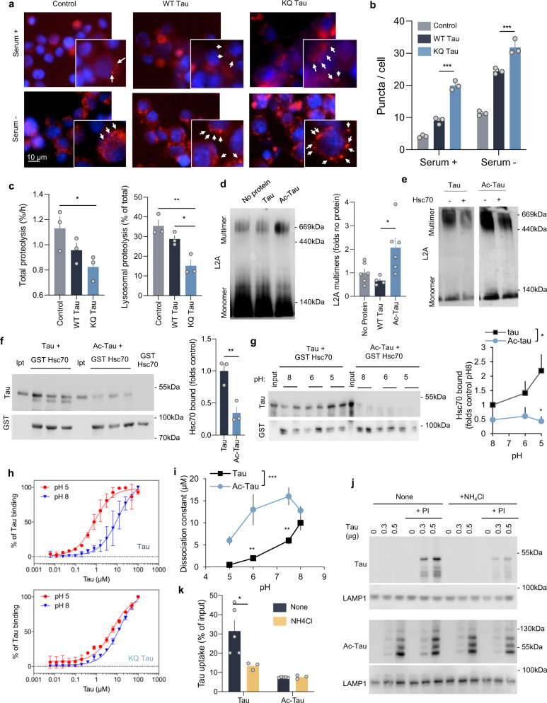
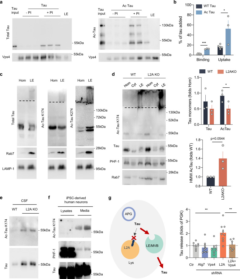
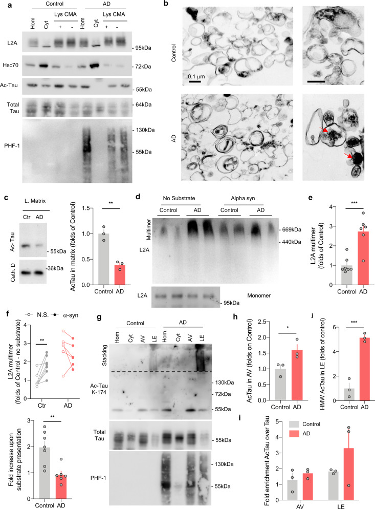
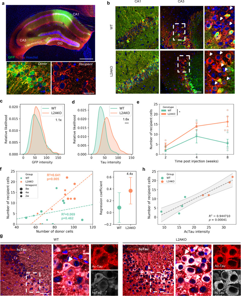
Disrupted homeostasis of the microtubule binding protein tau is a shared feature of a set of neurodegenerative disorders known as tauopathies. Acetylation of soluble tau is an early pathological event in neurodegeneration. In this work, we find that a large fraction of neuronal tau is degraded by chaperone-mediated autophagy (CMA) whereas, upon acetylation, tau is preferentially degraded by macroautophagy and endosomal microautophagy. Rerouting of acetylated tau to these other autophagic pathways originates, in part, from the inhibitory effect that acetylated tau exerts on CMA and results in its extracellular release. In fact, experimental blockage of CMA enhances cell-to-cell propagation of pathogenic tau in a mouse model of tauopathy. Furthermore, analysis of lysosomes isolated from brains of patients with tauopathies demonstrates similar molecular mechanisms leading to CMA dysfunction. This study reveals that CMA failure in tauopathy brains alters tau homeostasis and could contribute to aggravate disease progression.
Overview
The claims and statistics of this paper haven’t been checked yet.
Run the check — free, no account needed. It reads the open-access text and checks the sources it cites.
Integrity signals
Registry
Cites
Statistics
Figures
Verification
Claims the paper makes
The claims and statistics of this paper haven’t been checked yet.
Run the check — free, no account needed. It reads the open-access text and checks the sources it cites.
Claims other papers make about this one
Each is checked against this paper’s own text: does it say what the citing paper says it does?
The check wasn’t confident either way, so it gives no verdict — never disputed.
In the citing paper
“The balance between cell proliferation and programmed cell death is altered in LSDs as well as neurodegenerative disorders.26,27”
From The lysosome and proteostatic stress at the intersection of pediatric neurological disorders and adult neurodegenerative diseases (2025) · cites this paper as [27]
Experimental — accuracy not yet measuredIn the citing paper
“In addition, the acetylation of tau is an early pathological feature of neurodegeneration. Acetylated tau inhibits its degradation, promotes pathological aggregation and propagation, and contributes to tauopathy [46–51].”
From Tau and neuroinflammation in Alzheimer’s disease: interplay mechanisms and clinical translation (2023) · cites this paper as [50]
In the citing paper
“Blockage of CMA leads to rerouting of some of the pathogenic forms of MAPT/tau toward endosomal microautophagy, as both pathways share the same chaperone, HSPA8, and this promotes fusion of late endosomes with the plasma membrane and subsequent extraneuronal release of the MAPT/tau variants, thus contributing to MAPT/tau propagation (Caballero et al, 2021).”
From Autophagy in major human diseases (2021) · cites this paper as Caballero et al, 2021
In the citing paper
“However, mutations and posttranslational modifications of this protein, such as acetylation, not only prevent MAPT/tau degradation by CMA but also inhibit normal CMA functioning (Caballero et al, 2018; Caballero et al, 2021).”
From Autophagy in major human diseases (2021) · cites this paper as Caballero et al, 2021
In the citing paper
“Chaperone‐mediated autophagy also contributes to degradation of a large fraction of neuronal MAPT/tau under physiological conditions (Caballero et al, 2018; Caballero et al, 2021).”
From Autophagy in major human diseases (2021) · cites this paper as Caballero et al, 2021
In the citing paper
“Reduction in neuronal CMA activity has been recently shown in AD patient's brains (Bourdenx et al, 2021; Caballero et al, 2021)”
From Autophagy in major human diseases (2021) · cites this paper as Caballero et al, 2021
Self-check
Percentages, sums and reported differences re-computed from the paper’s own numbers.
The self-check needs the paper’s full text.
Citations
Cites
PROTEIN MEASUREMENT WITH THE FOLIN PHENOL REAGENT
Lowry, O. H., Rosebrough, N., Farr, A. L., & Randall, R. (1951) · Journal of Biological Chemistry · doi:10.1016/s0021-9258(19)52451-6 · Cited by 316,394
Analysis of Relative Gene Expression Data Using Real-Time Quantitative PCR and the 2−ΔΔCT Method
Livak, K. J., & Schmittgen, T. D. (2001) · Methods · doi:10.1006/meth.2001.1262 · Cited by 180,860
Towbin, H., Staehelin, T., & Gordon, J. (1979) · Proceedings of the National Academy of Sciences · doi:10.1073/pnas.76.9.4350 · Cited by 53,218
Raza, H., John, A., & Nemmar, A. (2013) · Cellular Physiology and Biochemistry · doi:10.1159/000350087 · Cited by 47,311
Repertoires of Autophagy in the Pathogenesis of Ocular Diseases
Li, Y., Jiang, Q. L., Cao, G.-F., Yao, J., & Yan, B. (2015) · Cellular Physiology and Biochemistry · doi:10.1159/000373980 · Cited by 16,351
Neuropathological stageing of Alzheimer-related changes.
Braak, H., & Braak, E. (1991) · Acta Neuropathologica · doi:10.1007/bf00308809 · Cited by 16,123
Amyloid oligomers exacerbate tau pathology in a mouse model of tauopathy
Selenica, M.-L. B., Brownlow, M., Jimenez, J. P., Lee, D. C., Pena, G., Dickey, C. A., Gordon, M. N., & Morgan, D. (2013) · Neurodegenerative Diseases · doi:10.1159/000337230 · Cited by 10,359
Autophagy: Renovation of Cells and Tissues
Mizushima, N., & Komatsu, M. (2011) · Cell · doi:10.1016/j.cell.2011.10.026 · Cited by 6,598
Cui, C., Cui, J., Jin, F., Cui, Y., Li, R., Jiang, X., Tian, Y., Wang, R. C., Jiang, P., & Gao, J. (2017) · Cellular Physiology and Biochemistry · doi:10.1159/000479571 · Cited by 5,214
Mutation of the mouse klotho gene leads to a syndrome resembling ageing
Kuro-o, M., Matsumura, Y., Aizawa, H., Kawaguchi, H., Suga, T., Utsugi, T., Ohyama, Y., Kurabayashi, M., Kaname, T., Kume, E., et al. (1997) · Nature · doi:10.1038/36285 · Cited by 3,837
Loss of autophagy in the central nervous system causes neurodegeneration in mice
Komatsu, M., Waguri, S., Chiba, T., Murata, S., Iwata, J.-I., Tanida, I., Ueno, T., Koike, M., Uchiyama, Y., Kominami, E., et al. (2006) · Nature · doi:10.1038/nature04723 · Cited by 3,500
Kimura, S., Noda, T., & Yoshimori, T. (2007) · Autophagy · doi:10.4161/auto.4451 · Cited by 2,278
The role of autophagy in neurodegenerative disease
Nixon, R. A. (2013) · Nature Medicine · doi:10.1038/nm.3232 · Cited by 2,059
Impaired Degradation of Mutant α-Synuclein by Chaperone-Mediated Autophagy
Cuervo, A. M., Stefanis, L., Fredenburg, R., Lansbury, P. T., & Sulzer, D. (2004) · Science · doi:10.1126/science.1101738 · Cited by 2,024
Self-citationTransmission and spreading of tauopathy in transgenic mouse brain
Clavaguera, F., Bolmont, T., Crowther, R. A., Abramowski, D., Frank, S., Probst, A., Fraser, G., Stalder, A. K., Beibel, M., Staufenbiel, M., et al. (2009) · Nature Cell Biology · doi:10.1038/ncb1901 · Cited by 1,803
Self-propagation of pathogenic protein aggregates in neurodegenerative diseases
Jucker, M., & Walker, L. C. (2013) · Nature · doi:10.1038/nature12481 · Cited by 1,663
Extensive Involvement of Autophagy in Alzheimer Disease: An Immuno-Electron Microscopy Study
Nixon, R. A., Wegiel, J., Kumar, A., Yu, W. H., Peterhoff, C., Cataldo, A., & Cuervo, A. M. (2005) · Journal of Neuropathology & Experimental Neurology · doi:10.1093/jnen/64.2.113 · Cited by 1,588
Self-citationThe coming of age of chaperone-mediated autophagy
Kaushik, S., & Cuervo, A. M. (2018) · Nature Reviews Molecular Cell Biology · doi:10.1038/s41580-018-0001-6 · Cited by 1,407
Self-citationLee, J.-H., Yu, W. H., Kumar, A., Lee, S., Mohan, P. S., Peterhoff, C. M., Wolfe, D. M., Martinez-Vicente, M., Massey, A. C., Sovak, G., et al. (2010) · Cell · doi:10.1016/j.cell.2010.05.008 · Cited by 1,212
Self-citationTau pathology and neurodegeneration
Spillantini, M. G., & Goedert, M. (2013) · The Lancet Neurology · doi:10.1016/s1474-4422(13)70090-5 · Cited by 1,133
A Role for a 70-Kilodalton Heat Shock Protein in Lysosomal Degradation of Intracellular Proteins
Chiang, H.-L., Terlecky, S. R., Plant, C. P., & Dice, J. F. (1989) · Science · doi:10.1126/science.2799391 · Cited by 982
A Receptor for the Selective Uptake and Degradation of Proteins by Lysosomes
Cuervo, A. M., & Dice, J. F. (1996) · Science · doi:10.1126/science.273.5274.501 · Cited by 979
Self-citationAcetylation of Tau Inhibits Its Degradation and Contributes to Tauopathy
Min, S.-W., Cho, S.-H., Zhou, Y., Schroeder, S., Haroutunian, V., Seeley, W. W., Huang, E. J., Shen, Y., Masliah, E., Mukherjee, C., et al. (2010) · Neuron · doi:10.1016/j.neuron.2010.08.044 · Cited by 949
Self-citationBramblett, G. T., Goedert, M., Jakes, R., Merrick, S. E., Trojanowski, J. Q., & Lee, V. M. Y. (1993) · Neuron · doi:10.1016/0896-6273(93)90057-x · Cited by 903
Microautophagy of Cytosolic Proteins by Late Endosomes
Sahu, R., Kaushik, S., Clement, C. C., Cannizzo, E. S., Scharf, B., Follenzi, A., Potolicchio, I., Nieves, E., Cuervo, A. M., & Santambrogio, L. (2011) · Developmental Cell · doi:10.1016/j.devcel.2010.12.003 · Cited by 902
Self-citationMorris, M., Maeda, S., Vossel, K., & Mucke, L. (2011) · Neuron · doi:10.1016/j.neuron.2011.04.009 · Cited by 899
Biernat, J., Gustke, N., Drewes, G., Mandelkow, E., & Mandelkow, E. (1993) · Neuron · doi:10.1016/0896-6273(93)90279-z · Cited by 796
Peptide sequences that target cytosolic proteins for lysosomal proteolysis
Dice, J. F. (1990) · Trends in Biochemical Sciences · doi:10.1016/0968-0004(90)90019-8 · Cited by 755
The acetylation of tau inhibits its function and promotes pathological tau aggregation
Cohen, T. J., Guo, J. L., Hurtado, D. E., Kwong, L. K., Mills, I. P., Trojanowski, J. Q., & Lee, V. M. Y. (2011) · Nature Communications · doi:10.1038/ncomms1255 · Cited by 724
The release and trans-synaptic transmission of Tau via exosomes
Wang, Y., Balaji, V., Kaniyappan, S., Krüger, L., Irsen, S., Tepper, K., Chandupatla, R., Maetzler, W., Schneider, A., Mandelkow, E., et al. (2017) · Molecular Neurodegeneration · doi:10.1186/s13024-016-0143-y · Cited by 674
Age-related decline in chaperone-mediated autophagy
Cuervo, A. M., & Dice, J. F. (2000) · Journal of Biological Chemistry · doi:10.1074/jbc.m002102200 · Cited by 665
Self-citationTau fragmentation, aggregation and clearance: the dual role of lysosomal processing
Wang, Y., Martinez-Vicente, M., Krüger, U., Kaushik, S., Wong, E., Mandelkow, E.-M., Cuervo, A. M., & Mandelkow, E. (2009) · Human Molecular Genetics · doi:10.1093/hmg/ddp367 · Cited by 617
Self-citationActivation of Chaperone-mediated Autophagy during Oxidative Stress
Kiffin, R., Christian, C., Knecht, E., & Cuervo, A. M. (2004) · Molecular Biology of the Cell · doi:10.1091/mbc.e04-06-0477 · Cited by 611
Self-citationInterplay of LRRK2 with chaperone-mediated autophagy
Orenstein, S. J., Kuo, S.-H., Tasset, I., Arias, E., Koga, H., Fernandez-Carasa, I., Cortes, E., Honig, L. S., Dauer, W., Consiglio, A., et al. (2013) · Nature Neuroscience · doi:10.1038/nn.3350 · Cited by 588
Self-citationBandyopadhyay, U., Kaushik, S., Varticovski, L., & Cuervo, A. M. (2008) · Molecular and Cellular Biology · doi:10.1128/mcb.02070-07 · Cited by 562
Self-citationConsequences of the selective blockage of chaperone-mediated autophagy
Massey, A. C., Kaushik, S., Sovak, G., Kiffin, R., & Cuervo, A. M. (2006) · Proceedings of the National Academy of Sciences · doi:10.1073/pnas.0507436103 · Cited by 529
Self-citationIdentification of oligomers at early stages of tau aggregation in Alzheimer's disease
Lasagna‐Reeves, C. A., Castillo‐Carranza, D. L., Sengupta, U., Sarmiento, J., Troncoso, J., Jackson, G. R., & Kayed, R. (2012) · The FASEB Journal · doi:10.1096/fj.11-199851 · Cited by 527
Ponpuak, M., Mandell, M. A., Kimura, T., Chauhan, S., Cleyrat, C., & Deretic, V. P. (2015) · Current Opinion in Cell Biology · doi:10.1016/j.ceb.2015.04.016 · Cited by 526
Caspase activation precedes and leads to tangles
de Calignon, A., Fox, L. M., Pitstick, R., Carlson, G. A., Bacskai, B. J., Spires-Jones, T. L., & Hyman, B. T. (2010) · Nature · doi:10.1038/nature08890 · Cited by 517
Self-citationCritical role of acetylation in tau-mediated neurodegeneration and cognitive deficits
Min, S.-W., Chen, X., Tracy, T. E., Li, Y., Zhou, Y., Wang, C., Shirakawa, K., Minami, S. S., Defensor, E., Mok, S. A., et al. (2015) · Nature Medicine · doi:10.1038/nm.3951 · Cited by 516
Self-citationTranscription factor-mediated differentiation of human iPSCs into neurons
Fernandopulle, M. S., Prestil, R., Grunseich, C., Wang, C., Gan, L., & Ward, M. E. (2018) · Current Protocols in Cell Biology · doi:10.1002/cpcb.51 · Cited by 461
Self-citationExosomes and Autophagy: Coordinated Mechanisms for the Maintenance of Cellular Fitness
Baixauli, F., López-Otı́n, C., & Mittelbrunn, M. (2014) · Frontiers in Immunology · doi:10.3389/fimmu.2014.00403 · Cited by 456
Acetylation Targets Mutant Huntingtin to Autophagosomes for Degradation
Jeong, H., Then, F., Melia, T. J., Mazzulli, J. R., Cui, L., Savas, J. N., Voisine, C., Paganetti, P., Tanese, N., Hart, A. C., et al. (2009) · Cell · doi:10.1016/j.cell.2009.03.018 · Cited by 438
A molecular chaperone complex at the lysosomal membrane is required for protein translocation
Agarraberes, F. A., & Dice, J. F. (2001) · Journal of Cell Science · doi:10.1242/jcs.114.13.2491 · Cited by 427
Posttranslational Modifications Mediate the Structural Diversity of Tauopathy Strains
Arakhamia, T., Lee, C. E., Carlomagno, Y., Kumar, M., Duong, D. M., Wesseling, H., Kundinger, S. R., Wang, K., Williams, D., Deture, M. A., et al. (2020) · Cell · doi:10.1016/j.cell.2020.01.027 · Cited by 410
Altered lipid content inhibits autophagic vesicular fusion
Koga, H., Kaushik, S., & Cuervo, A. M. (2010) · The FASEB Journal · doi:10.1096/fj.09-144519 · Cited by 395
Self-citationSelective autophagy as a potential therapeutic target for neurodegenerative disorders
Scrivo, A., Bourdenx, M., Pampliega, O., & Cuervo, A. M. (2018) · The Lancet Neurology · doi:10.1016/s1474-4422(18)30238-2 · Cited by 355
Self-citationStimulation of autophagy reduces neurodegeneration in a mouse model of human tauopathy
Schaeffer, V., Lavenir, I., Ozcelik, S., Tolnay, M., Winkler, D. T., & Goedert, M. (2012) · Brain · doi:10.1093/brain/aws143 · Cited by 343
Deficient Chaperone-Mediated Autophagy in Liver Leads to Metabolic Dysregulation
Schneider, J. L., Suh, Y., & Cuervo, A. M. (2014) · Cell Metabolism · doi:10.1016/j.cmet.2014.06.009 · Cited by 340
Self-citationTau degradation: the ubiquitin-proteasome system versus the autophagy-lysosome system
Lee, M. J., Lee, J. H., & Rubinsztein, D. C. (2013) · Progress in Neurobiology · doi:10.1016/j.pneurobio.2013.03.001 · Cited by 336
An Intralysosomal hsp70 Is Required for a Selective Pathway of Lysosomal Protein Degradation
Agarraberes, F. A., Terlecky, S. R., & Dice, J. F. (1997) · The Journal of Cell Biology · doi:10.1083/jcb.137.4.825 · Cited by 329
Cook, C. N., Carlomagno, Y., Gendron, T. F., Dunmore, J. H., Scheffel, K., Stetler, C. T., Davis, M., Dickson, D. W., Jarpe, M. B., Deture, M. A., et al. (2013) · Human Molecular Genetics · doi:10.1093/hmg/ddt402 · Cited by 324
Mechanisms of secretion and spreading of pathological tau protein
Brunello, C. A., Merezhko, M., Uronen, R.-L., & Huttunen, H. J. (2020) · Cellular and Molecular Life Sciences · doi:10.1007/s00018-019-03349-1 · Cited by 304
Chemomechanical Coupling without ATP: The Source of Energy for Motility and Chemotaxis in Bacteria
Larsen, S. H., Adler, J., Gargus, J. J., & Hogg, R. W. (1974) · Proceedings of the National Academy of Sciences · doi:10.1073/pnas.71.4.1239 · Cited by 296
Cuervo, A. M., Dice, J. F., & Knecht, E. (1997) · Journal of Biological Chemistry · doi:10.1074/jbc.272.9.5606 · Cited by 296
Self-citationHippocampal and entorhinal cortex atrophy in frontotemporal dementia and Alzheimer’s disease
Frisoni, G. B., Laakso, M. P., Beltramello, A., Geroldi, C., Bianchetti, A., Soininen, H. S., & Trabucchi, M. (1999) · Neurology · doi:10.1212/wnl.52.1.91 · Cited by 290
Hamano, T., Gendron, T. F., Causevic, E., Yen, S., Lin, W., Isidoro, C., DeTure, M., & Ko, L. (2008) · European Journal of Neuroscience · doi:10.1111/j.1460-9568.2008.06084.x · Cited by 283
Acetylated tau, a novel pathological signature in Alzheimer's disease and other tauopathies
Irwin, D. J., Cohen, T. J., Grossman, M., Arnold, S. E., Xie, S. X., Lee, V. M.-Y., & Trojanowski, J. Q. (2012) · Brain · doi:10.1093/brain/aws013 · Cited by 281
Tracy, T. E., Sohn, P. D., Minami, S. S., Wang, C., Min, S.-W., Li, Y., Zhou, Y., Le, D., Lo, I., Ponnusamy, R., et al. (2016) · Neuron · doi:10.1016/j.neuron.2016.03.005 · Cited by 277
Self-citationProteome-wide analysis of chaperone-mediated autophagy targeting motifs
Kirchner, P., Bourdenx, M., Madrigal-Matute, J., Tiano, S., Diaz, A., Bartholdy, B. A., Will, B., & Cuervo, A. M. (2019) · PLOS Biology · doi:10.1371/journal.pbio.3000301 · Cited by 257
Self-citationBakkour, A., Morris, J. C., Wolk, D. A., & Dickerson, B. C. (2013) · NeuroImage · doi:10.1016/j.neuroimage.2013.02.059 · Cited by 250
Identification of Regulators of Chaperone-Mediated Autophagy
Bandyopadhyay, U., Sridhar, S., Kaushik, S., Kiffin, R., & Cuervo, A. M. (2010) · Molecular Cell · doi:10.1016/j.molcel.2010.08.004 · Cited by 240
Self-citationConstitutive secretion of tau protein by an unconventional mechanism
Chai, X., Dage, J. L., & Citron, M. (2012) · Neurobiology of Disease · doi:10.1016/j.nbd.2012.05.021 · Cited by 238
Avrahami, L., Farfara, D., Shaham-Kol, M., Vassar, R. J., Frenkel, D., & Eldar-Finkelman, H. (2012) · Journal of Biological Chemistry · doi:10.1074/jbc.m112.409250 · Cited by 235
A photoconvertible fluorescent reporter to track chaperone-mediated autophagy
Koga, H., Martínez‐Vicente, M., Macián, F., Verkhusha, V. V., & Cuervo, A. M. (2011) · Nature Communications · doi:10.1038/ncomms1393 · Cited by 226
Self-citationSanchez-Varo, R., Trujillo-Estrada, L., Sanchez-Mejias, E., Torres, M., Baglietto-Vargas, D., Moreno-Gonzalez, I., De Castro, V., Jimenez, S., Ruano, D., Vizuete, M., et al. (2012) · Acta Neuropathologica · doi:10.1007/s00401-011-0896-x · Cited by 222
Gustke, N., Steiner, B., Mandelkow, E., Biernat, J., Meyer, H. E., Goedert, M., & Mandelkow, E. (1992) · FEBS Letters · doi:10.1016/0014-5793(92)80767-b · Cited by 219
Interplay of pathogenic forms of human tau with different autophagic pathways
Caballero, B., Wang, Y., Diaz, A., Tasset, I., Juste, Y. R., Stiller, B., Mandelkow, E., Mandelkow, E., & Cuervo, A. M. (2018) · Aging Cell · doi:10.1111/acel.12692 · Cited by 213
Self-citationLoss of hepatic chaperone‐mediated autophagy accelerates proteostasis failure in aging
Schneider, J. L., Villarroya, J., Díaz-Carretero, A. M., Patel, B., Urbanska, A. M., Thi, M. M., Villarroya, F., Santambrogio, L., & Cuervo, A. M. (2015) · Aging Cell · doi:10.1111/acel.12310 · Cited by 200
Self-citationCastellino, F., & Germain, R. N. (1995) · Immunity · doi:10.1016/1074-7613(95)90080-2 · Cited by 194
Barghorn, S., Biernat, J., & Mandelkow�, E. (2005) · Humana Press eBooks · doi:10.1385/1-59259-874-9:035 · Cited by 188
Rab5-dependent autophagosome closure by ESCRT
Zhou, F., Wu, Z., Zhao, M., Murtazina, R., Cai, J., Zhang, A., Li, R. Q., Sun, D., Li, W., Zhao, L. Q., et al. (2019) · The Journal of Cell Biology · doi:10.1083/jcb.201811173 · Cited by 179
O’Leary, J. C., Li, Q., Marinec, P. S., Blair, L. J., Congdon, E. E., Johnson, A. G., Jinwal, U. K., Koren, J., Jones, J. R., Kraft, C., et al. (2010) · Molecular Neurodegeneration · doi:10.1186/1750-1326-5-45 · Cited by 178
Self-citationMacroautophagy deficiency mediates age-dependent neurodegeneration through a phospho-tau pathway
Inoue, K., Rispoli, J., Kaphzan, H., Klann, E., Chen, E. I., Kim, J., Komatsu, M., & Abeliovich, A. (2012) · Molecular Neurodegeneration · doi:10.1186/1750-1326-7-48 · Cited by 168
Rauch, J. N., & Gestwicki, J. E. (2013) · Journal of Biological Chemistry · doi:10.1074/jbc.m113.521997 · Cited by 167
Self-citationIsolation of autophagic vacuoles from rat liver: morphological and biochemical characterization.
Marzella, L., Ahlberg, J., & Glaumann, H. (1982) · The Journal of Cell Biology · doi:10.1083/jcb.93.1.144 · Cited by 149
Experimental evidence for the age dependence of tau protein spread in the brain
Wegmann, S., Bennett, R. E., Delorme, L., Robbins, A. B., Hu, M., MacKenzie, D., Kirk, M. J., Schiantarelli, J., Tunio, N., Amaral, A. C., et al. (2019) · Science Advances · doi:10.1126/sciadv.aaw6404 · Cited by 144
Self-citationChang, L., Bertelsen, E. B., Wisén, S., Larsen, E. M., Zuiderweg, E. R. P., & Gestwicki, J. E. (2007) · Analytical Biochemistry · doi:10.1016/j.ab.2007.08.020 · Cited by 142
Self-citationCuervo, A. M., Terlecky, S. R., Dice, J. F., & Knecht, E. (1994) · Journal of Biological Chemistry · doi:10.1016/s0021-9258(18)47204-3 · Cited by 138
Self-citationChapter 19 Methods to Monitor Chaperone‐Mediated Autophagy
Kaushik, S., & Cuervo, A. M. (2009) · Methods in enzymology on CD-ROM/Methods in enzymology · doi:10.1016/s0076-6879(08)03619-7 · Cited by 134
Self-citationThe ubiquitin proteasome system in neurodegenerative diseases: Culprit, accomplice or victim?
Dennissen, F. J. A., Kholod, N., & van Leeuwen, F. W. (2012) · Progress in Neurobiology · doi:10.1016/j.pneurobio.2012.01.003 · Cited by 128
Acetylated Tau Neuropathology in Sporadic and Hereditary Tauopathies
Irwin, D. J., Cohen, T. J., Grossman, M., Arnold, S. E., McCarty-Wood, E., Van Deerlin, V. M., Lee, V. M.-Y., & Trojanowski, J. Q. (2013) · The American Journal of Pathology · doi:10.1016/j.ajpath.2013.04.025 · Cited by 125
Acetylation changes tau interactome to degrade tau in Alzheimer’s disease animal and organoid models
Choi, H., Kim, H. J., Yang, J., Chae, S., Lee, W., Chung, S., Kim, J., Choi, H., Song, H., Lee, C. K., et al. (2019) · Aging Cell · doi:10.1111/acel.13081 · Cited by 124
Propagation of Tau via Extracellular Vesicles
Pérez, M., Avila, J., & Hernández, F. (2019) · Frontiers in Neuroscience · doi:10.3389/fnins.2019.00698 · Cited by 118
Argyrophilic grain disease differs from other tauopathies by lacking tau acetylation
Grinberg, L. T., Wang, X., Wang, C., Sohn, P., Theofilas, P., Sidhu, M., Arevalo, J. B., Heinsen, H., Huang, E. J., Rosen, H. J., et al. (2013) · Acta Neuropathologica · doi:10.1007/s00401-013-1080-2 · Cited by 110
Self-citationChen, X., Li, Y., Wang, C., Tang, Y., Mok, S.-A., Tsai, R. M., Rojas, J. C., Karydas, A., Miller, B. L., Boxer, A. L., et al. (2020) · Molecular Neurodegeneration · doi:10.1186/s13024-019-0354-0 · Cited by 107
Self-citationAge-Related Oxidative Stress Compromises Endosomal Proteostasis
Cannizzo, E. S., Clement, C. C., Morozova, K., Valdor, R., Kaushik, S., Almeida, L. N., Follo, C., Sahu, R., Cuervo, A. M., Macian, F., et al. (2012) · Cell Reports · doi:10.1016/j.celrep.2012.06.005 · Cited by 95
Self-citationAn Improved Method for Collection of Cerebrospinal Fluid from Anesthetized Mice
Lim, N. K.-H., Moestrup, V., Zhang, X., Wang, W.-A., Møller, A., & Huang, F.-D. (2018) · Journal of Visualized Experiments · doi:10.3791/56774 · Cited by 87
Thompson, A. D., Scaglione, K. M., Prensner, J. R., Gillies, A. T., Chinnaiyan, A. M., Paulson, H. L., Jinwal, U. K., Dickey, C. A., & Gestwicki, J. E. (2012) · ACS Chemical Biology · doi:10.1021/cb3002599 · Cited by 83
Self-citationWisén, S., Androsavich, J. R., Evans, C. G., Chang, L., & Gestwicki, J. E. (2007) · Bioorganic & Medicinal Chemistry Letters · doi:10.1016/j.bmcl.2007.11.027 · Cited by 76
Self-citationStudying tau protein propagation and pathology in the mouse brain using adeno-associated viruses
Wegmann, S., Bennett, R. E., Amaral, A. C. S., & Hyman, B. T. (2017) · Methods in cell biology · doi:10.1016/bs.mcb.2017.06.014 · Cited by 29
Self-citationGreen, C. W., Reid, D. H., McCarn, J. E., Schepis, M. M., Phillips, J. F., & Parsons, M. B. (1986) · Applied Research in Mental Retardation · doi:10.1016/0270-3092(86)90016-0 · Cited by 21
Research works printed but not linked: 1
- Morris, M., Maeda, S., Vossel, K. & Mucke, L. The many faces of Tau. Neuron 70, 410–426 (2011). · DOI · Paper page
Cited by
Sorted by citations
Autophagy in major human diseases
Klionsky, D. J., Petroni, G., Amaravadi, R. K., Baehrecke, E. H., Ballabio, A., Boya, P., Bravo‐San Pedro, J. M., Cadwell, K., Cecconi, F., Choi, A. M. K., et al. (2021) · The EMBO Journal · doi:10.15252/embj.2021108863 · Cited by 1,585
Self-citationModulation of cellular processes by histone and non-histone protein acetylation
Shvedunova, M., & Akhtar, A. (2022) · Nature Reviews Molecular Cell Biology · doi:10.1038/s41580-021-00441-y · Cited by 1,007
Tau and neuroinflammation in Alzheimer’s disease: interplay mechanisms and clinical translation
Chen, Y., & Yu, Y. (2023) · Journal of Neuroinflammation · doi:10.1186/s12974-023-02853-3 · Cited by 516
The different autophagy degradation pathways and neurodegeneration
Fleming, A., Bourdenx, M., Fujimaki, M., Karabiyik, C., Krause, G. J., Lopez, A., Martín‐Segura, A., Puri, C., Scrivo, A., Skidmore, J., et al. (2022) · Neuron · doi:10.1016/j.neuron.2022.01.017 · Cited by 500
Self-citationThe emerging mechanisms and functions of microautophagy
Wang, L., Klionsky, D. J., & Shen, H. (2022) · Nature Reviews Molecular Cell Biology · doi:10.1038/s41580-022-00529-z · Cited by 490
Metadata
- Authors
- Caballero, B., Bourdenx, M., Luengo, E., Diaz, A., Sohn, P. D., Chen, X., Wang, C., Juste, Y. R., Wegmann, S., Patel, B., Young, Z. T., Kuo, S. Y., Rodriguez-Navarro, J. A., Shao, H., Lopez, M. G., Karch, C. M., Goate, A. M., Gestwicki, J. E., Hyman, B. T., Gan, L., & Cuervo, A. M.
- Affiliations
- Albert Einstein College of Medicine (US)
- Centre National de la Recherche Scientifique (FR)
- Weill Cornell Medicine (US)
- Harvard University (US)
- University of California, San Francisco (US)
- Washington University in St. Louis (US)
- Icahn School of Medicine at Mount Sinai (US)
- ORCID
- Venue
- Nature Communications
- Published
- 2021-04-14
- Volume
- 12
- Issue
- 1
- Pages
- 2238-2238
- ISSN
- 2041-1723 (ISSN-L)
- PMC
- PMC8047017
- OpenAlex
- W3154991192
- Cited by
- 223
- References
- 92
- Citations by year
- 2026: 29 · 2025: 61 · 2024: 30 · 2023: 47 · 2022: 42 · 2021: 14
- FWCI
- 20.89
- Type
- journal article
- Language
- en
- Open access
- gold
- doi.org
- Locations
- Nature Communications (publishedVersion, cc-by)
- PubMed Central (publishedVersion, cc-by)
- PubMed (publishedVersion)
- Digital Commons@Becker (Washington University School of Medicine) (submittedVersion, cc-by)
- RePEc: Research Papers in Economics (submittedVersion)
- DOAJ (DOAJ: Directory of Open Access Journals) (submittedVersion, cc-by-sa)
- eScholarship (California Digital Library) (submittedVersion)
- eScholarship (California Digital Library) (submittedVersion, cc-by)
- DZNE Pub (submittedVersion, other-oa)
- Funders
- Fundación Tatiana Pérez de Guzmán el Bueno (Fellowship)
- U.S. Department of Health & Human Services | National Institutes of Health (T32 GM007491)
- Charles H. Revson Foundation (CP13-00234)
- Rainwaters Foundation
- U.S. Department of Health & Human Services | NIH | National Institute on Aging (AG05681, AG058674, AG036884, AG021904, AG054108, AG031782, AG038072)
- Rainwaters Foundation JPB Foundation
- U.S. Department of Health & Human Services | NIH | National Institute of Neurological Disorders and Stroke (NS100717)
- Rainwaters Foundation Backus Foundation JPB Foundation
- Topics
- Life Sciences › Neuroscience › Cellular and Molecular Neuroscience › Alzheimer's disease research and treatments
- Life Sciences › Biochemistry, Genetics and Molecular Biology › Cell Biology › Autophagy in Disease and Therapy
- Life Sciences › Biochemistry, Genetics and Molecular Biology › Molecular Biology › Heat shock proteins research
- Keywords
- tau acetylation, chaperone-mediated autophagy, tauopathy, tau degradation, macroautophagy, microautophagy, lysosomes, cell-to-cell transmission, neurodegeneration
- MeSH terms
- Chaperone-Mediated Autophagy, Acetylation, Animals, Brain, Female, Humans, Male, Mice, Inbred C57BL, Neurons, tau Proteins, Tauopathies, Mice
About these checks
Registry, retraction, Cites and statistics results are computed, not judged, with one exception: a printed reference that no index lists is read once by a model before it counts as a citation error. Claims are extracted by a model and kept only when their quote appears verbatim in the text; each cited source is then checked, with a model’s help, for whether it says what the claim relies on. Those verdicts can be wrong — read the claim’s quote and the cited source before relying on one.
Statistics and arithmetic are recomputed, not judged.
Confidence bands are shown only for fields calibrated on our annotated test set. None is calibrated yet, so no band is shown.
What counts as a claim ›How we measure confidence ›How we check this ›
Are you an author, or do you see a problem with this page? Request a review
