

Osteocyte transcriptome mapping identifies a molecular landscape controlling skeletal homeostasis and susceptibility to skeletal disease
Open accessPublished version of a preprint
The DOI matches its Crossref record; 133 of the 134 references it cites were found in a registry or an index; 14 of the 48 sources its claims cite were checked for support.
Abstract
Osteocytes are master regulators of the skeleton. We mapped the transcriptome of osteocytes from different skeletal sites, across age and sexes in mice to reveal genes and molecular programs that control this complex cellular-network. We define an osteocyte transcriptome signature of 1239 genes that distinguishes osteocytes from other cells. 77% have no previously known role in the skeleton and are enriched for genes regulating neuronal network formation, suggesting this programme is important in osteocyte communication. We evaluated 19 skeletal parameters in 733 knockout mouse lines and reveal 26 osteocyte transcriptome signature genes that control bone structure and function. We showed osteocyte transcriptome signature genes are enriched for human orthologs that cause monogenic skeletal disorders ( P = 2.4 × 10 −22 ) and are associated with the polygenic diseases osteoporosis ( P = 1.8 × 10 −13 ) and osteoarthritis ( P = 1.6 × 10 −7 ). Thus, we reveal the molecular landscape that regulates osteocyte network formation and function and establish the importance of osteocytes in human skeletal disease.
Overview
CoChat Summary
TL;DR
The paper maps the mouse osteocyte transcriptome across skeletal sites, ages, and sexes and defines a 1,239-gene osteocyte transcriptome signature (OTS) enriched for neuronal/axon-guidance programs. It links OTS genes to skeletal control in knockout mice and to human monogenic skeletal disorders, osteoporosis/eBMD, and osteoarthritis.
Key findings
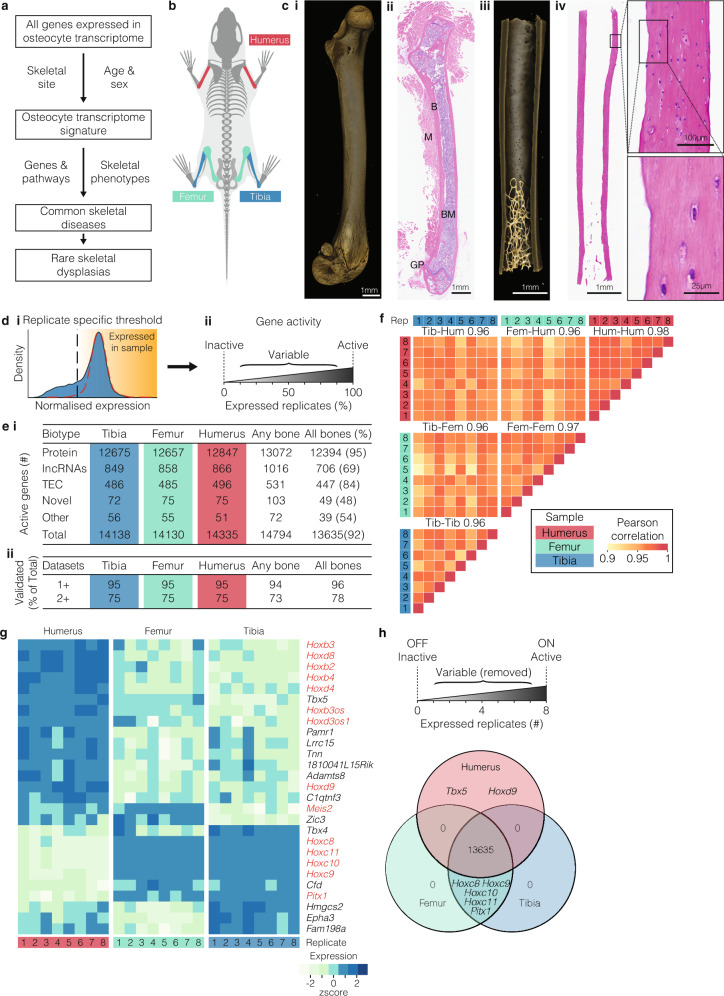
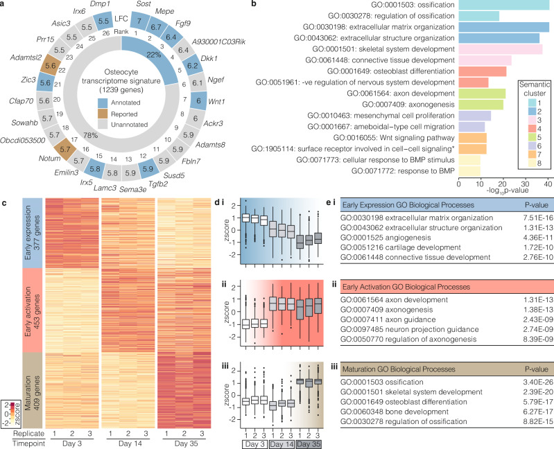
- 14,794 genes were actively expressed in osteocytes from tibiae, femora, and humeri; 92% were expressed at all sites and 96% were validated in orthogonal datasets. The OTS comprised 1,239 genes; 77% had no previously known role in the skeleton, and the signature was enriched for genes regulating neuronal network formation/axon guidance.
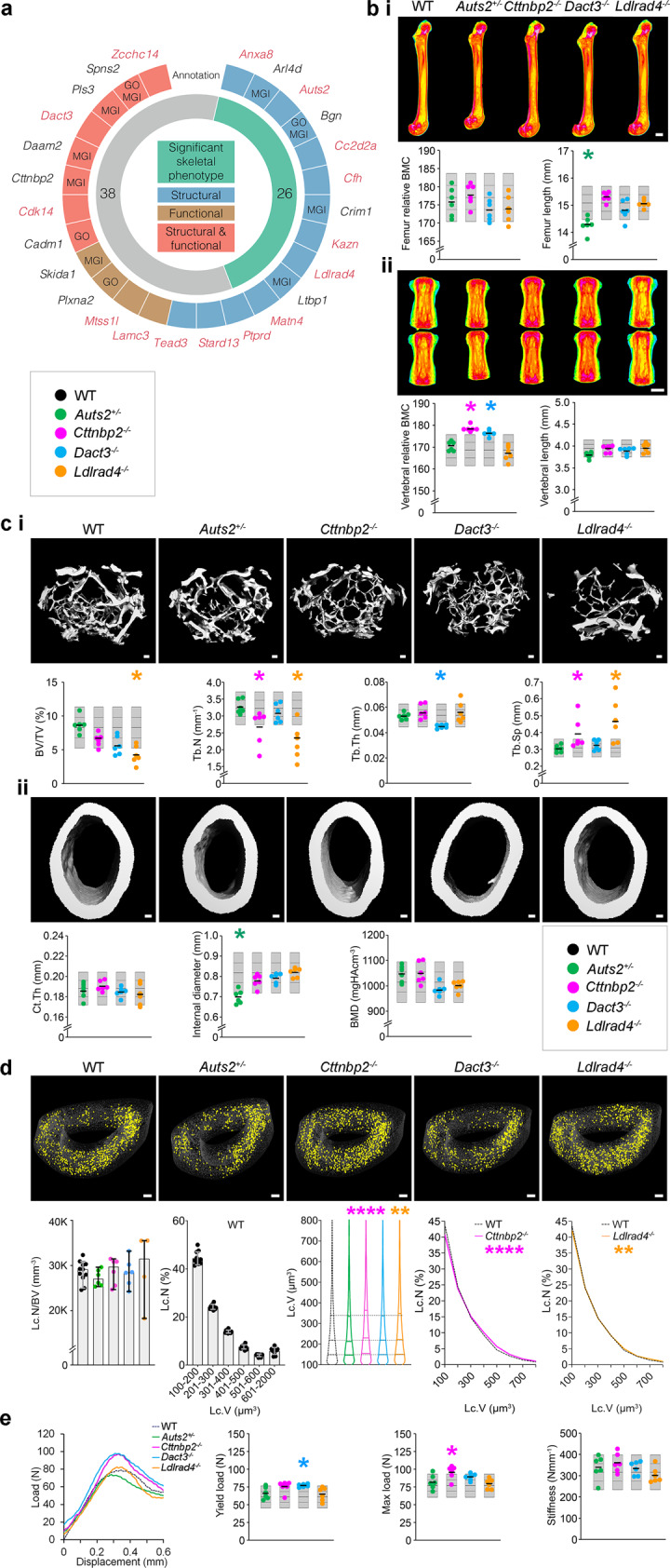
- In 733 knockout mouse lines screened, 64 had deletions of OTS genes and 26 (41%) had structural and/or functional skeletal phenotypes; 15 were in genes not annotated with skeletal terms or phenotypes. Examples included Auts2+/− with decreased femoral bone length and cortical diameter, Cttnbp2−/− with increased vertebral BMC and maximum load, Dact3−/− with increased vertebral BMC and yield load, and Ldlrad4−/− with decreased BV/TV and trabecular number and increased trabecular separation.
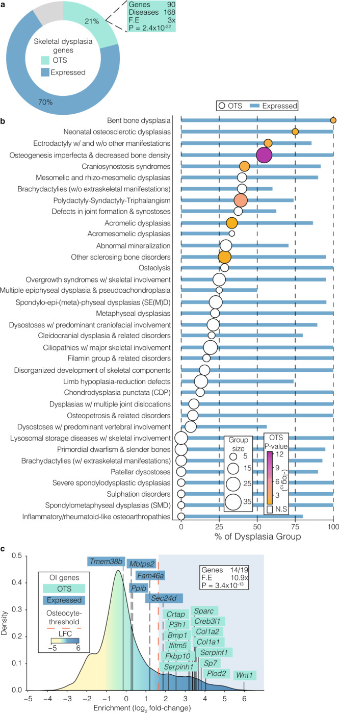
- OTS genes were enriched for human orthologs causing monogenic skeletal disorders: 90 OTS genes cause 168 of 612 annotated skeletal disorders (3-fold enrichment, P = 2.4 × 10⁻²²). In the osteogenesis imperfecta/decreased bone density group, 18/33 causal genes were in the signature (P = 7 × 10⁻¹³); all 19 known OI genes were actively expressed in osteocytes and 14 were in the OTS.
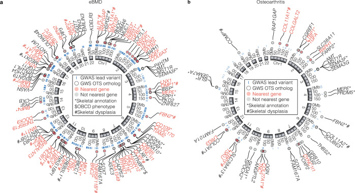
- OTS orthologs were associated with eBMD and osteoarthritis: LDSC-SEG showed OTS gene regions explained ~12.5% of eBMD SNP heritability (P = 1.6 × 10⁻⁷; 2.2-fold enrichment), competitive gene-set analysis P = 1.8 × 10⁻¹³, and 259/992 OTS orthologs had significant gene-level eBMD associations. For OA at any site, P = 1.6 × 10⁻⁷, ~11.6% of SNP heritability, and 40/992 OTS orthologs had significant gene-level associations.
Methods
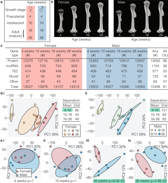
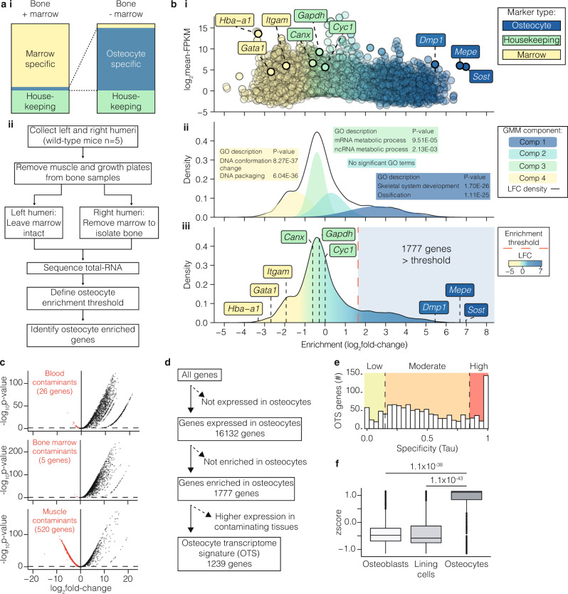
- Mouse RNA-seq cohorts: bone comparison cohort (16-week-old male tibiae, femora, humeri; n = 8 per bone type), skeletal maturation cohort (4-, 10-, 16-, and 26-week-old male and female humeri; n = 5 per sample type), and osteocyte enrichment cohort (10-week-old male humeri; n = 5 per condition). Osteocyte-enriched bone samples were >80% osteocytes; analyses included de novo transcriptome assembly, activity thresholds, Gaussian mixture modeling, WGCNA, and orthogonal dataset validation.
- Functional skeletal phenotyping used the Origins of Bone and Cartilage Disease (OBCD)/IMPC resource: 733 knockout mouse lines, 19 structural and functional skeletal parameters, DXA, micro-CT, and biomechanical testing; novel CRISPR-generated knockout lines were also phenotyped.
- Human genetic analyses used UK Biobank data for eBMD (362,924 individuals) and OA summary statistics (77,052 cases and 378,169 controls), with LDSC-SEG, MAGMA competitive gene-set analysis, and nearest-gene enrichment analysis.
Limitations
- The paper states: only long bones were studied, not spine or calvaria; cancellous bone was only 1–2% of sampled tissue, so the transcriptome mainly represents cortical osteocytes; contaminating cells cannot be fully excluded; and the study measured transcriptome, not protein expression.
- Evident from the text: skeletal phenotypes come from mouse knockouts, and human disease links are genetic associations rather than direct causal demonstrations; temporal osteocyte differentiation clusters rely on the in vitro IDG-SW3 cell-line model.
Model-written summary. Check it against the paper before relying on it.
Integrity signals
Registry
Cites
Statistics
Figures
Verification
Claims the paper makes
8 Valid · 0 Not valid · 26 Not checked · 163 Own results and interpretations
Every source the paper cites for this claim was checked and supports it.
In the paper
“In total, 85% of these genes showed moderate to high expression specificity29, using the Tau measure, within the osteocyte network relative to 12 non-skeletal tissues”
§ Osteocyte transcriptome mapping identifies a molecular landscape controlling skeletal homeostasis and susceptibility to skeletal disease > Results > A unique transcriptome signature defines the osteocyte
What the sources say
- Supportedcites [29] Yanai, I. et al. Genome-wide midrange transcription profiles reveal expression level relationships in human tissue specification. Bioinformatics 21, 650–659 (2005). source
The citation is attached to expression specificity, and the cited paper’s “Tissue specificity index” section defines τ as a graded measure of expression specificity. The 85% figure for 1,239 osteocyte-network genes is the citing authors’ result, not a result attributed to this source. (LLM, conf=0.88)
plain- Asserted: passes — The authors put it forward as true.
- Checkable: passes — It can be checked against evidence.
- Load-bearing: passes — The paper’s conclusions depend on it.
- Based: passes — The paper gives a reason why it holds.
- Supported
Every source the paper cites for this claim was checked and supports it.
In the paper
“As an additional level of stringency 538 genes enriched in bone marrow or tissues that could contaminate the osteocyte enrichment strategy, such as blood or muscle (Fig. 3c and Supplementary Data 2)13, were excluded leaving 1239 genes significantly enriched for expression in osteocytes”
§ Osteocyte transcriptome mapping identifies a molecular landscape controlling skeletal homeostasis and susceptibility to skeletal disease > Results > A unique transcriptome signature defines the osteocyte
What the sources say
- Supportedcites [13] Ayturk, U. M. et al. An RNA-seq protocol to identify mRNA expression changes in mouse diaphyseal bone: applications in mice with bone property altering Lrp5 mutations. J. Bone Miner. Res. 28, 2081–2093 (2013). source
The citation is attached to the identification of bone marrow, blood and muscle as potential contaminating tissues, not to the citing authors’ counts of 538 and 1,239 genes. The Results section (‘Improved in silico filtering of contaminating transcripts’) describes filtering transcripts associated with those tissues from diaphyseal bone RNA-seq data. (LLM, conf=0.88)
plain- Asserted: passes — The authors put it forward as true.
- Checkable: passes — It can be checked against evidence.
- Load-bearing: passes — The paper’s conclusions depend on it.
- Based: passes — The paper gives a reason why it holds.
- Supported
The cited sources support only part of this claim — read each source’s verdict below.
In the paper
“Mutations in genes that have highly enriched expression in osteocytes cause rare bone and mineral disorders.”
§ Osteocyte transcriptome mapping identifies a molecular landscape controlling skeletal homeostasis and susceptibility to skeletal disease > Introduction
What the sources say
- Not checked yetcites [17] Brunkow, M. E. et al. Bone dysplasia sclerosteosis results from loss of the SOST gene product, a novel cystine knot-containing protein. Am. J. Hum. Genet. 68, 577–589 (2001). source
- Partly supportedcites [18] Balemans, W. et al. Increased bone density in sclerosteosis is due to the deficiency of a novel secreted protein (SOST). Hum. Mol. Genet. 10, 537–543 (2001). sourceExperimental — accuracy not yet measured
Balemans et al. (2001) do support the mutation–disease part of the sentence: the Mutation analysis section reports three loss-of-function mutations (Trp124X, Arg126X, IVS1+3 A→T) in the previously unidentified SOST gene in sclerosteosis patients, a rare progressive sclerosing bone dysplasia (Abstract; Discussion). However, the claim's qualifier 'highly enriched expression in osteocytes' is not what this paper shows: the Expression analysis of the SOST mRNA section reports highly restricted, low-level expression with the highest level in kidney (2.5% of GAPDH), then bone marrow (0.6%), some in lung/heart/pancreas, and expression in differentiated primary human osteoblasts (0.3%) — osteocytes are not measured (searched: 'osteocyte expression', 'SOST expression in osteoblasts'; no exact osteocyte passage in our copy). The paper also makes no general statement about mutations in osteocyte-enriched genes causing rare bone and mineral disorders as a class; its concluding point is limited to SOST deficiency causing increased bone density in sclerosteosis. (LLM, conf=0.72)
- Partly supportedcites [19] Balemans, W. et al. Identification of a 52 kb deletion downstream of the SOST gene in patients with van Buchem disease. J. Med. Genet. 39, 91–97 (2002). sourceExperimental — accuracy not yet measured
Our copy supports the generic core of the claim: it reports that loss of function mutations in SOST cause sclerosteosis (Introduction/Discussion: 'This gene harbours different loss of function mutations within SOST in two sclerosteosis families...'; the 52 kb deletion downstream of SOST is implicated in van Buchem disease), both described as rare ('very uncommon disorder', 'incidence of sclerosteosis is very low') sclerosing bone dysplasias. However, the distinctive qualifier of the claim — that the mutated genes are 'highly enriched expression in osteocytes' — is absent: the only expression statement in our copy is that 'SOST is not expressed in lymphocytes or other readily available tissue' (Discussion), and no passage mentions osteocytes or mineral disorders. So the osteocyte-enrichment part of what is attributed to this source is not supported by it. (LLM, conf=0.68)
- Supportedcites [20] Feng, J. Q. et al. Loss of DMP1 causes rickets and osteomalacia and identifies a role for osteocytes in mineral metabolism. Nat. Genet. 38, 1310–1315 (2006). source
The Abstract states that DMP1 'is highly expressed in osteocytes' and that affected individuals in two consanguineous kindreds carry biallelic DMP1 mutations (start-codon A1→G; 7-bp deletion 1484-1490) causing autosomal recessive hypophosphatemic rickets with rickets, osteomalacia and renal phosphate-wasting. The same Introduction/Abstract frames these as 'human disorders of phosphate (Pi) handling and skeletal mineralization' arising from mutations in PHEX (XLH) and FGF23 (ADHR), with the Hyp mouse showing increased osteocyte expression of FGF23, and the Summary attributes both the skeletal mineralization and phosphate-homeostasis defects to defective osteocyte function. Thus our copy supports the paraphrase that mutations in osteocyte-enriched genes cause rare bone and mineral metabolism disorders. (LLM, conf=0.85)
plainstates a cause- Asserted: passes — The authors put it forward as true.
- Checkable: passes — It can be checked against evidence.
- Load-bearing: passes — The paper’s conclusions depend on it.
- Based: passes — The paper gives a reason why it holds.
The cited sources support only part of this claim — read each source’s verdict below.
In the paper
“Eleven were in genes with established roles in the skeleton, including Daam225 and Pls351.”
§ Osteocyte transcriptome mapping identifies a molecular landscape controlling skeletal homeostasis and susceptibility to skeletal disease > Results > Osteocyte transcriptome signature genes control bone structure and function
What the sources say
- Partly supportedcites [25] Morris, J. A. et al. An atlas of genetic influences on osteoporosis in humans and mice. Nat. Genet. 51, 258–266 (2019). sourceExperimental — accuracy not yet measured
The Results section on the mouse knockout screen reports skeletal phenotyping of 126 target-gene knockout lines, and the Discussion says the target genes were enriched for genes with known roles in bone biology. Our copy does not report that 11 of 26 knockout lines involved genes with established skeletal roles. The In-Depth Characterization of DAAM2 section instead describes DAAM2 as not previously implicated in osteoporosis. (LLM, conf=0.78)
- Supportedcites [51] Balasubramanian, M. et al. Novel PLS3 variants in X-linked osteoporosis: exploring bone material properties. Am. J. Med. Genet. A 176, 1578–1586 (2018). source
The citation is attached to Pls3 as an example of a gene with an established skeletal role, not to the count of eleven knockout lines. The paper describes PLS3 variants associated with reduced bone mineral density (p. 6) and concludes that PLS3 has a role in bone mineralisation (p. 5). (LLM, conf=0.91)
plain- Asserted: passes — The authors put it forward as true.
- Checkable: passes — It can be checked against evidence.
- Load-bearing: unclear — It is unclear whether the paper’s conclusions depend on it.
- Based: passes — The paper gives a reason why it holds.
- Partly supported
Every source the paper cites for this claim was checked and supports it.
In the paper
“Competitive gene-set analysis62 also showed that human orthologs of osteocyte transcriptome signature genes had stronger associations with OA than non-osteocyte genes (OA at any site (P = 9.1.6 × 10−3), at the knee (P = 7.2 × 10⁻³), hip (P = 3.0 × 10⁻³) and hip and/or knee (P = 6.6 × 10⁻²).”
§ Osteocyte transcriptome mapping identifies a molecular landscape controlling skeletal homeostasis and susceptibility to skeletal disease > Results > The osteocyte transcriptome signature is enriched for human orthologs associated with skeletal disease susceptibility
What the sources say
- Supportedcites [62] de Leeuw, C. A., Mooij, J. M., Heskes, T. & Posthuma, D. MAGMA: generalized gene-set analysis of GWAS data. PLoS Comput. Biol. 11, 1–19 (2015). source
Citation 62 is attached to “Competitive gene-set analysis,” identifying the method rather than attributing the osteoarthritis results or P-values to MAGMA. The source’s Materials and Methods section describes a competitive gene-set test that compares associations in a gene set with those in other genes. (LLM, conf=0.93)
plainstates an associationn = 77,052- Asserted: passes — The authors put it forward as true.
- Checkable: passes — It can be checked against evidence.
- Load-bearing: passes — The paper’s conclusions depend on it.
- Based: passes — The paper gives a reason why it holds.
- Supported
117 more claims · 1 with their cited sources checked
Create a free account to see all 197 claims and to verify the 34 references still open.
- It may state two separate claims as one. 47
- Probably not a substantial claim. 33
- The quote only partly states this claim. 2
Claims other papers make about this one
Each is checked against this paper’s own text: does it say what the citing paper says it does?
The cited source says what the claim attributes to it.
In the citing paper
“They are both expressed specifically in osteocytes 38 .”
From Sex-specific gene expression signatures of calcific aortic valve stenosis in a large collection of human tissues (2026) · cites this paper as [38]
Self-check
Percentages, sums and reported differences re-computed from the paper’s own numbers.
Each contradiction between two passages listed here was confirmed by an independent check.
Rated major: it would touch a result — a value a result rests on, reported and recomputed p-values on opposite sides of the significance level, two passages that conflict with no sign which is right, or a cited work retracted before this paper was published.
In the paper
§ Osteocyte transcriptome mapping identifies a molecular landscape controlling skeletal homeostasis and susceptibility to skeletal disease > Methods > Transcriptome sequencing mouse cohorts
“Left and right humeri were collected from 4, 10, 16 and 26-week-old female and male C57BL6/NTac mice (n = 5 per sample type, 80 samples in total).”
§ Osteocyte transcriptome mapping identifies a molecular landscape controlling skeletal homeostasis and susceptibility to skeletal disease > Methods > Morphological analysis of bone samples
“Whole femoral length, bone mineral density (BMD) and bone mineral content (BMC) were measured in excised left femora using a Lunar Piximus II dual X-ray absorptiometer (DXA) (GE Medical Systems).”
Rated minor: it would not touch a result — a value no result rests on, a p-value that stays on the same side of the significance level, or a conflict that is clerical, resolved elsewhere in the paper, or outside its own findings.
In the paper
“The osteocyte transcriptome signature gene annotation (±20 kb) spanned 5.1% of the genome (i.e. 65,332/1,142,435 SNPs)”
§ Osteocyte transcriptome mapping identifies a molecular landscape controlling skeletal homeostasis and susceptibility to skeletal disease > Results > The osteocyte transcriptome signature is enriched for human orthologs associated with skeletal disease susceptibility
Reported 5.1 · recomputed 5.718662
Rated minor: it would not touch a result — a value no result rests on, a p-value that stays on the same side of the significance level, or a conflict that is clerical, resolved elsewhere in the paper, or outside its own findings.
In the paper
§ Osteocyte transcriptome mapping identifies a molecular landscape controlling skeletal homeostasis and susceptibility to skeletal disease > Abstract
“77% have no previously known role in the skeleton”
§ Osteocyte transcriptome mapping identifies a molecular landscape controlling skeletal homeostasis and susceptibility to skeletal disease > Results > The majority of osteocyte transcriptome signature genes have no known function in the skeleton
“The majority of osteocyte transcriptome signature genes (78%, n = 968) had not previously been shown to have a role in the skeleton”
Citations
Cites
Fiji: an open-source platform for biological-image analysis
Schindelin, J., Arganda-Carreras, I., Frise, E., Kaynig, V., Longair, M., Pietzsch, T., Preibisch, S., Rueden, C., Saalfeld, S., Schmid, B., et al. (2012) · Nature Methods · doi:10.1038/nmeth.2019 · Cited by 66,890
STAR: ultrafast universal RNA-seq aligner
Dobin, A., Davis, C. A., Schlesinger, F., Drenkow, J., Zaleski, C., Jha, S., Batut, P., Chaisson, M., & Gingeras, T. R. (2013) · Bioinformatics · doi:10.1093/bioinformatics/bts635 · Cited by 51,709
Gene Ontology: tool for the unification of biology
Ashburner, M., Ball, C. A., Blake, J. A., Botstein, D., Butler, H., Cherry, J. M., Davis, A. P., Dolinski, K., Dwight, S. S., Eppig, J. T., et al. (2000) · Nature Genetics · doi:10.1038/75556 · Cited by 43,206
limma powers differential expression analyses for RNA-sequencing and microarray studies
Ritchie, M. E., Phipson, B., Wu, D., Hu, Y., Law, C. W., Shi, W., & Smyth, G. K. (2015) · Nucleic Acids Research · doi:10.1093/nar/gkv007 · Cited by 40,458
clusterProfiler: an R Package for Comparing Biological Themes Among Gene Clusters
Yu, G., Wang, L.-G., Han, Y., & He, Q.-Y. (2012) · OMICS: A Journal of Integrative Biology · doi:10.1089/omi.2011.0118 · Cited by 36,259
Wickham, H. (2016) · Use R! · doi:10.1007/978-3-319-24277-4 · Cited by 33,115
KEGG: Kyoto Encyclopedia of Genes and Genomes
Ogata, H., Goto, S., Sato, K., Fujibuchi, W., Bono, H., & Kanehisa, M. (1999) · Nucleic Acids Research · doi:10.1093/nar/27.1.29 · Cited by 31,232
WGCNA: an R package for weighted correlation network analysis
Langfelder, P., & Horvath, S. (2008) · BMC Bioinformatics · doi:10.1186/1471-2105-9-559 · Cited by 27,252
RSEM: accurate transcript quantification from RNA-Seq data with or without a reference genome
Li, B., & Dewey, C. N. (2011) · BMC Bioinformatics · doi:10.1186/1471-2105-12-323 · Cited by 22,179
Ror2 signaling regulates Golgi structure and transport through IFT20 for tumor invasiveness
Nishita, M., Park, S.-Y., Nishio, T., Kamizaki, K., Wang, Z., Tamada, K., Takumi, T., Hashimoto, R., Otani, H., Pazour, G. J., et al. (2017) · Scientific Reports · doi:10.1038/s41598-016-0028-x · Cited by 15,947
Research works printed but not linked: 1
- Warnes, G. R. et al. gplots: various R programming tools for plotting data. R. Package version 2, 1 (2009).
Cited by
Osteoclasts recycle via osteomorphs during RANKL-stimulated bone resorption
McDonald, M. M., Khoo, W. H., Ng, P. Y., Xiao, Y., Zamerli, J., Thatcher, P., Kyaw, W., Pathmanandavel, K., Grootveld, A. K., Moran, I., et al. (2021) · Cell · doi:10.1016/j.cell.2021.02.002 · Cited by 433
“This separation between Acp5 gene and TRAP protein expression has also been described in osteocytes (Youlten et al., 2020).”
Self-citationThe osteocyte as a signaling cell
Delgado-Calle, J., & Bellido, T. (2022) · Physiological Reviews · doi:10.1152/physrev.00043.2020 · Cited by 261
Tang, Y., Peng, B., Liu, J., Liu, Z., Xia, Y., & Geng, B. (2022) · Frontiers in Immunology · doi:10.3389/fimmu.2022.975400 · Cited by 188
Osteocytes in bone aging: Advances, challenges, and future perspectives
Cui, J., Shibata, Y., Zhu, T., Zhou, J., & Zhang, J. (2022) · Ageing Research Reviews · doi:10.1016/j.arr.2022.101608 · Cited by 171
Control of osteocyte dendrite formation by Sp7 and its target gene osteocrin
Wang, J. S., Kamath, T., Mazur, C. M., Mirzamohammadi, F., Rotter, D., Hojo, H., Castro, C. D., Tokavanich, N., Patel, R., Govea, N., et al. (2021) · Nature Communications · doi:10.1038/s41467-021-26571-7 · Cited by 99
Metadata
- Authors
- Youlten, S. E., Kemp, J. P., Logan, J. G., Ghirardello, E. J., Sergio, C. M., Dack, M. R. G., Guilfoyle, S. E., Leitch, V. D., Butterfield, N. C., Komla-Ebri, D., Chai, R. C., Corr, A. P., Smith, J. T., Mohanty, S. T., Morris, J. A., McDonald, M. M., Quinn, J. M. W., McGlade, A. R., Bartonicek, N., Jansson, M., Hatzikotoulas, K., Irving, M. D., Beleza-Meireles, A., Rivadeneira, F., Duncan, E., Richards, J. B., Adams, D. J., Lelliott, C. J., Brink, R., Phan, T. G., Eisman, J. A., Evans, D. M., Zeggini, E., Baldock, P. A., Bassett, J. H. D., Williams, G. R., & Croucher, P. I.
- Affiliations
- Garvan Institute of Medical Research (AU)
- The University of Queensland (AU)
- Imperial College London (GB)
- New York Genome Center (US)
- Guy's Hospital (GB)
- Wellcome Sanger Institute (GB)
- Erasmus MC (NL)
- Queensland University of Technology (AU)
- King's College London (GB)
- Corresponding author
- Bassett, J. H. Duncan
- Williams, Graham R.
- Croucher, Peter I.
- ORCID
- Youlten, Scott E.
- Kemp, John P.
- Logan, John G.
- Ghirardello, Elena J.
- Sergio, Claudio M.
- Dack, Michael R. G.
- Guilfoyle, Siobhan E.
- Leitch, Victoria D.
- Butterfield, Natalie C.
- Komla-Ebri, Davide
- Chai, Ryan C.
- Corr, Alexander P.
- Smith, James T.
- Mohanty, Sindhu T.
- Morris, John A.
- McDonald, Michelle M.
- Quinn, Julian M. W.
- Hatzikotoulas, Konstantinos
- Irving, Melita D.
- Rivadeneira, Fernando
- Duncan, Emma
- Richards, J. Brent
- Adams, David J.
- Lelliott, Christopher J.
- Brink, Robert
- Phan, Tri Giang
- Eisman, John A.
- Evans, David M.
- Zeggini, Eleftheria
- Bassett, J. H. Duncan
- Williams, Graham R.
- Croucher, Peter I.
- Venue
- Nature Communications
- Published
- 2021-05-05
- Volume
- 12
- Issue
- 1
- Pages
- 2444-2444
- ISSN
- 2041-1723 (ISSN-L)
- PMC
- PMC8100170
- OpenAlex
- W3157344667
- Semantic Scholar
- 7bef6229f2fb943e57cb887d9b76bef463adf4c0
- Cited by
- 133
- References
- 134
- Citations by year
- 2026: 27 · 2025: 24 · 2024: 21 · 2023: 28 · 2022: 13 · 2021: 14
- FWCI
- 5.93
- Type
- journal article
- Language
- en
- Open access
- gold
- doi.org
- Licence
- cc-by
- creativecommons.org/licenses/by/4.0 (tdm)
- creativecommons.org/licenses/by/4.0 (vor)
- Locations
- Nature Communications (publishedVersion, cc-by)
- bioRxiv (Cold Spring Harbor Laboratory) (acceptedVersion, cc-by-nc-nd)
- PubMed Central (publishedVersion, cc-by)
- PubMed (publishedVersion)
- QUT ePrints (Queensland University of Technology) (submittedVersion, cc-by)
- ResearchOnline@ND (The University of Notre Dame) (submittedVersion)
- EUR Research Repository (Erasmus University Rotterdam) (submittedVersion, other-oa)
- RePub (Erasmus University Rotterdam) (submittedVersion, other-oa)
- RePEc: Research Papers in Economics (submittedVersion)
- RMIT Research Repository (RMIT University Library) (submittedVersion, other-oa)
- DOAJ (DOAJ: Directory of Open Access Journals) (submittedVersion, cc-by-sa)
- Figshare (submittedVersion, cc-by)
- Research Portal (King's College London) (submittedVersion, other-oa)
- Site cant be reached (submittedVersion, cc-by)
- PuSH - Publication Server of Helmholtz Zentrum München (submittedVersion, cc-by)
- Spiral (Imperial College London) (submittedVersion, cc-by)
- UNSWorks (University of New South Wales, Sydney, Australia) (submittedVersion, cc-by-nc-nd)
- Apollo (University of Cambridge) (submittedVersion, cc-by)
- Apollo (cc-by)
- Funders
- Wellcome Trust (101123/Z/13/A)
- Mrs Janice Gibson and the Ernest Heine Family Foundation
- Topics
- Life Sciences › Biochemistry, Genetics and Molecular Biology › Molecular Biology › Bone Metabolism and Diseases
- Health Sciences › Medicine › Orthopedics and Sports Medicine › Bone health and osteoporosis research
- Life Sciences › Biochemistry, Genetics and Molecular Biology › Biochemistry › Bone and Dental Protein Studies
- Keywords
- osteocytes, transcriptome, skeletal homeostasis, neuronal network formation, osteoporosis, osteoarthritis, bone structure, sex differences, aging
- MeSH terms
- Age Factors, Animals, Bone and Bones, Bone Diseases, Female, Homeostasis, Humans, Male, Osteocytes, Osteoporosis, Sex Factors, Sequence Analysis, RNA, Mice, Knockout, Computational Biology, Mice, Transcriptome
About these checks
Registry, retraction, Cites and statistics results are computed, not judged, with one exception: a printed reference that no index lists is read once by a model before it counts as a citation error. Claims are extracted by a model and kept only when their quote appears verbatim in the text; each cited source is then checked, with a model’s help, for whether it says what the claim relies on. Those verdicts can be wrong — read the claim’s quote and the cited source before relying on one.
The claim verdicts this report counts are experimental: their accuracy is not yet measured.
A language model drew the claims from the paper on 2026-10-04, under version 3 of the claim definition (model deepseek-v4.1-flash, prompt extract-v3).
A second model labelled each claim on 2026-10-04 (model jev-1.13-20260917, definition revision gold-guideline-v0.2).
Cited sources are checked with a language model’s help (gpt-6-sol and qwen3.8-omni-flash).
Statistics and arithmetic are recomputed, not judged.
Confidence bands are shown only for fields calibrated on our annotated test set. None is calibrated yet, so no band is shown.
What counts as a claim ›How we measure confidence ›How we check this ›
Are you an author, or do you see a problem with this page? Request a review
How to cite
Cite CoChat for the checks on this page, and the paper for its own findings.
- This page
- CoChat. (2026). Citation check: Osteocyte transcriptome mapping identifies a molecular landscape controlling skeletal homeostasis and susceptibility to skeletal disease. https://app.cochat.ai/papers/10.1038/s41467-021-22517-1
- The paper
- Youlten, S. E., Kemp, J. P., Logan, J. G., Ghirardello, E. J., Sergio, C. M., Dack, M. R. G., Guilfoyle, S. E., Leitch, V. D., Butterfield, N. C., Komla-Ebri, D., Chai, R. C., Corr, A. P., Smith, J. T., Mohanty, S. T., Morris, J. A., McDonald, M. M., Quinn, J. M. W., McGlade, A. R., Bartonicek, N., . . . Croucher, P. I. (2021). Osteocyte transcriptome mapping identifies a molecular landscape controlling skeletal homeostasis and susceptibility to skeletal disease. Nature Communications. https://doi.org/10.1038/s41467-021-22517-1
Data sources
Bibliographic data: Semantic Scholar, OpenAlex, Crossref.
Contains information from the Semantic Scholar Academic Graph Datasets, made available under the ODC Attribution License (ODC-BY 1.0).
Semantic Scholar’s data is described in: Kinney, R., et al. (2023). The Semantic Scholar Open Data Platform. arXiv:2301.10140.
