

The DOI matches its Crossref record; 11 of the 11 references it cites were found in a registry or an index.
Dual targeted extracellular vesicles regulate oncogenic genes in advanced pancreatic cancer
Abstract
Pancreatic ductal adenocarcinoma (PDAC) tumours carry multiple gene mutations and respond poorly to treatments. There is currently an unmet need for drug carriers that can deliver multiple gene cargoes to target high solid tumour burden like PDAC. Here, we report a dual targeted extracellular vesicle (dtEV) carrying high loads of therapeutic RNA that effectively suppresses large PDAC tumours in mice. The EV surface contains a CD64 protein that has a tissue targeting peptide and a humanized monoclonal antibody. Cells sequentially transfected with plasmid DNAs encoding for the RNA and protein of interest by Transwell®-based asymmetric cell electroporation release abundant targeted EVs with high RNA loading. Together with a low dose chemotherapy drug, Gemcitabine, dtEVs suppress large orthotopic PANC-1 and patient derived xenograft tumours and metastasis in mice and extended animal survival. Our work presents a clinically accessible and scalable way to produce abundant EVs for delivering multiple gene cargoes to large solid tumours.
Overview
The claims and statistics of this paper haven’t been checked yet.
Run the check — free, no account needed. It reads the open-access text and checks the sources it cites.
Integrity signals
Registry
Cites
Statistics
Figures
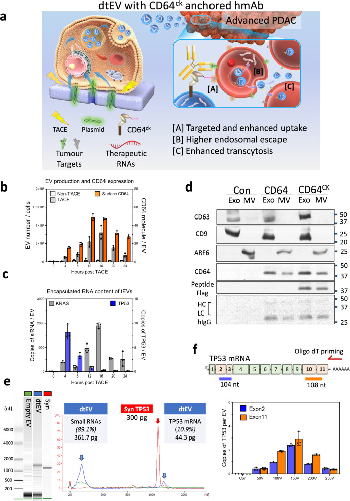
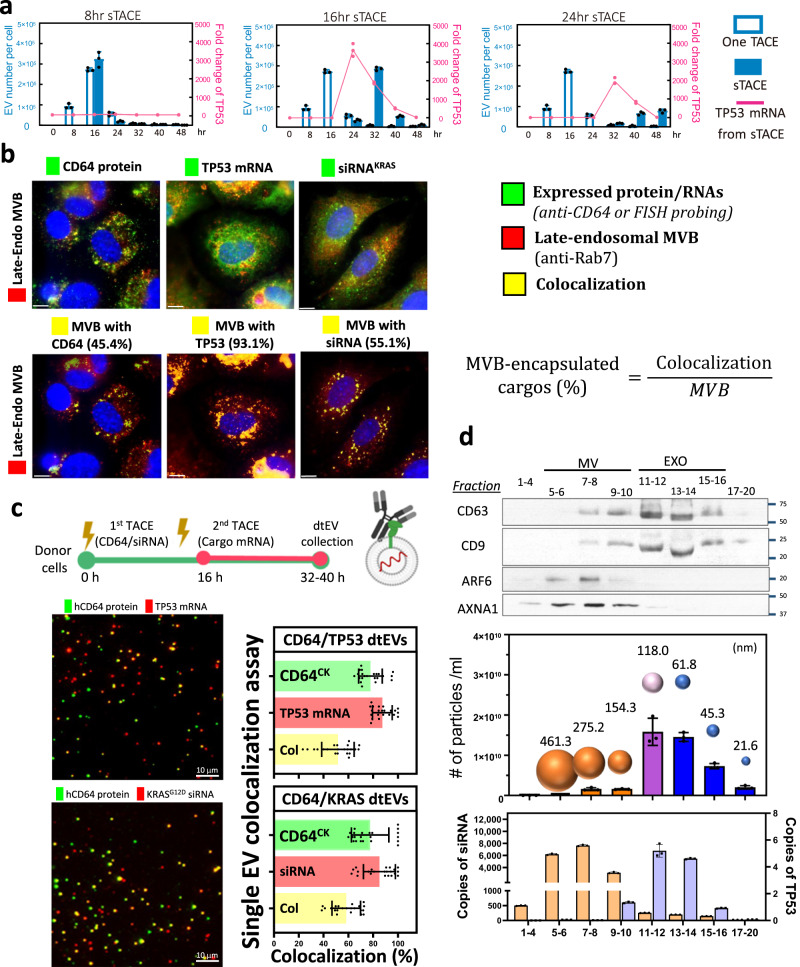
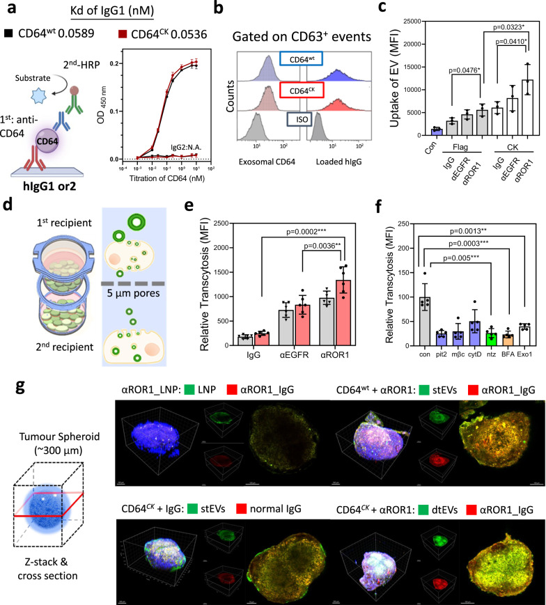
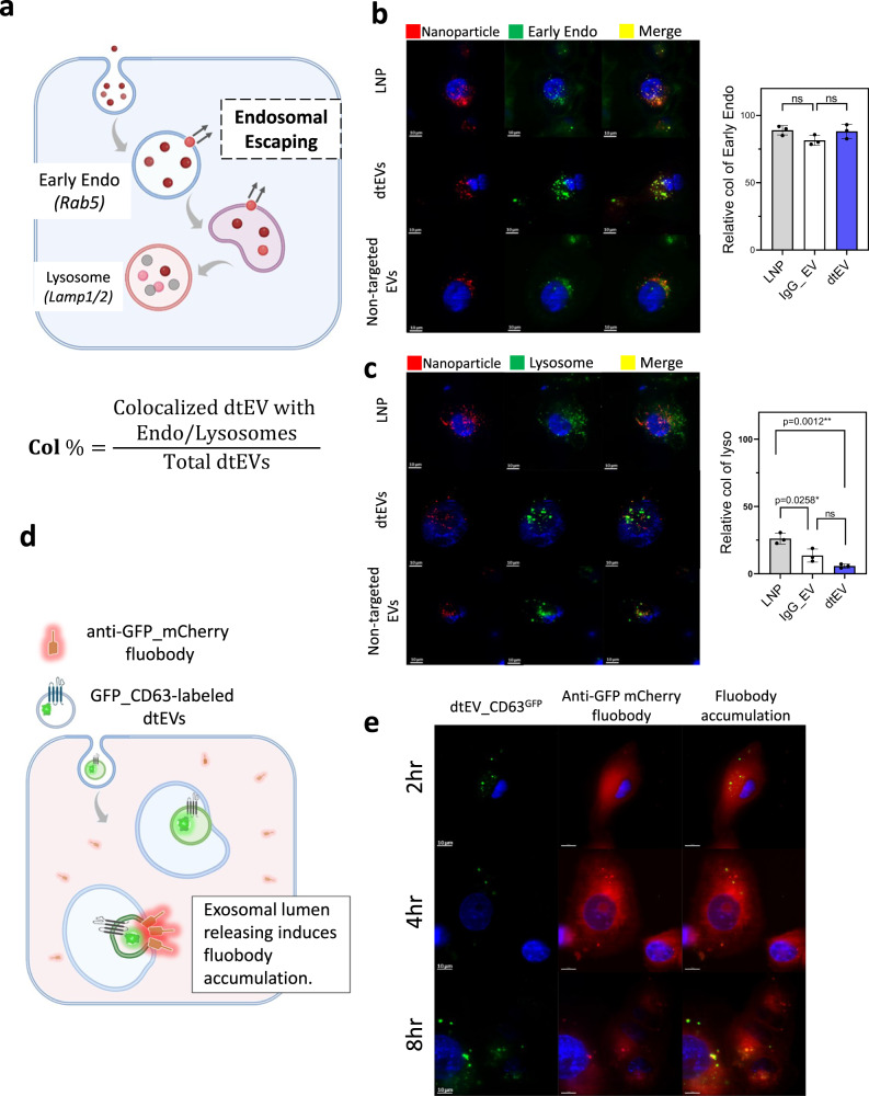
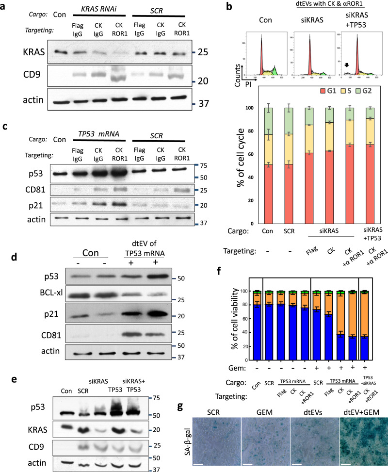
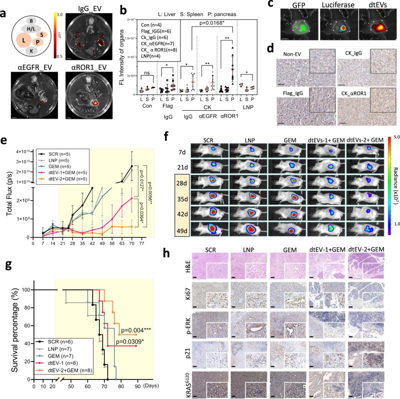
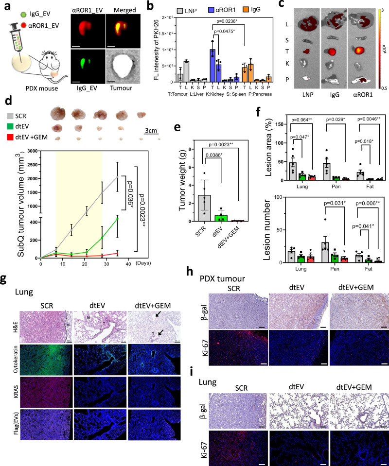
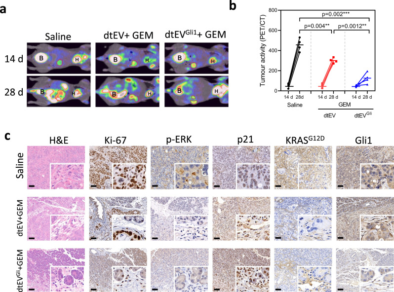
Verification
Claims the paper makes
The claims and statistics of this paper haven’t been checked yet.
Run the check — free, no account needed. It reads the open-access text and checks the sources it cites.
Claims other papers make about this one
Each is checked against this paper’s own text: does it say what the citing paper says it does?
In the citing paper
“| Pancreatic cancer | dtEVs | Effectively suppressed large solid PDAC | [141] |”
From Emerging role of exosomes in cancer therapy: progress and challenges (2025) · cites this paper as [141]
In the citing paper
“This study showcases an easy, cost-effective method for producing plentiful targeted EVs loaded with high levels of genetic cargo, demonstrating their potential for effectively treating advanced cancers in animal models [141].”
From Emerging role of exosomes in cancer therapy: progress and challenges (2025) · cites this paper as [141]
Self-check
Percentages, sums and reported differences re-computed from the paper’s own numbers.
The self-check needs the paper’s full text.
Citations
Cites
Exosomes facilitate therapeutic targeting of oncogenic KRAS in pancreatic cancer
Kamerkar, S., LeBleu, V. S., Sugimoto, H., Yang, S., Ruivo, C. F., Melo, S. A., Lee, J. J., & Kalluri, R. (2017) · Nature · doi:10.1038/nature22341 · Cited by 2,639
Hidalgo, M. (2010) · New England Journal of Medicine · doi:10.1056/nejmra0901557 · Cited by 2,570
Sotorasib for Lung Cancers with KRAS p.G12C Mutation
Skoulidis, F., Li, B. T., Dy, G. K., Price, T. J., Falchook, G. S., Wolf, J., Italiano, A., Schuler, M., Borghaei, H., Barlesi, F., et al. (2021) · New England Journal of Medicine · doi:10.1056/nejmoa2103695 · Cited by 1,849
RNA delivery by extracellular vesicles in mammalian cells and its applications
O’Brien, K., Breyne, K., Ughetto, S., Laurent, L. C., & Breakefield, X. O. (2020) · Nature Reviews Molecular Cell Biology · doi:10.1038/s41580-020-0251-y · Cited by 1,811
Senescence in tumours: evidence from mice and humans
Collado, M., & Serrano, M. (2010) · Nature Reviews Cancer · doi:10.1038/nrc2772 · Cited by 1,124 · cited 1×
“It is well-established that aberrant activation of mutant KRAS can override oncogene-induced senescence (OIS), thereby promoting the survival and proliferation of cancer cells 36,37 .”
BackgroundKRAS: The Critical Driver and Therapeutic Target for Pancreatic Cancer
Waters, A. M., & Der, C. J. (2018) · Cold Spring Harbor Perspectives in Medicine · doi:10.1101/cshperspect.a031435 · Cited by 875
Boosting Brain Uptake of a Therapeutic Antibody by Reducing Its Affinity for a Transcytosis Target
Yu, Y. J., Zhang, Y., Kenrick, M., Hoyte, K., Luk, W., Lu, Y., Atwal, J., Elliott, J. M., Prabhu, S., Watts, R. J., et al. (2011) · Science Translational Medicine · doi:10.1126/scitranslmed.3002230 · Cited by 794
Large-scale generation of functional mRNA-encapsulating exosomes via cellular nanoporation
Yang, Z., Shi, J., Xie, J., Wang, Y., Sun, J., Liu, T., Zhao, Y., Zhao, X., Wang, X., Ma, Y., et al. (2019) · Nature Biomedical Engineering · doi:10.1038/s41551-019-0485-1 · Cited by 744
Self-citationGonzalez-Quintela, A., Alende, R., Gude, F., Campos, J., Rey, J., Meijide, L. M., Fernandez-Merino, C., & Vidal, C. (2007) · Clinical and Experimental Immunology · doi:10.1111/j.1365-2249.2007.03545.x · Cited by 630
Kong, N., Tao, W., Ling, X., Wang, J., Xiao, Y., Shi, S., Ji, X., Shajii, A., Gan, S. T., Kim, N. Y., et al. (2019) · Science Translational Medicine · doi:10.1126/scitranslmed.aaw1565 · Cited by 314
Clathrin adaptor AP1B controls adenovirus infectivity of epithelial cells
Diaz, F., Gravotta, D., Deora, A., Schreiner, R., Schoggins, J., Falck-Pedersen, E., & Rodriguez-Boulan, E. (2009) · Proceedings of the National Academy of Sciences · doi:10.1073/pnas.0811227106 · Cited by 73
Research works printed but not linked: 35
- Siegel, R. L., Miller, K. D., Fuchs, H. E. & Jemal, A. Cancer Statistics, 2021. CA Cancer J. Clin. 71, 7–33 (2021). · DOI · Paper page
- Kim, M. P. et al. Oncogenic KRAS recruits an expansive transcriptional network through mutant p53 to drive pancreatic cancer metastasis. Cancer Discov. 11, 2094–2111 (2021). · DOI
- Shirley, J. L., de Jong, Y. P., Terhorst, C. & Herzog, R. W. Immune responses to viral gene therapy vectors. Mol. Ther. 28, 709–722 (2020). · DOI · Paper page
- Witwer, K. W. & Wolfram, J. Extracellular vesicles versus synthetic nanoparticles for drug delivery. Nat. Rev. Mater. 6, 103–106 (2021). · DOI
- Herrmann, I. K., Wood, M. J. A. & Fuhrmann, G. Extracellular vesicles as a next-generation drug delivery platform. Nat. Nanotechnol. 16, 748–759 (2021). · DOI · Paper page
- Alvarez-Erviti, L. et al. Delivery of siRNA to the mouse brain by systemic injection of targeted exosomes. Nat. Biotechnol. 29, 341–345 (2011). · DOI · Paper page
- Kojima, R. et al. Designer exosomes produced by implanted cells intracerebrally deliver therapeutic cargo for Parkinson’s disease treatment. Nat. Commun. 9, 1305 (2018). · DOI · Paper page
- Joyce, J. A. et al. Stage-specific vascular markers revealed by phage display in a mouse model of pancreatic islet tumorigenesis. Cancer Cell 4, 393–403 (2003). · DOI · Paper page
- Dekkers, G. et al. Affinity of human IgG subclasses to mouse Fc gamma receptors. MAbs 9, 767–773 (2017). · DOI · Paper page
- Cui, B. et al. Targeting ROR1 inhibits epithelial-mesenchymal transition and metastasis. Cancer Res. 73, 3649–3660 (2013). · DOI · Paper page
- Boukany, P. E. et al. Nanochannel electroporation delivers precise amounts of biomolecules into living cells. Nat. Nanotechnol. 6, 747–754 (2011). · DOI
- Nguyen, L. T. H. et al. An immunogold single extracellular vesicular RNA and protein ((Au) SERP) biochip to predict responses to immunotherapy in non-small cell lung cancer patients. J. Extracell. Vesicles 11, e12258 (2022). · DOI
- Hu, J. et al. A signal-amplifiable biochip quantifies extracellular vesicle-associated RNAs for early cancer detection. Nat. Commun. 8, 1683 (2017).
- Li, M. et al. Structural basis of dual Ca(2+)/pH regulation of the endolysosomal TRPML1 channel. Nat. Struct. Mol. Biol. 24, 205–213 (2017). · DOI
- Morad, G. et al. Tumor-derived extracellular vesicles breach the intact blood-brain barrier via transcytosis. ACS Nano 13, 13853–13865 (2019). · DOI
- Ghetie, V. & Ward, E. S. Transcytosis and catabolism of antibody. Immunol. Res. 25, 97–113 (2002). · DOI
- Dutta, D., Williamson, C. D., Cole, N. B. & Donaldson, J. G. Pitstop 2 is a potent inhibitor of clathrin-independent endocytosis. PLoS ONE 7, e45799 (2012). · DOI
- Le, P. U., Guay, G., Altschuler, Y. & Nabi, I. R. Caveolin-1 is a negative regulator of caveolae-mediated endocytosis to the endoplasmic reticulum. J. Biol. Chem. 277, 3371–3379 (2002). · DOI
- Datta, A. et al. High-throughput screening identified selective inhibitors of exosome biogenesis and secretion: a drug repurposing strategy for advanced cancer. Sci. Rep. 8, 8161 (2018). · DOI
- Joshi, B. S., de Beer, M. A., Giepmans, B. N. G. & Zuhorn, I. S. Endocytosis of extracellular vesicles and release of their cargo from endosomes. ACS Nano 14, 4444–4455 (2020). · DOI
- Bonsergent, E. et al. Quantitative characterization of extracellular vesicle uptake and content delivery within mammalian cells. Nat. Commun. 12, 1864 (2021). · DOI
- Frank, P. G., Pedraza, A., Cohen, D. E. & Lisanti, M. P. Adenovirus-mediated expression of caveolin-1 in mouse liver increases plasma high-density lipoprotein levels. Biochemistry 40, 10892–10900 (2001). · DOI
- Di Pasquale, G. & Chiorini, J. A. AAV transcytosis through barrier epithelia and endothelium. Mol. Ther. 13, 506–516 (2006). · DOI · Paper page
- Xiao, P. J. & Samulski, R. J. Cytoplasmic trafficking, endosomal escape, and perinuclear accumulation of adeno-associated virus type 2 particles are facilitated by microtubule network. J. Virol. 86, 10462–10473 (2012). · DOI
- Vincent, A., Herman, J., Schulick, R., Hruban, R. H. & Goggins, M. Pancreatic cancer. Lancet 378, 607–620 (2011). · DOI
- Lee, K. E. & Bar-Sagi, D. Oncogenic KRas suppresses inflammation-associated senescence of pancreatic ductal cells. Cancer Cell 18, 448–458 (2010). · DOI
- Shirole, N. H. et al. TP53 exon-6 truncating mutations produce separation of function isoforms with pro-tumorigenic functions. Elife 6, e25532 (2017). · DOI
- Rajurkar, M. et al. The activity of Gli transcription factors is essential for Kras-induced pancreatic tumorigenesis. Proc. Natl Acad. Sci. USA 109, E1038–1047 (2012). · DOI
- Ji, Z., Mei, F. C., Xie, J. & Cheng, X. Oncogenic KRAS activates hedgehog signaling pathway in pancreatic cancer cells. J. Biol. Chem. 282, 14048–14055 (2007). · DOI
- Wang, Y. et al. Oncogenic functions of gli1 in pancreatic adenocarcinoma are supported by its PRMT1-mediated methylation. Cancer Res. 76, 7049–7058 (2016). · DOI
- Jia, Y. et al. The role of GLI-SOX2 signaling axis for gemcitabine resistance in pancreatic cancer. Oncogene 38, 1764–1777 (2019). · DOI
- Alvarez-Erviti, L. et al. Delivery of siRNA to the mouse brain by systemic injection of targeted exosomes. Nat. Biotechnol. 29, 341–U179 (2011). · DOI · Paper page
- Kim, M. S. et al. Engineering macrophage-derived exosomes for targeted paclitaxel delivery to pulmonary metastases: in vitro and in vivo evaluations. Nanomedicine 14, 195–204 (2018). · DOI · Paper page
- Gabizon, A. & Papahadjopoulos, D. Liposome formulations with prolonged circulation time in blood and enhanced uptake by tumors. Proc. Natl Acad. Sci. USA 85, 6949–6953 (1988). · DOI
- Chiang, C. L. et al. ROR1-targeted delivery of miR-29b induces cell cycle arrest and therapeutic benefit in vivo in a CLL mouse model. Blood 134, 432–444 (2019). · DOI
Cited by
Sorted by citations
Emerging role of exosomes in cancer therapy: progress and challenges
Li, J., Wang, J., & Chen, Z. (2025) · Molecular Cancer · doi:10.1186/s12943-024-02215-4 · Cited by 297
Metadata
- Authors
- Chiang, C.-L., Ma, Y., Hou, Y.-C., Pan, J., Chen, S.-Y., Chien, M.-H., Zhang, Z.-X., Hsu, W.-H., Wang, X., Zhang, J., Li, H., Sun, L., Fallen, S., Lee, I., Chen, X.-Y., Chu, Y.-S., Zhang, C., Cheng, T.-S., Jiang, W., Kim, B. Y. S., Reategui, E., Lee, R., Yuan, Y., Liu, H.-C., Wang, K., Hsiao, M., Huang, C.-Y. F., Shan, Y.-S., Lee, A. S., & James Lee, L.
- Venue
- Nature Communications
- Published
- 2023-10-23
- Volume
- 14
- Issue
- 1
- ISSN
- 2041-1723
- PMC
- PMC10593751
- Cited by
- 63
- References
- 11
- Language
- en
- Funders
- National Science and Technology Council
About these checks
Registry, retraction, Cites and statistics results are computed, not judged, with one exception: a printed reference that no index lists is read once by a model before it counts as a citation error. Claims are extracted by a model and kept only when their quote appears verbatim in the text; each cited source is then checked, with a model’s help, for whether it says what the claim relies on. Those verdicts can be wrong — read the claim’s quote and the cited source before relying on one.
Statistics and arithmetic are recomputed, not judged.
Confidence bands are shown only for fields calibrated on our annotated test set. None is calibrated yet, so no band is shown.
What counts as a claim ›How we measure confidence ›How we check this ›
Are you an author, or do you see a problem with this page? Request a review
