

The DOI matches its Crossref record; 77 of the 77 references it cites were found in a registry or an index.
The Human Microglia Atlas (HuMicA) unravels changes in disease-associated microglia subsets across neurodegenerative conditions
Open accessPublished version of a preprint
Abstract
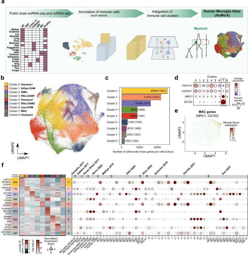
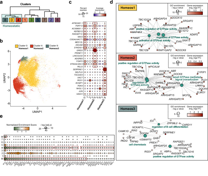
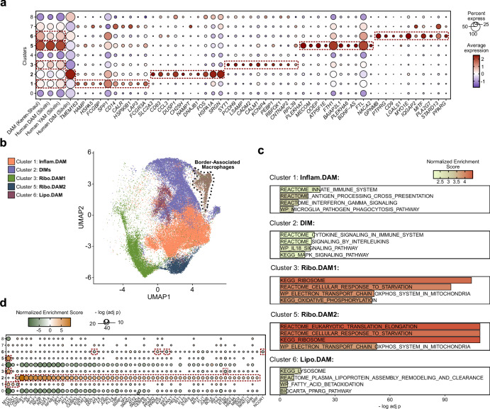
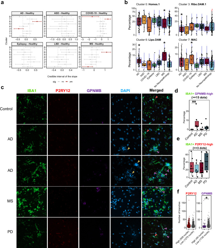
ABSTRACT Dysregulated microglia activation, leading to neuroinflammation, is crucial in neurodegenerative disease development and progression. The initial M1/M2 dual activation classification for microglia is outdated. Even the ‘disease-associated microglia’ (DAM) phenotype, firstly described in mice, falls short in representing the diverse microglia phenotypes in pathology. In this study, we have constructed a transcriptomic atlas of human brain immune cells by integrating single-nucleus (sn)RNA-seq datasets from multiple neurodegenerative conditions. Sixteen datasets were included, comprising 295 samples from patients with Alzheimer’s disease, autism spectrum disorder, epilepsy, multiple sclerosis, Lewy body diseases, COVID-19, and healthy controls. The integrated Human Microglia Atlas ( HuMicA ) dataset included 60,557 nuclei and revealed 11 microglial subpopulations distributed across all pathological and healthy conditions. Among these, we identified four different homeostatic clusters as well as pathological phenotypes. These included two stages of early and late activation of the DAM phenotype and the disease-inflammatory macrophage (DIM) phenotype, which was recently described in mice, and is also present in human microglia, as indicated by our analysis. The high versatility of microglia is evident through changes in subset distribution across various pathologies, suggesting their contribution in shaping pathological phenotypes. Our analysis showed overall depletion of four substates of homeostatic microglia, and expansion of niche subpopulations within the DAM and DIM spectrum across distinct neurodegenerative pathologies. The HuMicA is invaluable in advancing the study of microglia biology in both healthy and disease settings.
Overview
The claims and statistics of this paper haven’t been checked yet.
Run the check — free, no account needed. It reads the open-access text and checks the sources it cites.
Integrity signals
Registry
Cites
Statistics
Figures
Verification
Claims the paper makes
The claims and statistics of this paper haven’t been checked yet.
Run the check — free, no account needed. It reads the open-access text and checks the sources it cites.
Claims other papers make about this one
Each is checked against this paper’s own text: does it say what the citing paper says it does?
The source backs part of the claim, not all of it — or backs it for a different population, condition or comparison.
In the citing paper
“Many disease-associated microglial programs are spatially enriched around plaques, which suggests that microglial state transitions are not only disease-stage-dependent but also anatomically localized.”
From Lymphoid-like Suppressive Microglia in Alzheimer’s Disease: A New Neuroimmune Regulatory Axis? (2026) · cites this paper as [47]
Experimental — accuracy not yet measuredThe source backs part of the claim, not all of it — or backs it for a different population, condition or comparison.
In the citing paper
“Large human microglial atlases and state-dynamics studies are beginning to show that microglia in Alzheimer’s disease occupy multiple transcriptional trajectories rather than discrete and permanent categories [22,47].”
From Lymphoid-like Suppressive Microglia in Alzheimer’s Disease: A New Neuroimmune Regulatory Axis? (2026) · cites this paper as [47]
Experimental — accuracy not yet measuredThe cited source says what the claim attributes to it.
In the citing paper
“Instead, microglia in Alzheimer’s disease appear to acquire hybrid programs: they remain tissue-resident myeloid cells, but they can express genes normally associated with professional antigen presentation, immune communication, and inflammatory coordination.”
From Lymphoid-like Suppressive Microglia in Alzheimer’s Disease: A New Neuroimmune Regulatory Axis? (2026) · cites this paper as [47]
The cited source says what the claim attributes to it.
In the citing paper
“Single-cell and single-nucleus studies have repeatedly identified Alzheimer ’s-associated microglial states enriched for immune activation, antigen-presentation genes, lipid-processing pathways, and inflammatory mediators.”
From Lymphoid-like Suppressive Microglia in Alzheimer’s Disease: A New Neuroimmune Regulatory Axis? (2026) · cites this paper as [47]
Self-check
Percentages, sums and reported differences re-computed from the paper’s own numbers.
The self-check needs the paper’s full text.
Citations
Cites
Moderated estimation of fold change and dispersion for RNA-seq data with DESeq2
Love, M. I., Huber, W., & Anders, S. (2014) · Genome Biology · doi:10.1186/s13059-014-0550-8 · Cited by 95,106
Integrated analysis of multimodal single-cell data
Hao, Y., Hao, S., Andersen-Nissen, E., Mauck, W. M., Zheng, S., Butler, A., Lee, M. J., Wilk, A. J., Darby, C., Zagar, M., et al. (2020) · doi:10.1101/2020.10.12.335331 · Cited by 15,508
Comprehensive Integration of Single-Cell Data
Stuart, T., Butler, A., Hoffman, P., Hafemeister, C., Papalexi, E., Mauck, W. M., Hao, Y., Stoeckius, M., Smibert, P., & Satija, R. (2019) · Cell · doi:10.1016/j.cell.2019.05.031 · Cited by 15,453
clusterProfiler 4.0: A universal enrichment tool for interpreting omics data
(2021) · doi:10.1016/j.xinn.2021.100141 · Cited by 12,455
Complex heatmaps reveal patterns and correlations in multidimensional genomic data
Gu, Z., Eils, R., & Schlesner, M. (2016) · Bioinformatics · doi:10.1093/bioinformatics/btw313 · Cited by 10,037
Massively parallel digital transcriptional profiling of single cells
Zheng, G. X. Y., Terry, J. M., Belgrader, P., Ryvkin, P., Bent, Z. W., Wilson, R., Ziraldo, S. B., Wheeler, T. D., McDermott, G. P., Zhu, J., et al. (2017) · Nature Communications · doi:10.1038/ncomms14049 · Cited by 7,353
Dictionary learning for integrative, multimodal and scalable single-cell analysis
Hao, Y., Stuart, T., Kowalski, M. H., Choudhary, S., Hoffman, P., Hartman, A., Srivastava, A., Molla, G., Madad, S., Fernandez-Granda, C., et al. (2024) · Nature Biotechnology · doi:10.1038/s41587-023-01767-y · Cited by 5,631
A Unique Microglia Type Associated with Restricting Development of Alzheimer’s Disease
Keren-Shaul, H., Spinrad, A., Weiner, A., Matcovitch-Natan, O., Dvir-Szternfeld, R., Ulland, T. K., David, E., Baruch, K., Lara-Astaiso, D., Toth, B., et al. (2017) · Cell · doi:10.1016/j.cell.2017.05.018 · Cited by 5,493
Fate Mapping Analysis Reveals That Adult Microglia Derive from Primitive Macrophages
Ginhoux, F., Greter, M., Leboeuf, M., Nandi, S., See, P., Gokhan, S., Mehler, M. F., Conway, S. J., Ng, L. G., Stanley, E. R., et al. (2010) · Science · doi:10.1126/science.1194637 · Cited by 5,175
McGinnis, C. S., Murrow, L. M., & Gartner, Z. J. (2019) · Cell Systems · doi:10.1016/j.cels.2019.03.003 · Cited by 4,521
Cited by
Sorted by citations
Cell-type-directed network-correcting combination therapy for Alzheimer’s disease
Li, Y., Serras, C. P., Blumenfeld, J. R., Xie, M., Hao, Y., Deng, E., Chun, Y. Y., Holtzman, J., An, A., Yoon, S. Y., et al. (2025) · Cell · doi:10.1016/j.cell.2025.06.035 · Cited by 66
Lipid and lipoprotein metabolism in microglia: Alzheimer’s disease mechanisms and interventions
Sprenger, K. G., Lietzke, E. E., Melchior, J. T., & Bruce, K. D. (2025) · Journal of Lipid Research · doi:10.1016/j.jlr.2025.100872 · Cited by 42
He, C., Chen, B., Yang, H., & Zhou, X. (2025) · Frontiers in Aging Neuroscience · doi:10.3389/fnagi.2025.1554398 · Cited by 40
Functional diversity of disorder-specific macrophages involved in various diseases
Mitsui, Y., & Satoh, T. (2025) · Inflammation and Regeneration · doi:10.1186/s41232-025-00390-5 · Cited by 38
Microglia-astrocyte crosstalk following ischemic stroke
Yang, S., Chen, Y., Tang, J., Cui, Y., Wei, W., Hao, Z., Xiao, Z., Pan, Y., Tian, Q., Xin, W., et al. (2025) · Molecular Brain · doi:10.1186/s13041-025-01244-4 · Cited by 29
Microglia across evolution: from conserved origins to functional divergence
Shimizu, T., & Prinz, M. (2025) · Cellular and Molecular Immunology · doi:10.1038/s41423-025-01368-6 · Cited by 28
Miller, C. R., Ealy, A., Gregory, A., Janarthanam, C., Albers, W. G., Richardson, G., Jin, H., Zenitsky, G. D., Anantharam, V., Kanthasamy, A., et al. (2025) · Cell Reports · doi:10.1016/j.celrep.2025.115618 · Cited by 21
Chamberlin, J., Lee, Y., Marth, G., & Quinlan, A. R. (2024) · Genome Research · doi:10.1101/gr.278253.123 · Cited by 19
Recent advances in colony stimulating factor-1 receptor (CSF1R) inhibitors
Peng, L., Fang, Z., Zhang, W., Rao, G.-W., & Zheng, Q. (2025) · Biochemical Pharmacology · doi:10.1016/j.bcp.2025.117187 · Cited by 19
Shultz, A., Vincze, A., Yau, T. T., Poirier, E. L., Demanche, H., & Rauch, J. N. (2025) · Neurobiology of Disease · doi:10.1016/j.nbd.2025.107058 · Cited by 13
Jiao, H., Ge, Y., Huang, L.-Y., Liu, Y., Wu, B., Lian, P., Hao, Y., Han, S., Li, Y., Wu, K., et al. (2025) · Molecular Neurodegeneration · doi:10.1186/s13024-025-00887-0 · Cited by 11
Acute neuroinflammation induces prolonged transcriptional reprogramming in microglia
Moon, S., Kim, C. G., Kim, Y.-K., Yun, C., Shin, M., & Seo, H. (2025) · Journal of Neuroinflammation · doi:10.1186/s12974-025-03572-7 · Cited by 11
The regulation of microglia by aging and autophagy in multiple sclerosis
Wang, X., Sun, Y., Yu, H., Xue, C., Pei, X., Chen, Y., & Guan, Y. (2025) · Pharmacological Research · doi:10.1016/j.phrs.2025.107786 · Cited by 11
Negredo, P. N., You, J., Hauptschein, M., Schroer, A. B., Richard, D. J., Abhiraman, G. C., Tsai, A. P., Sun, E., Notarangelo, G., Ramirez-Matias, J., et al. (2026) · Immunity · doi:10.1016/j.immuni.2026.01.016 · Cited by 12
Maria Jose Perez, J., Lam, A., Weissleder, C., Bertoli, F., Raji, H., Bosch, M., Nemazanyy, I., Kalb, S., Kehili, M., Hirschberg, I., et al. (2026) · Nature Neuroscience · doi:10.1038/s41593-026-02320-1 · Cited by 15
Penning, A., Snoeck, S., Garritsen, O., Tosoni, G., Hof, A., de Boer, F., van Hasenbroek, J., Zhang, L., Thrupp, N., Craessaerts, K., et al. (2024) · Scientific Reports · doi:10.1038/s41598-024-72096-6 · Cited by 11
Glial Cells and Aging: From the CNS to the Cerebellum
Sala, G. L., & Farini, D. (2025) · International Journal of Molecular Sciences · doi:10.3390/ijms26157553 · Cited by 12
Transcriptional and neuroprotective effects of hexokinase-2 inhibitors administered after stroke
Won, S. J., Uruk, G., Mai, N., Tu, C., Ogut, D., Asatryan, S., Mocanu, E., Shon, R., Kim, K. S., Awakoaiye, K., et al. (2025) · Journal of Neuroinflammation · doi:10.1186/s12974-025-03594-1 · Cited by 9
El-Hamri, M.-A., & Abboussi, O. (2026) · Ageing Research Reviews · doi:10.1016/j.arr.2026.103138 · Cited by 9
Emerging Insights into Brain Inflammation: Stem-Cell-Based Approaches for Regenerative Medicine
Karam, M., Ortega-Gascó, A., & Tornero, D. (2025) · International Journal of Molecular Sciences · doi:10.3390/ijms26073275 · Cited by 10
He, Z., Liu, S., Shi, W., Yang, Y., Wang, J., Wang, J., Yin, J., Shen, S., Zhou, D., & Li, J. (2025) · Brain Research · doi:10.1016/j.brainres.2025.149885 · Cited by 10
Neuroimmune Dysregulation and AI-Driven Therapeutic Strategies in Alzheimer’s Disease
Ghosh, S., Bhaskar, R., Singh, D. K. K., Sharma, S., Yarlagadda, B., Sinha, J. K., & Han, S. S. (2025) · Cellular and Molecular Neurobiology · doi:10.1007/s10571-025-01651-0 · Cited by 11
Zhu, Q., Song, Y., Qian, Y., Zhang, R., Xue, J., & Hou, Y. (2025) · Ageing and Neurodegenerative Diseases · doi:10.20517/and.2025.25 · Cited by 6
Expanding the neuroimmune research toolkit with in vivo brain organoid technologies
Tian, A., Bhattacharya, A., Muffat, J., & Li, Y. (2025) · Disease Models & Mechanisms · doi:10.1242/dmm.052200 · Cited by 6
Chimeric brain models to study human glial-neuronal and macroglial-microglial interactions
Jin, M., Ma, Z., Zhang, H., Dang, R., Papetti, A. V., Stillitano, A. C., Zou, L., Goldman, S. A., & Jiang, P. (2026) · Cell Reports · doi:10.1016/j.celrep.2025.116794 · Cited by 6
Comprehensive single-cell transcriptomic atlas of microglia in Alzheimer’s disease mouse models
Liu, P., Sun, T., Yang, J., Li, H., Min, W., Zhou, X., Xiong, R., Wu, J., Huang, Y., Li, Y., et al. (2026) · Molecular Psychiatry · doi:10.1038/s41380-026-03529-z · Cited by 6
Microglia in autism spectrum disorder: heterogeneity, immunometabolism, and synapse-related pathways
Lian, A., He, M., Zhang, H., & Yang, Y. (2026) · Frontiers in Immunology · doi:10.3389/fimmu.2026.1783755 · Cited by 7
Çetin, S., Uysal, S., Girgin, D., Alp, A., Kiliç, E., & Çıray, R. O. (2025) · Neuroglia · doi:10.3390/neuroglia6030034 · Cited by 4
Microglia Single‐Cell RNA ‐Seq Enables Robust and Applicable Markers of Biological Aging
Stanley, N., Dhawka, L., Jaikumar, S., Huang, Y., & Zannas, A. S. (2025) · Aging Cell · doi:10.1111/acel.70095 · Cited by 6
Li, H., Xie, Z., Tian, Y., Zhou, R., Yang, Y., Lin, B., Chen, S., Wu, J., Deng, Z., Li, J., et al. (2025) · Molecular Psychiatry · doi:10.1038/s41380-025-03318-0 · Cited by 7
Ishii, T., Wang, T., Shibata, K., Nishitani, S., Yamanashi, T., Wahba, N. E., Seki, T., Thompson, K. J., Yamanishi, K., Nishiguchi, T., et al. (2025) · bioRxiv (Cold Spring Harbor Laboratory) · doi:10.1101/2025.03.13.643155 · Cited by 5
Evans, J. R., Grant‐Peters, M., Beckwith, J. S., Toomey, C. E., Popovic, R., Breiter, J. C., Brenton, J., Fairbrother-Browne, A., Strohbuecker, S., Lachica, J., et al. (2025) · bioRxiv (Cold Spring Harbor Laboratory) · doi:10.1101/2025.08.25.671921 · Cited by 5
Zawadzki, M. E., Hehnly, C., Benson, J. C., Velez, D. X. F., Gelb, S., Stanley, C., Byer, L. I. J., Xu, H., Sadegh, C., Pragana, A., et al. (2025) · bioRxiv (Cold Spring Harbor Laboratory) · doi:10.1101/2025.10.02.679906 · Cited by 7
Zhao, P., Li, S., Liu, Q., Peng, X.-C., Liu, L., Yang, F.-Y., Wang, C., Qian, F., & Tang, F.-R. (2026) · Frontiers in Cellular Neuroscience · doi:10.3389/fncel.2026.1726194 · Cited by 6
Liu, Y., Zhang, Z., Zhao, Y., Jiang, R., Geng, Z., Tao, Y., Zhang, J., & Tao, W. (2025) · Journal of Ethnopharmacology · doi:10.1016/j.jep.2025.119628 · Cited by 4
Swindell, W. R. (2025) · NeuroSci · doi:10.3390/neurosci6030065 · Cited by 3
Alcazar‐Félix, R. J., Cheng, L., Srinath, A., Bindal, A., Tieri, D. A., Cruz, D. V., Yuan, M., Allaw, S., Muntu, N. A., Montas, D. M., et al. (2025) · Acta Neuropathologica Communications · doi:10.1186/s40478-025-02112-x · Cited by 3
Zhang, T., Wang, C., Li, Y., Fei, X., Dou, Y., Liu, J., Wang, J., Zhang, D., Jiang, L., Zhao, L., et al. (2025) · Neuroscience Research · doi:10.1016/j.neures.2025.104988 · Cited by 4
Uemura, K., Hiro, S., Attachaipanich, S., Du, R., Yusof, N. I. S. M., Kinoshita, M., Hikosaka, M., & Ohtsuki, G. (2026) · Frontiers in Cellular Neuroscience · doi:10.3389/fncel.2025.1669272 · Cited by 5
Henze, D. E., Tsai, A. P., Wyss‐Coray, T., & Quake, S. R. (2026) · Nature Aging · doi:10.1038/s43587-026-01089-z · Cited by 8
CD11c+ microglia: From basic research to clinical application
Zhou, Z., Zhao, Y., Fan, X., Zhang, J., Niu, R., Ma, Y., Xie, F., Tang, P., Mei, X., Zhang, L., et al. (2025) · Neural Regeneration Research · doi:10.4103/nrr.nrr-d-25-00868 · Cited by 2
Hu, Y., Tang, K., Hu, H., 米红波, Li, X., Wang, H., Liu, J., Xia, Y., Huang, W., Kirchhoff, F., et al. (2026) · Journal of Neuroinflammation · doi:10.1186/s12974-026-03782-7 · Cited by 4
Wang, S., Liu, M., Shi, X., Xiong, T., Li, R., Zheng, W., Huang, Q., Nan, Y., & Ai, K. (2026) · 中国科学通报:英文版 · doi:10.1016/j.scib.2026.05.020 · Cited by 2
A proteogenomic tool uncovers protein markers for human microglial statesPreprint
Haage, V. C., Bautista, A. R., Tuddenham, J. F., Marshe, V. S., Chiu, R., Liu, Y., Lama, T., Kelly, S. S., Parghi, N., Park, J., et al. (2025) · bioRxiv (Cold Spring Harbor Laboratory) · doi:10.1101/2025.03.31.646212 · Cited by 3
Integrative analysis reveals generalizable human neurodegenerative disease-associated glial statesPreprint
Horan-Portelance, L., Acri, D. J., Mann, A., Brooks, J., Bailey, H. M., Gibbs, J. R., DeCasien, A. R., & Cookson, M. (2025) · bioRxiv (Cold Spring Harbor Laboratory) · doi:10.1101/2025.09.25.678630 · Cited by 3
Spatial patterning of transcriptional and regulatory programs in the primate subcortexPreprint
Dan, S., Turner, M. A., DeBerardine, M., Caceres, L., Nieto-Caballero, V. E., Schembri, J., Levandowski, K. M., Cardenas, C., Wang, N., Li, C., et al. (2025) · bioRxiv (Cold Spring Harbor Laboratory) · doi:10.1101/2025.11.22.689869 · Cited by 4
Kudo, T., Lopez, R., Meireles, A. M., Rios, A., Coelho, P. P., Chandrasekar, V., Lam, D. C., Huetter, J.-C., Ota, M., Rozenblatt–Rosen, O., et al. (2026) · bioRxiv (Cold Spring Harbor Laboratory) · doi:10.64898/2026.05.27.728345 · Cited by 2
Weyer, M.-P., Hahnefeld, L., Franck, L., Angioni, C., Klein, M., Geißlinger, G., Schäfer, M. K. E., & Tegeder, I. (2026) · Journal of Neuroinflammation · doi:10.1186/s12974-025-03665-3 · Cited by 1
Oxytocin Modulates Microglial IL-17-Linked Inflammatory Pathways Through the IL-6/COX-2
Hwang, W., Jang, Y. H., Hong, J., Kang, S., Hur, J. K., & Lee, H. J. (2026) · Life · doi:10.3390/life16010105 · Cited by 1
TSC-associated microglial hyperactivity: enhanced calcium signaling, metabolism, and phagocytosis
Kalf, R. S., Luinenburg, M. J., Dematteis, G., Scheper, M., Anink, J. J., Cavallo, G., Mattarei, A., Hecke, W. V., Mühlebner, A., Tapella, L., et al. (2026) · Acta Neuropathologica · doi:10.1007/s00401-026-02986-8 · Cited by 2
Gaudet, A. D., & Fonken, L. K. (2026) · Trends in Neurosciences · doi:10.1016/j.tins.2026.01.002 · Cited by 3
Li, J., & Zhao, Y. (2026) · Frontiers in Cellular Neuroscience · doi:10.3389/fncel.2026.1718657 · Cited by 1
Wu, J., Zhou, X., Luo, T., Wang, X., Zuo, H., Yang, R., Tian, R., Wu, Y., Wu, Y. F., Jiang, W., et al. (2026) · Molecular Psychiatry · doi:10.1038/s41380-026-03545-z · Cited by 1
Abi3 S212F Alzheimer's disease variant alters plaque structure and disrupts microglia
Butler, C. A., Gee, M. S., O'Gara, K., Milinkeviciute, G., Cunha, C. D., Kawauchi, S., Lu, Y.-H., Kwang, N. E., Tsourmas, K. I., Prekopa, C. A., et al. (2026) · Alzheimer s & Dementia · doi:10.1002/alz.71452 · Cited by 2
Immune landscape of the affected brain in Rasmussen encephalitis
Quinones-Valdez, G., Chang, J. W., Magaki, S., Vinters, H. V., Salamon, N., Wang, A. H., Fallah, A., & Owens, G. C. (2026) · Scientific Reports · doi:10.1038/s41598-026-51295-3 · Cited by 1
Microglial state transitions are constrained by developmental and metabolic checkpoints
Mesaoudi, A. E., & Kim, D. W. (2026) · Frontiers in Cellular Neuroscience · doi:10.3389/fncel.2026.1852176 · Cited by 1
Song, C., Lee, Y., & Kim, M. S. (2026) · Frontiers in Immunology · doi:10.3389/fimmu.2026.1813048 · Cited by 1
HMC3 revealed: how much do these “Microglia” really tell us?
Buccarello, L., Privitera, A., Partsinevelos, K., Serwa, K., Carota, G., Pietro, L. D., Cardaci, V., Mangione, R., Sibbitts, J., Lazzarino, G., et al. (2026) · Frontiers in Immunology · doi:10.3389/fimmu.2026.1778798 · Cited by 2
Distéfano‐Gagné, F., Belhamiti, N., Saxon, W., Bitarafan, S., Xavier, A. M., Fiola, S., Rivest, S., & Gosselin, D. (2026) · Cell Reports · doi:10.1016/j.celrep.2026.117569 · Cited by 1
Olschewski, D. N., Koenig, A. M., Nerone, H. M., Groß, E., Šarić, T., Vay, S. U., Schroeter, M., Fink, G. R., & Rueger, M. A. (2026) · Frontiers in Neuroscience · doi:10.3389/fnins.2026.1785992 · Cited by 1
Human Microglial Molecular Alterations in Aging and Alzheimer’s Disease
Baldo, K. A. T., Gozlan, E., Chidebe, E. O., Awogbindin, I. O., Prieto, M. O., Dalmacio, L. M. M., Ben-Azu, B., Frenkel, D., & Tremblay, M. (2026) · Cells · doi:10.3390/cells15131159 · Cited by 1
Li, Z., Ming, Q., Fu, L., Yang, J., Lv, Y., Chen, B., & Lu, Z. (2026) · Frontiers in Nutrition · doi:10.3389/fnut.2026.1886340 · Cited by 1
Lei, Y., Liu, L., & Tang, Y. (2026) · Ageing Research Reviews · doi:10.1016/j.arr.2026.103287 · Cited by 1
Ishii, T., Wang, T., Shibata, K., Nishitani, S., Yamanashi, T., Wahba, N. E., Seki, T., Thompson, K. J., Yamanishi, K., Nishiguchi, T., et al. (2026) · Molecular Psychiatry · doi:10.1038/s41380-026-03804-z · Cited by 6
The auditory nerve glial transition zone is a focal site of age-related immune-myelin interactions
Payne, S. A., Anderson, H. R., Brown, I. M., Chai, J., Chen, P., Yao, H., Barth, J. L., & Lang, H. (2026) · Frontiers in Immunology · doi:10.3389/fimmu.2026.1757234 · Cited by 1
Cui, Q., Zheng, K., Liu, Q., Wang, L., Liu, Y., Bai, R., Zhang, W., Guo, J., Chang, X., & Wang, J. (2026) · Frontiers in Molecular Neuroscience · doi:10.3389/fnmol.2026.1921079 · Cited by 1
Fawzy, M. N. (2026) · Inflammopharmacology · doi:10.1007/s10787-026-02363-7 · Cited by 1
Sensitive detection of rare HIV-infected cells in the human brain by single-nucleus RNA sequencing
Cai, Y.-Y., Jacobs, N., Gabuzda, D. H., Corley, M. J., Herb, B. R., Volsky, D. J., Receveur, J. P., Matkhanov, D., White, O. R., Spudich, S., et al. (2026) · Virology Journal · doi:10.1186/s12985-026-03285-0 · Cited by 1
Kawamura, Y., Johnson, C. W., DeLong, J. H., Camillo, L. P. D. L., Velazquez, S. E., Takahashi, M., Beatty, H. E., Herbert, R., Cord, B. J., Matouk, C. C., et al. (2024) · bioRxiv (Cold Spring Harbor Laboratory) · doi:10.1101/2024.12.24.630187 · Cited by 1
Kamińska, M., Dudzinska, N. A. M., Klapper, A., Geißler, S., Taudte, N., Greiser, S., Potempa, J., Cynis, H., & Mydel, P. M. (2025) · bioRxiv (Cold Spring Harbor Laboratory) · doi:10.1101/2025.06.06.658266 · Cited by 1
Kikuchi, M., Miyashita, A., Hirota, Y., Hara, N., Hasegawa, M., Sakakibara, Y., Sekiya, M., Ozaki, K., Niida, S., Morishima, M., et al. (2025) · medRxiv · doi:10.1101/2025.10.06.25336925 · Cited by 1
Kumari, R., Bowen, C. A., Srivastava, U., Brandelli, A. D., Kumar, P., Kour, D., Malepati, S., Jang, W. E., Bromwich, M., Zeng, H., et al. (2025) · bioRxiv (Cold Spring Harbor Laboratory) · doi:10.64898/2025.12.25.696456 · Cited by 2
Cell-type specific impact of opioid use disorder and HIV on the human forebrain and cerebellumPreprint
Green, A. A., Vashist, T. D., Jakhmola, S., Chen, X., Baidwan, G., Buchanan, J., Tiwari, S. K., Griffin, E. N., Howell, A., Lee, Y., et al. (2026) · bioRxiv (Cold Spring Harbor Laboratory) · doi:10.64898/2026.03.01.708876 · Cited by 1
Chen, T., Li, X., & Dolga, A. M. (2026) · bioRxiv (Cold Spring Harbor Laboratory) · doi:10.64898/2026.04.28.721424 · Cited by 1
Lymphoid-like Suppressive Microglia in Alzheimer’s Disease: A New Neuroimmune Regulatory Axis?
Chmiel, J., Gawełczyk, W., Soczyńska, J., & Leszek, J. (2026) · Cells · doi:10.3390/cells15131151
“Reviews of single-cell microglial biology in Alzheimer’s disease have therefore highlighted MHC-II-positive and antigen-presentation-associated microglial populations as part of the broader disease-state spectrum [18,46,47].”
InfluentialA proteogenomic tool uncovers protein markers for human microglial statesPreprint
Jager, P. L. D., Haage, V. C., Bautista, A. R., Tuddenham, J. F., Marshe, V. S., Chiu, R., Liu, Y., Lama, T., Kelly, S. S., Parghi, N., et al. (2025) · Research Square · doi:10.21203/rs.3.rs-6380446/v1
Distéfano‐Gagné, F., Belhamiti, N., Saxon, W., Bitarafan, S., Xavier, A. M., Fiola, S., Rivest, S., & Gosselin, D. (2025) · bioRxiv (Cold Spring Harbor Laboratory) · doi:10.1101/2025.08.21.670551
Alvandipour, K., Weiss, A., Mathews, M., Besemer, B., Segschneider, M., Hanifehlou, Z., Felski, C., Peitz, M., Ogier, A., Sommer, P., et al. (2026) · iScience · doi:10.1016/j.isci.2026.116575
Transcriptional Activation of the TREM2 Gene by ZEB2 in a Zinc Finger-Dependent Manner
Yanaizu, M., Takata, Y., Kato, M., Fujiwara, H., & Kino, Y. (2025) · Genes · doi:10.3390/genes16111329
Lu, J., Jia, M., Luan, X., Zhang, Z., Wu, J., Huang, X., Yang, L., Zhao, X., Zhou, M., Fu, Y., et al. (2025) · Frontiers in Pharmacology · doi:10.3389/fphar.2025.1656503
Kumar, S., Amaral, A. C. S., Agüero, C., Klein, C. Z., Chong, M. S. T., Ramanan, P., Scapellato, M., Sinha, R., Schneider, J. A., Bennett, D., et al. (2025) · medRxiv · doi:10.64898/2025.12.15.25341752
Kim, H., Ardi, F. R., Spinicci, K., & Kim, J. K. (2026) · Computer Methods and Programs in Biomedicine Update · doi:10.1016/j.cmpbup.2026.100232
Protein folding stress shapes microglial phenotype in progressive supranuclear palsyPreprint
Tanikawa, S., Kuwabara, T., Tanaka, S., & Kovács, G. G. (2026) · bioRxiv (Cold Spring Harbor Laboratory) · doi:10.64898/2026.01.26.700084 · Cited by 1
Han, B., Chen, Y., Ouyang, W., Chen, D., Li, J., Liang, W., Zhang, X., Wei, C., Liu, L., Yan, S., et al. (2026) · Scientific Data · doi:10.1038/s41597-026-06882-4
From mechanisms to therapeutics: The expanding role of cell-based strategies in Alzheimer's disease
Sun, X., Deng, W., Yu, J., & Xu, X. (2026) · European Journal of Pharmacology · doi:10.1016/j.ejphar.2026.178867
P2X4 Drives Sex-Specific neuroprotection in autoimmune neuroinflammation
Mata, P., Bosch-Juan, M., Luengo-Arias, S., Montilla, A., Astiz, M., Battefeld, A., Carracedo, S., Sánchez‐Gómez, M. V., Matute, C., Pérez‐Samartín, A., et al. (2026) · Brain Behavior and Immunity · doi:10.1016/j.bbi.2026.106771 · Cited by 1
Tran, C., Reddy, N., Thomas, J. K., Venugopal, V., & Bowser, R. P. (2026) · BMJ Neurology Open · doi:10.1136/bmjno-2025-001225 · Cited by 2
Satellite microglia-like cells in human dorsal root ganglia and changes with diabetic neuropathyPreprint
Mazhar, K., O'Brien, J. A., Wilde, M. A., Srikanth, H., Wangzhou, A., Pastor, V., Maina, C. W., Arefin, N. S., Moreno, M. M., Sankaranarayanan, I., et al. (2026) · bioRxiv (Cold Spring Harbor Laboratory) · doi:10.64898/2026.05.12.724479
Xie, S., Wang, H., Huang, S., He, Y., Zhang, Y., Yang, S., Huang, X., Ren, Y., Fan, X., Hu, Z., et al. (2026) · Pathogens · doi:10.3390/pathogens15050531
Zhang, B., Zhao, R., Li, X.-Y., Wei, Z.-H., Wang, D.-M., & Lu, M.-H. (2026) · Experimental Neurology · doi:10.1016/j.expneurol.2026.115845
Kim, J. S., Hicks, S. C., Maynard, K. R., Pavlidis, P., & Ciernia, A. V. (2026) · bioRxiv (Cold Spring Harbor Laboratory) · doi:10.64898/2026.06.05.730425
Expression of GPR34 in microglia remains stable in human Alzheimer’s disease
Seiffer, S., Rotter, J., Brendler, J., Ricken, A. M., Detzer, Z., Braune, M., Schöneberg, T., Schulz, A., Winter, K., & Bechmann, I. (2026) · Acta Neuropathologica · doi:10.1007/s00401-026-03035-0
Ding, X., Liu, H., Zhang, W., Shen, J., Zhang, Y., Zhao, D., Guo, M., Yuan, X., Wang, X., & Feng, D. (2026) · Cell Communication and Signaling · doi:10.1186/s12964-026-02998-7 · Cited by 1
Akela, M. A., Qamar, M. T. U., & Alamri, M. A. (2026) · Molecular Diversity · doi:10.1007/s11030-026-11612-4
Microglia-centered cellular crosstalk in neurodegeneration
Huang, H., & Mass, E. (2026) · Advances in immunology · doi:10.1016/bs.ai.2026.06.001
Li, Q., Ding, M., He, R., Cheng, J., & Yuan, Z. (2026) · Brain Behavior and Immunity Integrative · doi:10.1016/j.bbii.2026.100179
Microglia in Alzheimer’s disease: Insights into disease progression and therapy
Saini, V., Mukherjee, S., Vora, C., Jindal, H., & Jha, H. C. (2026) · Advances in immunology · doi:10.1016/bs.ai.2026.06.004
Screening of cell-type-specific meta-programs for drug repurposing in Alzheimer’s disease
Zhang, C., Zhang, Y., Wu, Z., Zhang, Y., Xue, F., Tan, Q., Zhong, X., Zhang, Y., Zhao, Z., Peng, Y., et al. (2026) · Briefings in Bioinformatics · doi:10.1093/bib/bbag411
Glutamate, Immune Cells and Alzheimer’s Disease: An Attempt to Link the Triad
Chinas, N. A., & Alexopoulos, H. (2026) · Cellular and Molecular Neurobiology · doi:10.1007/s10571-026-01784-w
Chu, M., Tan, M., Gan, X., Cui, S., & Shi, L. (2026) · Molecular Neurobiology · doi:10.1007/s12035-026-06091-5
Metadata
- Authors
- Martins-Ferreira, R., Calafell-Segura, J., Leal, B., Rodríguez-Ubreva, J., Martínez-Saez, E., Mereu, E., Pinho E Costa, P., Laguna, A., & Ballestar, E.
- Affiliations
- Universidade do Porto (PT)
- Josep Carreras Leukaemia Research Institute (ES)
- Universitat Autònoma de Barcelona (ES)
- Corresponding author
- Ballestar, Esteban
- ORCID
- Venue
- Nature Communications
- Published
- 2025-01-16
- Volume
- 16
- Issue
- 1
- Pages
- 739-739
- ISSN
- 2041-1723 (ISSN-L)
- PMC
- PMC11739505
- OpenAlex
- W4406437527
- Cited by
- 123
- References
- 77
- Citations by year
- 2026: 79 · 2025: 41
- FWCI
- 43.29
- Type
- journal article
- Language
- en
- Open access
- gold
- doi.org
- Licence
- Locations
- Nature Communications (publishedVersion, cc-by-nc-nd)
- bioRxiv (Cold Spring Harbor Laboratory) (acceptedVersion)
- Research Square (acceptedVersion, cc-by)
- PubMed Central (publishedVersion, cc-by-nc-nd)
- PubMed (publishedVersion)
- Dipòsit Digital de Documents de la UAB (Universitat Autònoma de Barcelona) (submittedVersion, cc-by-nc-nd)
- RePEc: Research Papers in Economics (submittedVersion)
- DOAJ (DOAJ: Directory of Open Access Journals) (submittedVersion, cc-by-sa)
- Portuguese National Funding Agency for Science, Research and Technology (RCAAP Project by FCT) (submittedVersion, other-oa)
- Funders
- Centres de Recerca de Catalunya (501100011033, MCIN/AEI/10.13039/501100011033)
- European Commission (MCIN /AEI/ 10.13039/501100011033, 10.13039/501100011033, 13039/501100011033, 501100011033, /AEI/10.13039/501100011033)
- Generalitat de Catalunya (10.13039/501100011033, 501100011033, MCIN /AEI/10.13039/501100011033)
- Ministerio de Ciencia e Innovación (MCIN/AEI/10, .13039/501100011033, 10.13039, / AEI/10, MCIN/AEI/10.13039/501100011033,, 10.13039/501100011033/, PID2020117212RB-I00, 501100011033, RYC2021, MCIN /AEI /10.13039, /AEI/10.13039/ 501100011033)
- Fundació Institut de Recerca Hospital Universitari Vall d’Hebron
- Centro de Investigación Biomédica en Red sobre Enfermedades Neurodegenerativas
- Fundação para a Ciência e a Tecnologia (UIDB/00215/2020 and UIDP/00215/2020, MCIN/AEI/10.13039/501100011033/, LA/P/006/2020, SFRH/BD/137900/2018, 10.13039/501100011033, UIDB/00215/2020, UIDP/00215/2020)
- Agència de Gestió d'Ajuts Universitaris i de Recerca (MCIN/AEI/10.13039/501100011033/, 10.13039/501100011033, 13039/501100011033)
- Instituto de Salud Carlos III (MCIN/ AEI /10.13039/501100011033, /10.13039/501100011033, PI21/01603, AEI/10.13039/501100011033)
- Agencia Estatal de Investigación (AEI//10.13039/501100011033/, 501100011033, 10.13039/501100011033, MCIN/AEI/10.13039/501 100 011 033, 13039, .13039/501100011033, 10.13039, AEI/10)
- Instituto de Ciências Biomédicas Abel Salazar, Universidade do Porto
- Institut de Recerca contra la Leucèmia Josep Carreras
- NextGenerationEU
- Topics
- Life Sciences › Neuroscience › Neurology › Neuroinflammation and Neurodegeneration Mechanisms
- Life Sciences › Biochemistry, Genetics and Molecular Biology › Molecular Biology › Single-cell and spatial transcriptomics
- Health Sciences › Medicine › Clinical Neurology › Multiple Sclerosis Research Studies
- Keywords
- microglia, disease-associated microglia, microglial subtypes, neuroinflammation, neurodegenerative diseases, Alzheimer's disease, multiple sclerosis, autism spectrum disorder, epilepsy, Lewy body disease, COVID-19 pandemic, GPNMB, single-nucleus RNA sequencing, single-cell RNA sequencing
- MeSH terms
- Autism Spectrum Disorder, RNA-Seq, COVID-19, Aged, Alzheimer Disease, Animals, Atlases as Topic, Brain, Female, Humans, Macrophages, Male, Middle Aged, Multiple Sclerosis, Microglia, Neurodegenerative Diseases, Lewy Body Disease, Single-Cell Analysis
About these checks
Registry, retraction, Cites and statistics results are computed, not judged, with one exception: a printed reference that no index lists is read once by a model before it counts as a citation error. Claims are extracted by a model and kept only when their quote appears verbatim in the text; each cited source is then checked, with a model’s help, for whether it says what the claim relies on. Those verdicts can be wrong — read the claim’s quote and the cited source before relying on one.
Statistics and arithmetic are recomputed, not judged.
Confidence bands are shown only for fields calibrated on our annotated test set. None is calibrated yet, so no band is shown.
What counts as a claim ›How we measure confidence ›How we check this ›
Are you an author, or do you see a problem with this page? Request a review
