Cited by 124Neuroinflammation and Neurodegeneration MechanismsSingle-cell and spatial transcriptomicsMultiple Sclerosis Research Studies

The DOI matches its Crossref record; 77 of the 77 references it cites were found in a registry or an index.

The Human Microglia Atlas (HuMicA) unravels changes in disease-associated microglia subsets across neurodegenerative conditions

Open access
Martins-Ferreira, Ricardo, Calafell-Segura, Josep, Leal, Bárbara, Rodríguez-Ubreva, Javier, Martínez-Saez, Elena, Mereu, Elisabetta, Pinho E Costa, Paulo, Laguna, Ariadna, Ballestar, Esteban·2025·Nature Communications

Published version of a preprint

Abstract

ABSTRACT Dysregulated microglia activation, leading to neuroinflammation, is crucial in neurodegenerative disease development and progression. The initial M1/M2 dual activation classification for microglia is outdated. Even the ‘disease-associated microglia’ (DAM) phenotype, firstly described in mice, falls short in representing the diverse microglia phenotypes in pathology. In this study, we have constructed a transcriptomic atlas of human brain immune cells by integrating single-nucleus (sn)RNA-seq datasets from multiple neurodegenerative conditions. Sixteen datasets were included, comprising 295 samples from patients with Alzheimer’s disease, autism spectrum disorder, epilepsy, multiple sclerosis, Lewy body diseases, COVID-19, and healthy controls. The integrated Human Microglia Atlas ( HuMicA ) dataset included 60,557 nuclei and revealed 11 microglial subpopulations distributed across all pathological and healthy conditions. Among these, we identified four different homeostatic clusters as well as pathological phenotypes. These included two stages of early and late activation of the DAM phenotype and the disease-inflammatory macrophage (DIM) phenotype, which was recently described in mice, and is also present in human microglia, as indicated by our analysis. The high versatility of microglia is evident through changes in subset distribution across various pathologies, suggesting their contribution in shaping pathological phenotypes. Our analysis showed overall depletion of four substates of homeostatic microglia, and expansion of niche subpopulations within the DAM and DIM spectrum across distinct neurodegenerative pathologies. The HuMicA is invaluable in advancing the study of microglia biology in both healthy and disease settings.

Overview

The claims and statistics of this paper haven’t been checked yet.

Run the check — free, no account needed. It reads the open-access text and checks the sources it cites.

A few free checks a day · papers already verified open instantly · results are public

Integrity signals

Registry

Crossref · 10.1038/s41467-025-56124-1

Cites

77 of 77 references verified0 citation errors · 0 not checkable

Statistics

Figures

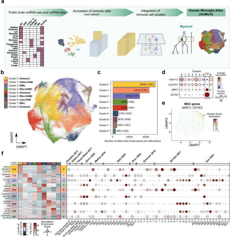
Fig. 1 The Human Microglia Atlas (HuMicA). a Schematic overview of the pipeline used to obtain the integrated HuMicA object. b UMAP visualization of the integrated and clustered HuMicA object, annotated by the nine obtained clusters. c Barplot representation of the number of nuclei/cells and the mean genes detected per nucleus/cell (between parentheses) in each cluster. d Dot plot representation of the expression of canonical markers of microglia (P2RY12, CX3CR1) and macrophages (MRC1, CD163) across all clusters. e UMAPs showing the module score (MSc) expression of macrophage (MRC1, CD163) markers. The gene expression values correspond to the normalized “RNA” assay of the integrated Seurat object. f Heatmap representation of the average expression values by cluster of the significant markers of all HuMicA clusters. Gene expression is represented as the z-score of the averaged normalized “RNA” counts. The number of significant markers for each cluster is represented, as well as the most significant and biologically relevant genes from each list. The dot plot represents a gene set enrichment analysis, performed using fgsea with one-tailed test, depicting the significance of the overlap between the lists of genes from the literature and the expression patterns in each HuMicA cluster. Significance is represented by the negative of the log of the adjusted p value (Benjamini-Hochberg) and the normalized enrichment score. The dots highlighted in bold are considered statistically significant (FDR < 0.05). Source data are provided as a Source Data file.
Fig. 1 The Human Microglia Atlas (HuMicA). a Schematic overview of the pipeline used to obtain the integrated HuMicA object. b UMAP visualization of the integrated and clustered HuMicA object, annotated by the nine obtained clusters. c Barplot representation of the number of nuclei/cells and the mean genes detected per nucleus/cell (between parentheses) in each cluster. d Dot plot representation of the expression of canonical markers of microglia (P2RY12, CX3CR1) and macrophages (MRC1, CD163) across all clusters. e UMAPs showing the module score (MSc) expression of macrophage (MRC1, CD163) markers. The gene expression values correspond to the normalized “RNA” assay of the integrated Seurat object. f Heatmap representation of the average expression values by cluster of the significant markers of all HuMicA clusters. Gene expression is represented as the z-score of the averaged normalized “RNA” counts. The number of significant markers for each cluster is represented, as well as the most significant and biologically relevant genes from each list. The dot plot represents a gene set enrichment analysis, performed using fgsea with one-tailed test, depicting the significance of the overlap between the lists of genes from the literature and the expression patterns in each HuMicA cluster. Significance is represented by the negative of the log of the adjusted p value (Benjamini-Hochberg) and the normalized enrichment score. The dots highlighted in bold are considered statistically significant (FDR < 0.05). Source data are provided as a Source Data file.CC BY-NC-ND · PubMed Central
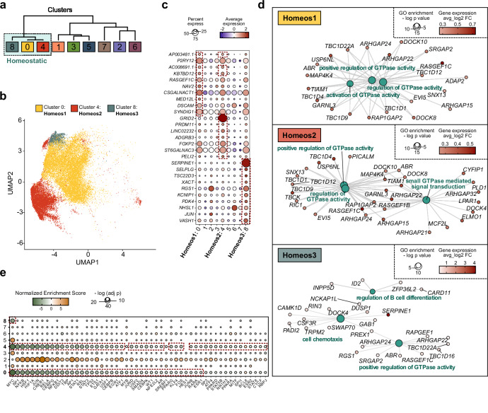
Fig. 2 Homeostatic microglia clusters. a Cluster dendrogram calculated considering the average expression values of all cells/nuclei for each cluster and the unique gene markers for all clusters (distance: “spearman”; clustering: “ward.D”). The box highlights the three grouped clusters that show homeostatic microglia profiles. b UMAP representation of the three clusters annotated as homeostatic microglia. Cluster 0, Homeos1; Cluster 4, Homeos2; Cluster 8, Homeos3. c Dot plot representation of the expression of the top 10 most significant upregulated markers (ordered by avg_log2FC) for the three homeostatic cluster, within the full HuMicA object. The red dashed boxes highlight the top 10 markers for each cluster. The gene expression values correspond to the normalized “RNA” assay of the Seurat object. d Net plot representation of the three most significant gene ontology (GO) terms enriched for the respective markers of the three homeostatic clusters. GO was calculated using enrichGO package followed by the simplify function. The significance of the enrichment is represented in function of the negative of the log of the adjusted p-value (Benjamini-Horchberg). The range of expression of each gene associated with the GO terms is represented by the avg_log2FC. e Representation of the enrichment of the statistically significant (FDR < 0.05) transcription factors (TFs) relative to the comparison of differential expression of each HuMicA homeostatic clusters vs all other. Significance is represented by the normalized enrichment score and the negative of log of the adjusted p value (Benjamini-Horchberg). All presented TFs showed statistically significant enrichment, and the red dashed boxes highlight the TFs with an FDR < 0.05 for each of the homeostatic clusters. Source data are provided as a Source Data file.
Fig. 2 Homeostatic microglia clusters. a Cluster dendrogram calculated considering the average expression values of all cells/nuclei for each cluster and the unique gene markers for all clusters (distance: “spearman”; clustering: “ward.D”). The box highlights the three grouped clusters that show homeostatic microglia profiles. b UMAP representation of the three clusters annotated as homeostatic microglia. Cluster 0, Homeos1; Cluster 4, Homeos2; Cluster 8, Homeos3. c Dot plot representation of the expression of the top 10 most significant upregulated markers (ordered by avg_log2FC) for the three homeostatic cluster, within the full HuMicA object. The red dashed boxes highlight the top 10 markers for each cluster. The gene expression values correspond to the normalized “RNA” assay of the Seurat object. d Net plot representation of the three most significant gene ontology (GO) terms enriched for the respective markers of the three homeostatic clusters. GO was calculated using enrichGO package followed by the simplify function. The significance of the enrichment is represented in function of the negative of the log of the adjusted p-value (Benjamini-Horchberg). The range of expression of each gene associated with the GO terms is represented by the avg_log2FC. e Representation of the enrichment of the statistically significant (FDR < 0.05) transcription factors (TFs) relative to the comparison of differential expression of each HuMicA homeostatic clusters vs all other. Significance is represented by the normalized enrichment score and the negative of log of the adjusted p value (Benjamini-Horchberg). All presented TFs showed statistically significant enrichment, and the red dashed boxes highlight the TFs with an FDR < 0.05 for each of the homeostatic clusters. Source data are provided as a Source Data file.CC BY-NC-ND · PubMed Central
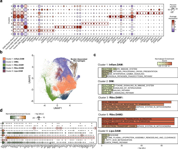
Fig. 3 DAM-related microglia clusters. a Dot plot representation of the expression of the module scores (MSc) of the original upregulated DAM signature from Keren-Shaul et al.8 and the refined human signatures for DAM, youth-associated microglia (YAM) and disease-inflammatory macrophages (DIMs) from Silvin et al.33, as well as the expression of top 10 most significant upregulated markers (ordered by avg_log2FC) for the five clusters considered within the “DAM-verse”. Cluster 1: Inflam.DAM, Cluster 2: DIMs, Cluster 3: Ribo.DAM1, Cluster 5: Ribo.DAM2 and Cluster 6: Lipo.DAM. The red dashed boxes highlight the expression of the MSc of the DAM, YAM and DIM signatures and the top 10 markers for each cluster. The expression values correspond to the normalized “RNA” assay of the Seurat object. b UMAP representation of the five DAM-related and DIM clusters and border-associated macrophages, MAC. c Barplot representing of selected gene set enrichment analysis (GSEA) terms from the REACTOME, WIKIPATHWAYS and KEGG repositories for the markers of the DAM-related and DIM clusters. Enrichment is represented as a function of the negative of the log of the adjusted p-value and the normalized enrichment score. d Representation of the enrichment of the statistically significant (adjusted p-value (Benjamini-Horchberg) < 0.05) transcription factors (TFs) relative to the comparison of differential expression of each HuMicA DAM and DIM clusters vs all other. Significance is represented by the normalized enrichment score and the negative of the log of the adjusted p-value (FDR). All presented TFs showed statistically significant enrichment, and the red dashed boxes highlight the TFs with an FDR < 0.05 for each of the DAM and DIM clusters. Source data are provided as a Source Data file.
Fig. 3 DAM-related microglia clusters. a Dot plot representation of the expression of the module scores (MSc) of the original upregulated DAM signature from Keren-Shaul et al.8 and the refined human signatures for DAM, youth-associated microglia (YAM) and disease-inflammatory macrophages (DIMs) from Silvin et al.33, as well as the expression of top 10 most significant upregulated markers (ordered by avg_log2FC) for the five clusters considered within the “DAM-verse”. Cluster 1: Inflam.DAM, Cluster 2: DIMs, Cluster 3: Ribo.DAM1, Cluster 5: Ribo.DAM2 and Cluster 6: Lipo.DAM. The red dashed boxes highlight the expression of the MSc of the DAM, YAM and DIM signatures and the top 10 markers for each cluster. The expression values correspond to the normalized “RNA” assay of the Seurat object. b UMAP representation of the five DAM-related and DIM clusters and border-associated macrophages, MAC. c Barplot representing of selected gene set enrichment analysis (GSEA) terms from the REACTOME, WIKIPATHWAYS and KEGG repositories for the markers of the DAM-related and DIM clusters. Enrichment is represented as a function of the negative of the log of the adjusted p-value and the normalized enrichment score. d Representation of the enrichment of the statistically significant (adjusted p-value (Benjamini-Horchberg) < 0.05) transcription factors (TFs) relative to the comparison of differential expression of each HuMicA DAM and DIM clusters vs all other. Significance is represented by the normalized enrichment score and the negative of the log of the adjusted p-value (FDR). All presented TFs showed statistically significant enrichment, and the red dashed boxes highlight the TFs with an FDR < 0.05 for each of the DAM and DIM clusters. Source data are provided as a Source Data file.CC BY-NC-ND · PubMed Central
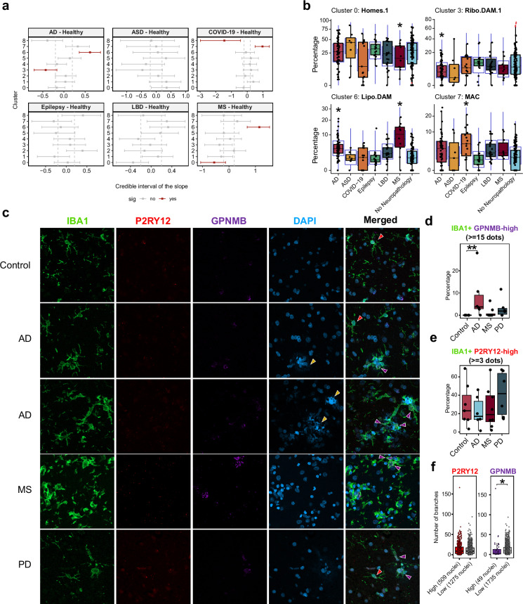
Fig. 4 Differential distribution of microglia populations. a Differential cluster composition in each group in relation to “No Neuropathology” individuals using sccomp represented by the mean of the posterior distribution for the composition parameter, and the 2.5 and 97.5% quantiles. (FDR < 0.05 is highlighted in red). b Proportion of the clusters showing altered distribution (Cluster 0: Homeos1; Cluster 3: Ribo.DAM1; Cluster 6: Lipo.DAM; Cluster 7: MAC), individualized by sample, per group. The blue boxes represent the posterior predictive check and the red triangles outliers (*FDR < 0.05). In panels (a, b) the groups include 60 Alzheimer’s Disease (AD), 7 autism spectrum disorder (ASD), 15 COVID-19, 17 epilepsy, 23 Lewy Body Diseases (LBD), 10 multiple sclerosis (MS) and 109 control samples. c Maximum projection of an immunostaining of the pan-macrophage marker IBA1 in green combined with RNAscope in situ hybridization for P2RY12 (red) and GPNMB (magenta), counterstained with DAPI. Images correspond to human postmortem temporal cortex tissue sections from controls (n = 7), AD (n = 6), MS (n = 8) and Parkinson’s disease (PD) (n = 6). The yellow arrows indicate the blue autofluorescence of amyloid senile plaques in AD samples. The red and magenta arrows indicate P2RY12-high and GPNMB-high cells, respectively. Scale 15 µm. d Fraction of IBA1 + GPNMB-high cells, considered for a number of GPNMB dots equal or superior to 15. Each dot represents a donor sample. e Fraction of IBA1 + P2RY12-high cells, considered for a number of P2RY12 dots equal or superior to 3. Each dot represents a donor sample. The significance in panels (d, e) was calculated using the non-parametric Kruskal-Wallis test followed by the Dunn’s test [*adjusted p (Bonferroni) < 0.05]. f Comparison of the mean number of branches between P2RY12-high (n = 509) and P2RY12-low IBA1+ cells (n = 1275) and between GPNMB-high (n = 49) and GPNMB-low IBA1+ cells (n = 1735). Each dot represents a cell. The significance was calculated using the non-parametric Mann-Whitney test (*p < 0.05). For box plots, the hinges correspond to the first and third quartiles, the whiskers denote minimum and maximum values (excluding outliers) and the horizontal line to the median. All tests used were two-sided. Source data are provided as a Source Data file.
Fig. 4 Differential distribution of microglia populations. a Differential cluster composition in each group in relation to “No Neuropathology” individuals using sccomp represented by the mean of the posterior distribution for the composition parameter, and the 2.5 and 97.5% quantiles. (FDR < 0.05 is highlighted in red). b Proportion of the clusters showing altered distribution (Cluster 0: Homeos1; Cluster 3: Ribo.DAM1; Cluster 6: Lipo.DAM; Cluster 7: MAC), individualized by sample, per group. The blue boxes represent the posterior predictive check and the red triangles outliers (*FDR < 0.05). In panels (a, b) the groups include 60 Alzheimer’s Disease (AD), 7 autism spectrum disorder (ASD), 15 COVID-19, 17 epilepsy, 23 Lewy Body Diseases (LBD), 10 multiple sclerosis (MS) and 109 control samples. c Maximum projection of an immunostaining of the pan-macrophage marker IBA1 in green combined with RNAscope in situ hybridization for P2RY12 (red) and GPNMB (magenta), counterstained with DAPI. Images correspond to human postmortem temporal cortex tissue sections from controls (n = 7), AD (n = 6), MS (n = 8) and Parkinson’s disease (PD) (n = 6). The yellow arrows indicate the blue autofluorescence of amyloid senile plaques in AD samples. The red and magenta arrows indicate P2RY12-high and GPNMB-high cells, respectively. Scale 15 µm. d Fraction of IBA1 + GPNMB-high cells, considered for a number of GPNMB dots equal or superior to 15. Each dot represents a donor sample. e Fraction of IBA1 + P2RY12-high cells, considered for a number of P2RY12 dots equal or superior to 3. Each dot represents a donor sample. The significance in panels (d, e) was calculated using the non-parametric Kruskal-Wallis test followed by the Dunn’s test [*adjusted p (Bonferroni) < 0.05]. f Comparison of the mean number of branches between P2RY12-high (n = 509) and P2RY12-low IBA1+ cells (n = 1275) and between GPNMB-high (n = 49) and GPNMB-low IBA1+ cells (n = 1735). Each dot represents a cell. The significance was calculated using the non-parametric Mann-Whitney test (*p < 0.05). For box plots, the hinges correspond to the first and third quartiles, the whiskers denote minimum and maximum values (excluding outliers) and the horizontal line to the median. All tests used were two-sided. Source data are provided as a Source Data file.CC BY-NC-ND · PubMed Central
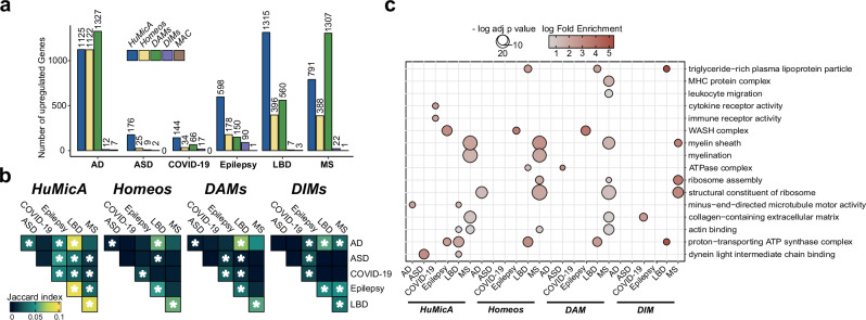
Fig. 5 Pseudobulk differentially expressed genes (DEGs). a Barplot representation of the number of pseudobulk DEGs calculated for each pathology in comparison to the control population. Upregulated DEGs were calculated using the complete HuMicA object and subsets consisting of Homeos subpopulations (clusters 0, 1, 4), DAMs (clusters 1, 3, 5, 6) and DIMs (cluster 2). Significant DEGs are considered for an adjusted p value (FDR) inferior to 0.05 and log2FoldChange superior to 1. b Heatmap representation of the significance of the overlap between the lists of pseudobulk DEGS, calculated with the GeneOverlap function. Overlaps were calculated between each pathology within each subset (HuMicA, Homeos, DAMs, DIMS). The degree of significance is depicted by the Jaccard index and a p-value inferior to 0.05 is highlighted by a white asterisk. c Dot plot representation of the gene ontology (GO) enrichment of all lists of pseudobulk DEGs between each pathology and controls accounting for the complete HuMicA object and subsets consisting of Homeos subpopulations (clusters 0, 1, 4), DAMs (clusters 1, 3, 5, 6), DIMs (cluster 2) and MAC (cluster 7). Selected statistically significant GO terms are represented (adjusted p-value (Benjamini-Horchberg) < 0.05). Significance is represented by the log of the fold change and the negative of the log of the adjusted p-value (FDR). Source data are provided as a Source Data file.
Fig. 5 Pseudobulk differentially expressed genes (DEGs). a Barplot representation of the number of pseudobulk DEGs calculated for each pathology in comparison to the control population. Upregulated DEGs were calculated using the complete HuMicA object and subsets consisting of Homeos subpopulations (clusters 0, 1, 4), DAMs (clusters 1, 3, 5, 6) and DIMs (cluster 2). Significant DEGs are considered for an adjusted p value (FDR) inferior to 0.05 and log2FoldChange superior to 1. b Heatmap representation of the significance of the overlap between the lists of pseudobulk DEGS, calculated with the GeneOverlap function. Overlaps were calculated between each pathology within each subset (HuMicA, Homeos, DAMs, DIMS). The degree of significance is depicted by the Jaccard index and a p-value inferior to 0.05 is highlighted by a white asterisk. c Dot plot representation of the gene ontology (GO) enrichment of all lists of pseudobulk DEGs between each pathology and controls accounting for the complete HuMicA object and subsets consisting of Homeos subpopulations (clusters 0, 1, 4), DAMs (clusters 1, 3, 5, 6), DIMs (cluster 2) and MAC (cluster 7). Selected statistically significant GO terms are represented (adjusted p-value (Benjamini-Horchberg) < 0.05). Significance is represented by the log of the fold change and the negative of the log of the adjusted p-value (FDR). Source data are provided as a Source Data file.CC BY-NC-ND · PubMed Central

Verification

Claims the paper makes

The claims and statistics of this paper haven’t been checked yet.

Run the check — free, no account needed. It reads the open-access text and checks the sources it cites.

A few free checks a day · papers already verified open instantly · results are public

Claims other papers make about this one

Each is checked against this paper’s own text: does it say what the citing paper says it does?

Self-check

Percentages, sums and reported differences re-computed from the paper’s own numbers.

The self-check needs the paper’s full text.

Citations

Cites

Show all 77 references

Cited by

Sorted by citations

See all 124

Metadata

Authors
Martins-Ferreira, R., Calafell-Segura, J., Leal, B., Rodríguez-Ubreva, J., Martínez-Saez, E., Mereu, E., Pinho E Costa, P., Laguna, A., & Ballestar, E.
Affiliations
  • Universidade do Porto (PT)
  • Josep Carreras Leukaemia Research Institute (ES)
  • Universitat Autònoma de Barcelona (ES)
Corresponding author
Ballestar, Esteban
Venue
Nature Communications
Published
2025-01-16
Volume
16
Issue
1
Pages
739-739
ISSN
2041-1723 (ISSN-L)
OpenAlex
W4406437527
Cited by
123
References
77
Citations by year
2026: 79 · 2025: 41
FWCI
43.29
Type
journal article
Language
en
Open access
Locations
  • Nature Communications (publishedVersion, cc-by-nc-nd)
  • bioRxiv (Cold Spring Harbor Laboratory) (acceptedVersion)
  • Research Square (acceptedVersion, cc-by)
  • PubMed Central (publishedVersion, cc-by-nc-nd)
  • PubMed (publishedVersion)
  • Dipòsit Digital de Documents de la UAB (Universitat Autònoma de Barcelona) (submittedVersion, cc-by-nc-nd)
  • RePEc: Research Papers in Economics (submittedVersion)
  • DOAJ (DOAJ: Directory of Open Access Journals) (submittedVersion, cc-by-sa)
  • Portuguese National Funding Agency for Science, Research and Technology (RCAAP Project by FCT) (submittedVersion, other-oa)
Funders
  • Centres de Recerca de Catalunya (501100011033, MCIN/AEI/10.13039/501100011033)
  • European Commission (MCIN /AEI/ 10.13039/501100011033, 10.13039/501100011033, 13039/501100011033, 501100011033, /AEI/10.13039/501100011033)
  • Generalitat de Catalunya (10.13039/501100011033, 501100011033, MCIN /AEI/10.13039/501100011033)
  • Ministerio de Ciencia e Innovación (MCIN/AEI/10, .13039/501100011033, 10.13039, / AEI/10, MCIN/AEI/10.13039/501100011033,, 10.13039/501100011033/, PID2020117212RB-I00, 501100011033, RYC2021, MCIN /AEI /10.13039, /AEI/10.13039/ 501100011033)
  • Fundació Institut de Recerca Hospital Universitari Vall d’Hebron
  • Centro de Investigación Biomédica en Red sobre Enfermedades Neurodegenerativas
  • Fundação para a Ciência e a Tecnologia (UIDB/00215/2020 and UIDP/00215/2020, MCIN/AEI/10.13039/501100011033/, LA/P/006/2020, SFRH/BD/137900/2018, 10.13039/501100011033, UIDB/00215/2020, UIDP/00215/2020)
  • Agència de Gestió d'Ajuts Universitaris i de Recerca (MCIN/AEI/10.13039/501100011033/, 10.13039/501100011033, 13039/501100011033)
  • Instituto de Salud Carlos III (MCIN/ AEI /10.13039/501100011033, /10.13039/501100011033, PI21/01603, AEI/10.13039/501100011033)
  • Agencia Estatal de Investigación (AEI//10.13039/501100011033/, 501100011033, 10.13039/501100011033, MCIN/AEI/10.13039/501 100 011 033, 13039, .13039/501100011033, 10.13039, AEI/10)
  • Instituto de Ciências Biomédicas Abel Salazar, Universidade do Porto
  • Institut de Recerca contra la Leucèmia Josep Carreras
  • NextGenerationEU
Topics
  • Life Sciences › Neuroscience › Neurology › Neuroinflammation and Neurodegeneration Mechanisms
  • Life Sciences › Biochemistry, Genetics and Molecular Biology › Molecular Biology › Single-cell and spatial transcriptomics
  • Health Sciences › Medicine › Clinical Neurology › Multiple Sclerosis Research Studies
Keywords
microglia, disease-associated microglia, microglial subtypes, neuroinflammation, neurodegenerative diseases, Alzheimer's disease, multiple sclerosis, autism spectrum disorder, epilepsy, Lewy body disease, COVID-19 pandemic, GPNMB, single-nucleus RNA sequencing, single-cell RNA sequencing
MeSH terms
Autism Spectrum Disorder, RNA-Seq, COVID-19, Aged, Alzheimer Disease, Animals, Atlases as Topic, Brain, Female, Humans, Macrophages, Male, Middle Aged, Multiple Sclerosis, Microglia, Neurodegenerative Diseases, Lewy Body Disease, Single-Cell Analysis

About these checks

Registry, retraction, Cites and statistics results are computed, not judged, with one exception: a printed reference that no index lists is read once by a model before it counts as a citation error. Claims are extracted by a model and kept only when their quote appears verbatim in the text; each cited source is then checked, with a model’s help, for whether it says what the claim relies on. Those verdicts can be wrong — read the claim’s quote and the cited source before relying on one.

Statistics and arithmetic are recomputed, not judged.

Confidence bands are shown only for fields calibrated on our annotated test set. None is calibrated yet, so no band is shown.

What counts as a claim ›How we measure confidence ›How we check this ›

Are you an author, or do you see a problem with this page? Request a review