

The DOI matches its Crossref record; 77 of the 77 references it cites were found in a registry or an index.
The Human Microglia Atlas (HuMicA) unravels changes in disease-associated microglia subsets across neurodegenerative conditions
Open accessPublished version of a preprint
Abstract
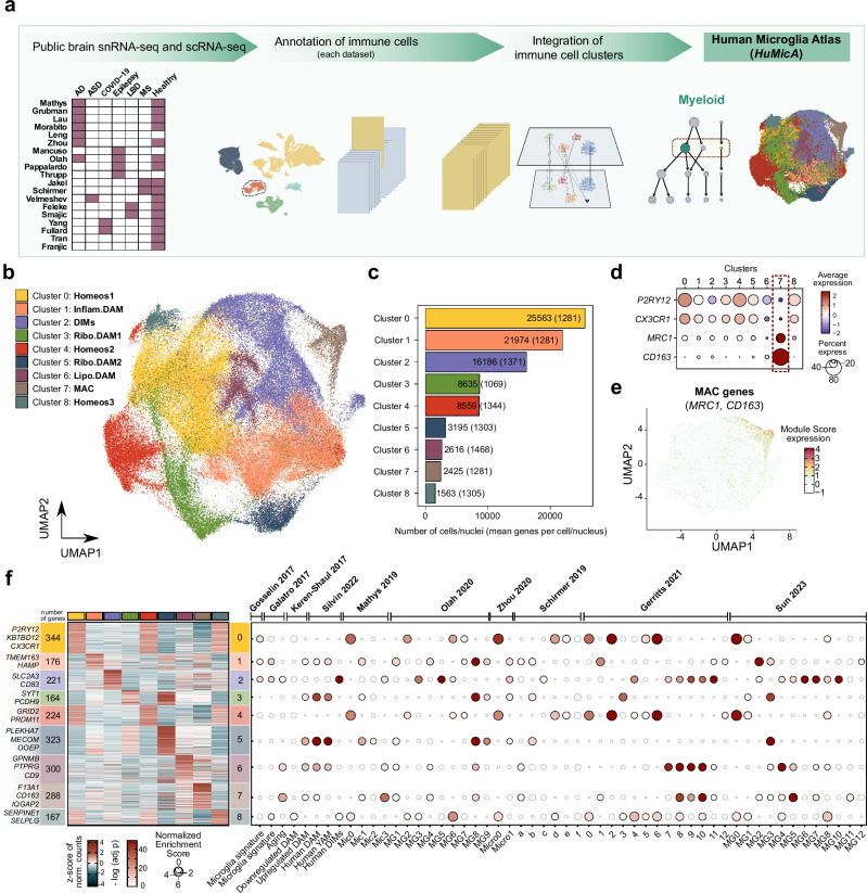
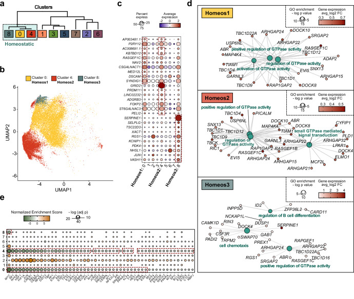
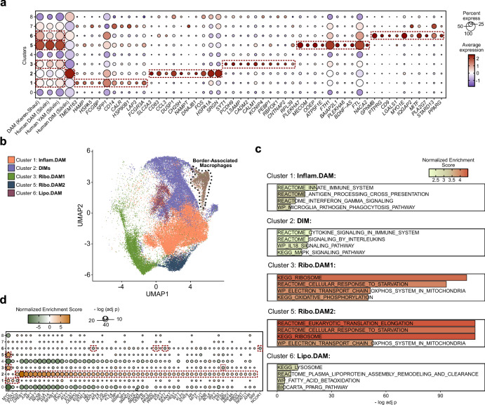
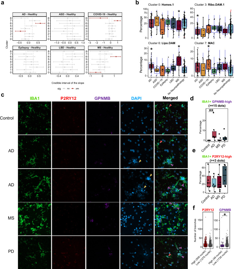
ABSTRACT Dysregulated microglia activation, leading to neuroinflammation, is crucial in neurodegenerative disease development and progression. The initial M1/M2 dual activation classification for microglia is outdated. Even the ‘disease-associated microglia’ (DAM) phenotype, firstly described in mice, falls short in representing the diverse microglia phenotypes in pathology. In this study, we have constructed a transcriptomic atlas of human brain immune cells by integrating single-nucleus (sn)RNA-seq datasets from multiple neurodegenerative conditions. Sixteen datasets were included, comprising 295 samples from patients with Alzheimer’s disease, autism spectrum disorder, epilepsy, multiple sclerosis, Lewy body diseases, COVID-19, and healthy controls. The integrated Human Microglia Atlas ( HuMicA ) dataset included 60,557 nuclei and revealed 11 microglial subpopulations distributed across all pathological and healthy conditions. Among these, we identified four different homeostatic clusters as well as pathological phenotypes. These included two stages of early and late activation of the DAM phenotype and the disease-inflammatory macrophage (DIM) phenotype, which was recently described in mice, and is also present in human microglia, as indicated by our analysis. The high versatility of microglia is evident through changes in subset distribution across various pathologies, suggesting their contribution in shaping pathological phenotypes. Our analysis showed overall depletion of four substates of homeostatic microglia, and expansion of niche subpopulations within the DAM and DIM spectrum across distinct neurodegenerative pathologies. The HuMicA is invaluable in advancing the study of microglia biology in both healthy and disease settings.
Overview
The claims and statistics of this paper haven’t been checked yet.
Run the check — free, no account needed. It reads the open-access text and checks the sources it cites.
Integrity signals
Registry
Cites
Statistics
Figures
Verification
Claims the paper makes
The claims and statistics of this paper haven’t been checked yet.
Run the check — free, no account needed. It reads the open-access text and checks the sources it cites.
Claims other papers make about this one
Each is checked against this paper’s own text: does it say what the citing paper says it does?
The source backs part of the claim, not all of it — or backs it for a different population, condition or comparison.
In the citing paper
“Many disease-associated microglial programs are spatially enriched around plaques, which suggests that microglial state transitions are not only disease-stage-dependent but also anatomically localized.”
From Lymphoid-like Suppressive Microglia in Alzheimer’s Disease: A New Neuroimmune Regulatory Axis? (2026) · cites this paper as [47]
Experimental — accuracy not yet measuredThe source backs part of the claim, not all of it — or backs it for a different population, condition or comparison.
In the citing paper
“Large human microglial atlases and state-dynamics studies are beginning to show that microglia in Alzheimer’s disease occupy multiple transcriptional trajectories rather than discrete and permanent categories [22,47].”
From Lymphoid-like Suppressive Microglia in Alzheimer’s Disease: A New Neuroimmune Regulatory Axis? (2026) · cites this paper as [47]
Experimental — accuracy not yet measuredThe cited source says what the claim attributes to it.
In the citing paper
“Instead, microglia in Alzheimer’s disease appear to acquire hybrid programs: they remain tissue-resident myeloid cells, but they can express genes normally associated with professional antigen presentation, immune communication, and inflammatory coordination.”
From Lymphoid-like Suppressive Microglia in Alzheimer’s Disease: A New Neuroimmune Regulatory Axis? (2026) · cites this paper as [47]
The cited source says what the claim attributes to it.
In the citing paper
“Single-cell and single-nucleus studies have repeatedly identified Alzheimer ’s-associated microglial states enriched for immune activation, antigen-presentation genes, lipid-processing pathways, and inflammatory mediators.”
From Lymphoid-like Suppressive Microglia in Alzheimer’s Disease: A New Neuroimmune Regulatory Axis? (2026) · cites this paper as [47]
Self-check
Percentages, sums and reported differences re-computed from the paper’s own numbers.
The self-check needs the paper’s full text.
Citations
Cites
Moderated estimation of fold change and dispersion for RNA-seq data with DESeq2
Love, M. I., Huber, W., & Anders, S. (2014) · Genome Biology · doi:10.1186/s13059-014-0550-8 · Cited by 95,106
Integrated analysis of multimodal single-cell data
Hao, Y., Hao, S., Andersen-Nissen, E., Mauck, W. M., Zheng, S., Butler, A., Lee, M. J., Wilk, A. J., Darby, C., Zagar, M., et al. (2020) · doi:10.1101/2020.10.12.335331 · Cited by 15,508
Comprehensive Integration of Single-Cell Data
Stuart, T., Butler, A., Hoffman, P., Hafemeister, C., Papalexi, E., Mauck, W. M., Hao, Y., Stoeckius, M., Smibert, P., & Satija, R. (2019) · Cell · doi:10.1016/j.cell.2019.05.031 · Cited by 15,453
clusterProfiler 4.0: A universal enrichment tool for interpreting omics data
(2021) · doi:10.1016/j.xinn.2021.100141 · Cited by 12,455
Complex heatmaps reveal patterns and correlations in multidimensional genomic data
Gu, Z., Eils, R., & Schlesner, M. (2016) · Bioinformatics · doi:10.1093/bioinformatics/btw313 · Cited by 10,037
Massively parallel digital transcriptional profiling of single cells
Zheng, G. X. Y., Terry, J. M., Belgrader, P., Ryvkin, P., Bent, Z. W., Wilson, R., Ziraldo, S. B., Wheeler, T. D., McDermott, G. P., Zhu, J., et al. (2017) · Nature Communications · doi:10.1038/ncomms14049 · Cited by 7,353
Dictionary learning for integrative, multimodal and scalable single-cell analysis
Hao, Y., Stuart, T., Kowalski, M. H., Choudhary, S., Hoffman, P., Hartman, A., Srivastava, A., Molla, G., Madad, S., Fernandez-Granda, C., et al. (2024) · Nature Biotechnology · doi:10.1038/s41587-023-01767-y · Cited by 5,631
A Unique Microglia Type Associated with Restricting Development of Alzheimer’s Disease
Keren-Shaul, H., Spinrad, A., Weiner, A., Matcovitch-Natan, O., Dvir-Szternfeld, R., Ulland, T. K., David, E., Baruch, K., Lara-Astaiso, D., Toth, B., et al. (2017) · Cell · doi:10.1016/j.cell.2017.05.018 · Cited by 5,493
Fate Mapping Analysis Reveals That Adult Microglia Derive from Primitive Macrophages
Ginhoux, F., Greter, M., Leboeuf, M., Nandi, S., See, P., Gokhan, S., Mehler, M. F., Conway, S. J., Ng, L. G., Stanley, E. R., et al. (2010) · Science · doi:10.1126/science.1194637 · Cited by 5,175
McGinnis, C. S., Murrow, L. M., & Gartner, Z. J. (2019) · Cell Systems · doi:10.1016/j.cels.2019.03.003 · Cited by 4,521
The single-cell transcriptional landscape of mammalian organogenesis
Cao, J., Spielmann, M., Qiu, X., Huang, X., Ibrahim, D. M., Hill, A. J., Zhang, F., Mundlos, S., Christiansen, L., Steemers, F. J., et al. (2019) · Nature · doi:10.1038/s41586-019-0969-x · Cited by 4,490
Hafemeister, C., & Satija, R. (2019) · Genome Biology · doi:10.1186/s13059-019-1874-1 · Cited by 4,479 · cited 1×
“The doubletFinder_v3 function 59 was used to estimate doublets from each individual subject, which were removed from each dataset ’ s Seurat object, followed by SCTransform normalization 60 with default 3000 variable genes.”
Fast gene set enrichment analysisPreprint
Korotkevich, G., Sukhov, V., Budin, N., Shpak, B., Artyomov, M. N., & Sergushichev, A. (2016) · bioRxiv (Cold Spring Harbor Laboratory) · doi:10.1101/060012 · Cited by 3,742
More than 50 long-term effects of COVID-19: a systematic review and meta-analysis
Lopez-Leon, S., Wegman-Ostrosky, T., Perelman, C., Sepulveda, R., Rebolledo, P. A., Cuapio, A., & Villapol, S. (2021) · Scientific Reports · doi:10.1038/s41598-021-95565-8 · Cited by 2,757
Single-cell transcriptomic analysis of Alzheimer’s disease
Mathys, H., Davila-Velderrain, J., Peng, Z., Gao, F., Mohammadi, S., Young, J. Z., Menon, M., He, L., Abdurrob, F., Jiang, X., et al. (2019) · Nature · doi:10.1038/s41586-019-1195-2 · Cited by 2,568
Hammond, T. R., Dufort, C., Dissing-Olesen, L., Giera, S., Young, A., Wysoker, A., Walker, A. J., Segel, M., Nemesh, J., Saunders, A., et al. (2018) · doi:10.1101/406140 · Cited by 2,225
Microglia and macrophages in brain homeostasis and disease
Li, Q., & Barres, B. A. (2018) · Nature Reviews Immunology · doi:10.1038/nri.2017.125 · Cited by 1,999
McCarthy, D. J., Campbell, K. R., Lun, A. T. L., & Wills, Q. F. (2017) · Bioinformatics · doi:10.1093/bioinformatics/btw777 · Cited by 1,852
SARS-CoV-2 is associated with changes in brain structure in UK Biobank
Douaud, G., Lee, S., Alfaro-Almagro, F., Arthofer, C., Wang, C., McCarthy, P., Lange, F., Andersson, J. L. R., Griffanti, L., Duff, E., et al. (2022) · Nature · doi:10.1038/s41586-022-04569-5 · Cited by 1,646
Spatial and temporal heterogeneity of mouse and human microglia at single-cell resolution
Masuda, T., Sankowski, R., Staszewski, O., Böttcher, C., Amann, L., Sagar, Scheiwe, C., Nessler, S., Kunz, P., van Loo, G., et al. (2019) · Nature · doi:10.1038/s41586-019-0924-x · Cited by 1,310
An environment-dependent transcriptional network specifies human microglia identity
Gosselin, D., Skola, D., Coufal, N. G., Holtman, I. R., Schlachetzki, J. C. M., Sajti, E., Jaeger, B. N., O’Connor, C., Fitzpatrick, C., Pasillas, M. P., et al. (2017) · Science · doi:10.1126/science.aal3222 · Cited by 1,220 · cited 2×
“These include standard human microglia transcriptomic signatures obtainedcomparingsortedmicrogliaandbulkcorticaltissue(Gosselin 2017 and Galatro 2017) 31,32 ; the original DAM signatures (Keren-Shaul 2017) 8 ; and markers of three populations within the DAM-verse, including the bona-fi de DAM,…”
Zhou, Y., Song, W. M., Andhey, P. S., Swain, A., Levy, T., Miller, K. R., Poliani, P. L., Cominelli, M., Grover, S., Gilfillan, S., et al. (2020) · Nature Medicine · doi:10.1038/s41591-019-0695-9 · Cited by 1,150
False discovery rates: a new deal
Stephens, M. (2016) · Biostatistics · doi:10.1093/biostatistics/kxw041 · Cited by 1,132
Li, Q., Cheng, Z., Zhou, L., Darmanis, S., Neff, N. F., Okamoto, J., Gulati, G., Bennett, M. L., Sun, L. O., Clarke, L. E., et al. (2019) · Neuron · doi:10.1016/j.neuron.2018.12.006 · Cited by 1,129
1-year outcomes in hospital survivors with COVID-19: a longitudinal cohort study.
Huang, L., Yao, Q., Gu, X., Wang, Q., Ren, L., Wang, Y., Hu, P., Guo, L., Liu, M., Xu, J., et al. (2021) · The Lancet · doi:10.1016/s0140-6736(21)01755-4 · Cited by 1,101
Massively parallel single-nucleus RNA-seq with DroNc-seq
Habib, N., Avraham-Davidi, I., Basu, A., Burks, T., Shekhar, K., Hofree, M., Choudhury, S. R., Aguet, F., Gelfand, E., Ardlie, K., et al. (2017) · Nature Methods · doi:10.1038/nmeth.4407 · Cited by 1,069
Clustering trees: a visualization for evaluating clusterings at multiple resolutions
Zappia, L., & Oshlack, A. (2018) · GigaScience · doi:10.1093/gigascience/giy083 · Cited by 1,064
Mrdjen, D., Pavlovic, A., Hartmann, F. J., Schreiner, B., Utz, S. G., Leung, B. P., Lelios, I., Heppner, F. L., Kipnis, J., Merkler, D., et al. (2018) · Immunity · doi:10.1016/j.immuni.2018.01.011 · Cited by 1,010
Alvarez, M. J., Shen, Y., Giorgi, F. M., Lachmann, A., Ding, B. B., Ye, B. H., & Califano, A. (2016) · Nature Genetics · doi:10.1038/ng.3593 · Cited by 966
Grubman, A., Chew, G., Ouyang, J. F., Sun, G., Choo, X. Y., McLean, C., Simmons, R. K., Buckberry, S., Vargas-Landin, D. B., Poppe, D., et al. (2019) · Nature Neuroscience · doi:10.1038/s41593-019-0539-4 · Cited by 956
Single-cell genomics identifies cell type–specific molecular changes in autism
Velmeshev, D., Schirmer, L., Jung, D., Haeussler, M., Perez, Y., Mayer, S., Bhaduri, A., Goyal, N., Rowitch, D. H., & Kriegstein, A. R. (2019) · Science · doi:10.1126/science.aav8130 · Cited by 927
Origin and differentiation of microglia
Ginhoux, F., Lim, S., Hoeffel, G., Low, D., & Huber, T. (2013) · Frontiers in Cellular Neuroscience · doi:10.3389/fncel.2013.00045 · Cited by 905 · cited 3×
“Furthermore, clusters 1, 2, 3, 5 and 6 (highlighted in Fig.”
Microglia-mediated neuroinflammation in neurodegenerative diseases
Subhramanyam, C. S., Wang, C., Hu, Q., & Dheen, S. T. (2019) · Seminars in Cell and Developmental Biology · doi:10.1016/j.semcdb.2019.05.004 · Cited by 897
Altered human oligodendrocyte heterogeneity in multiple sclerosis
Jäkel, S., Agirre, E., Mendanha Falcão, A., van Bruggen, D., Lee, K. W., Knuesel, I., Malhotra, D., ffrench-Constant, C., Williams, A., & Castelo-Branco, G. (2019) · Nature · doi:10.1038/s41586-019-0903-2 · Cited by 854
The Semantics of Microglia Activation: Neuroinflammation, Homeostasis, and Stress
Woodburn, S. C., Bollinger, J. L., & Wohleb, E. S. (2021) · Journal of Neuroinflammation · doi:10.1186/s12974-021-02309-6 · Cited by 783
Transcriptomic analysis of purified human cortical microglia reveals age-associated changes
Galatro, T. F., Holtman, I. R., Lerario, A. M., Vainchtein, I. D., Brouwer, N., Sola, P. R., Veras, M. M., Pereira, T. F., Leite, R. E. P., Möller, T., et al. (2017) · Nature Neuroscience · doi:10.1038/nn.4597 · Cited by 741
Single cell RNA sequencing of human microglia uncovers a subset associated with Alzheimer’s disease
Olah, M., Menon, V., Habib, N., Taga, M. F., Ma, Y., Yung, C. J., Cimpean, M., Khairallah, A., Coronas-Samano, G., Sankowski, R., et al. (2020) · Nature Communications · doi:10.1038/s41467-020-19737-2 · Cited by 722
Chiu, I. M., Morimoto, E. T. A., Goodarzi, H., Liao, J. T., O’Keeffe, S., Phatnani, H. P., Muratet, M., Carroll, M. C., Levy, S., Tavazoie, S., et al. (2013) · Cell Reports · doi:10.1016/j.celrep.2013.06.018 · Cited by 651
Single-nucleus chromatin accessibility and transcriptomic characterization of Alzheimer’s disease
Morabito, S., Miyoshi, E., Michael, N., Shahin, S., Martini, A. C., Head, E., Silva, J., Leavy, K., Perez-Rosendahl, M., & Swarup, V. (2021) · Nature Genetics · doi:10.1038/s41588-021-00894-z · Cited by 643
Neuronal vulnerability and multilineage diversity in multiple sclerosis
Schirmer, L., Velmeshev, D., Holmqvist, S., Kaufmann, M., Werneburg, S., Jung, D., Vistnes, S., Stockley, J. H., Young, A., Steindel, M., et al. (2019) · Nature · doi:10.1038/s41586-019-1404-z · Cited by 638
Dysregulation of brain and choroid plexus cell types in severe COVID-19
Yang, A. C., Kern, F., Losada, P. M., Agam, M. R., Maat, C. A., Schmartz, G. P., Fehlmann, T., Stein, J. A., Schaum, N., Lee, D. P., et al. (2021) · Nature · doi:10.1038/s41586-021-03710-0 · Cited by 620
Smajić, S., Prada-Medina, C. A., Landoulsi, Z., Ghelfi, J., Delcambre, S., Dietrich, C., Jarazo, J., Henck, J., Balachandran, S., Pachchek, S., et al. (2022) · Brain · doi:10.1093/brain/awab446 · Cited by 590
Mathys, H., Peng, Z., Boix, C. A., Victor, M. B., Leary, N., Babu, S., Abdelhady, G., Jiang, X., Ng, A. P., Ghafari, K., et al. (2023) · Cell · doi:10.1016/j.cell.2023.08.039 · Cited by 495
Molecular characterization of selectively vulnerable neurons in Alzheimer’s disease
Leng, K., Li, E., Eser, R., Piergies, A., Sit, R., Tan, M., Neff, N., Li, S. H., Rodriguez, R. D., Suemoto, C. K., et al. (2021) · Nature Neuroscience · doi:10.1038/s41593-020-00764-7 · Cited by 485
Lau, S.-F., Cao, H., Fu, A. K. Y., & Ip, N. Y. (2020) · Proceedings of the National Academy of Sciences · doi:10.1073/pnas.2008762117 · Cited by 476
Müller‐Dott, S., Tsirvouli, E., Vázquez, M., Flores, R. O. R., Badia-i-Mompel, P., Fallegger, R., Türei, D., Lægreid, A., & Sáez-Rodríguez, J. (2023) · Nucleic Acids Research · doi:10.1093/nar/gkad841 · Cited by 468
Human microglial state dynamics in Alzheimer’s disease progression
Sun, N., Victor, M. B., Park, Y. P., Xiong, X., Scannail, A. N., Leary, N., Prosper, S., Viswanathan, S., Luna, X., Boix, C. A., et al. (2023) · Cell · doi:10.1016/j.cell.2023.08.037 · Cited by 462
Distinct amyloid-β and tau-associated microglia profiles in Alzheimer’s disease
Gerrits, E., Brouwer, N., Kooistra, S. M., Woodbury, M. E., Vermeiren, Y., Lambourne, M., Mulder, J., Kummer, M., Möller, T., Biber, K., et al. (2021) · Acta Neuropathologica · doi:10.1007/s00401-021-02263-w · Cited by 441
Nebulosa recovers single-cell gene expression signals by kernel density estimation
Alquicira-Hernandez, J., & Powell, J. E. (2021) · Bioinformatics · doi:10.1093/bioinformatics/btab003 · Cited by 381
Silvin, A., Uderhardt, S., Piot, C., Da Mesquita, S., Yang, K., Geirsdottir, L., Mulder, K., Eyal, D., Liu, Z., Bridlance, C., et al. (2022) · Immunity · doi:10.1016/j.immuni.2022.07.004 · Cited by 368
Single-Nucleus RNA-Seq Is Not Suitable for Detection of Microglial Activation Genes in Humans
Thrupp, N., Sala Frigerio, C., Wolfs, L., Skene, N. G., Fattorelli, N., Poovathingal, S., Fourne, Y., Matthews, P. M., Theys, T., Mancuso, R., et al. (2020) · Cell Reports · doi:10.1016/j.celrep.2020.108189 · Cited by 329 · cited 4×
“…expression of the full list of human genes in the DAM signature ( n =210), based on Keren-Shaul et al. 8 , and as previously reported for human data by Thrupp et al. 44 , and the summarized human gene signatures for DAM, YAM and DIMs provided by Silvin et al. 33 reinforces these patterns (Fig.”
InfluentialFranjic, D., Skarica, M., Ma, S., Arellano, J. I., Tebbenkamp, A. T. N., Choi, J., Xu, C., Li, Q., Morozov, Y. M., Andrijevic, D., et al. (2022) · Neuron · doi:10.1016/j.neuron.2021.10.036 · Cited by 308
Stem-cell-derived human microglia transplanted in mouse brain to study human disease
Mancuso, R., Van Den Daele, J., Fattorelli, N., Wolfs, L., Balusu, S., Burton, O., Liston, A., Sierksma, A., Fourne, Y., Poovathingal, S., et al. (2019) · Nature Neuroscience · doi:10.1038/s41593-019-0525-x · Cited by 295
ShinyCell: simple and sharable visualization of single-cell gene expression data
Ouyang, J. F., Kamaraj, U. S., Cao, E. Y., & Rackham, O. J. L. (2021) · Bioinformatics · doi:10.1093/bioinformatics/btab209 · Cited by 245
Tran, M. N., Maynard, K. R., Spangler, A., Huuki, L. A., Montgomery, K. D., Sadashivaiah, V., Tippani, M., Barry, B. K., Hancock, D. B., Hicks, S. C., et al. (2021) · Neuron · doi:10.1016/j.neuron.2021.09.001 · Cited by 219
Transcription Factor Activities Enhance Markers of Drug Sensitivity in Cancer
García‐Alonso, L., Iorio, F., Matchan, A., Fonseca, N. A., Jaaks, P., Peat, G., Pignatelli, M., Falcone, F., Benes, C. H., Dunham, I., et al. (2017) · Cancer Research · doi:10.1158/0008-5472.can-17-1679 · Cited by 199
Microglia as Dynamic Cellular Mediators of Brain Function
Wright-Jin, E. C., & Gutmann, D. H. (2019) · Trends in Molecular Medicine · doi:10.1016/j.molmed.2019.08.013 · Cited by 197
Dolan, M.-J., Therrien, M., Jereb, S., Kamath, T., Gazestani, V., Atkeson, T., Marsh, S. E., Goeva, A., Lojek, N. M., Murphy, S., et al. (2023) · Nature Immunology · doi:10.1038/s41590-023-01558-2 · Cited by 185
Ramanan, S., Kooshki, M., Zhao, W., Hsu, F., & Robbins, M. E. C. (2008) · Free Radical Biology and Medicine · doi:10.1016/j.freeradbiomed.2008.09.002 · Cited by 162
STAT1 drives M1 microglia activation and neuroinflammation under hypoxia.
Butturini, E., Boriero, D., de Prati, A. C., & Mariotto, S. (2019) · Archives of Biochemistry and Biophysics · doi:10.1016/j.abb.2019.05.011 · Cited by 149
Transcriptomic and clonal characterization of T cells in the human central nervous system
Pappalardo, J. L., Zhang, L., Pecsok, M. K., Perlman, K., Zografou, C., Raddassi, K., Abulaban, A. A., Krishnaswamy, S., Antel, J. P., van Dijk, D., et al. (2020) · Science Immunology · doi:10.1126/sciimmunol.abb8786 · Cited by 134
Turnover of Rat Brain Perivascular Cells
Bechmann, I., Kwidzinski, E., Kovac, A. D., Simbürger, E., Horvath, T., Gimsa, U., Dirnagl, U., Priller, J., & Nitsch, R. (2001) · Experimental Neurology · doi:10.1006/exnr.2000.7618 · Cited by 134
Systematic analysis of gene expression patterns associated with postmortem interval in human tissues
Zhu, Y., Wang, L., Yin, Y., & Yang, E. (2017) · Scientific Reports · doi:10.1038/s41598-017-05882-0 · Cited by 129
Fullard, J. F., Lee, H.-C., Voloudakis, G., Suo, S., Javidfar, B., Shao, Z., Peter, C. J., Zhang, W., Jiang, S., Corvelo, A., et al. (2021) · Genome Medicine · doi:10.1186/s13073-021-00933-8 · Cited by 108
sccomp: Robust differential composition and variability analysis for single-cell data
Mangiola, S., Roth‐Schulze, A. J., Trussart, M., Zozaya‐Valdés, E., Ma, M., Gao, Z., Rubin, A. F., Speed, T. P., Shim, H., & Papenfuss, A. T. (2023) · Proceedings of the National Academy of Sciences · doi:10.1073/pnas.2203828120 · Cited by 85
Feleke, R., Reynolds, R. H., Smith, A. M., Tilley, B. S., Taliun, S. A. G., Hardy, J., Matthews, P. M., Gentleman, S., Owen, D. R., Johnson, M. R., et al. (2021) · Acta Neuropathologica · doi:10.1007/s00401-021-02343-x · Cited by 69
Microglial innate memory and epigenetic reprogramming in neurological disorders
Martins-Ferreira, R., Leal, B., Costa, P., & Ballestar, E. (2020) · Progress in Neurobiology · doi:10.1016/j.pneurobio.2020.101971 · Cited by 55
Self-citationTranscription factor p53 influences microglial activation phenotype
Jayadev, S., Nesser, N. K., Hopkins, S., Myers, S. J., Case, A., Lee, R. J., Seaburg, L., Uo, T., Murphy, S. P., Morrison, R. S., et al. (2011) · Glia · doi:10.1002/glia.21178 · Cited by 52
Ta, H. M., Le, T. M., Ishii, H., Takarada‐Iemata, M., Hattori, T., Hashida, K., Yamamoto, Y., Mori, K., Takahashi, R., Kitao, Y., et al. (2016) · Journal of Neurochemistry · doi:10.1111/jnc.13714 · Cited by 40
Li, Q., Zhou, L., Wang, L., Li, S., Xu, G., Gu, H., Li, D., Liu, M., Fang, L., Wang, Z., et al. (2020) · European Journal of Immunology · doi:10.1002/eji.201948299 · Cited by 38
Tan, W., Su, P. P., Leff, J., Gao, X., Chen, J., Guan, A. K., Kalyanasundaram, G., Ma, A. I., & Guan, Z. (2022) · Cell Discovery · doi:10.1038/s41421-022-00377-3 · Cited by 38
Olah, M., Menon, V., Habib, N., Taga, M. F., Ma, Y., Khairallah, A., Yung, C. J., Coronas‐Samano, G., Vonsattel, J. P., Teich, A. F., et al. (2020) · Alzheimer s & Dementia · doi:10.1002/alz.038589 · Cited by 30
Yuan, T., Dong, L., Pearsall, E., Zhou, K., Cheng, R., & Ma, J. (2022) · Cells · doi:10.3390/cells11233869 · Cited by 25
Miao, J., Chen, Z., Wu, Y. Z., Hu, Q., & Ji, T. (2022) · ACS Chemical Neuroscience · doi:10.1021/acschemneuro.2c00440 · Cited by 24
Chen, M., Chen, X., Hu, X., Dai, J., & Sun, J. (2021) · Neuroscience Letters · doi:10.1016/j.neulet.2021.136283 · Cited by 11
Harris, H. K., Nakayama, T., Lai, J. F., Zhao, B., Argyrou, N., Gubbels, C. S., Soucy, A., Genetti, C. A., Rodan, L. H., Tiller, G. E., et al. (2020) · medRxiv · doi:10.1101/2020.09.09.20187104 · Cited by 8
Cited by
Sorted by citations
Cell-type-directed network-correcting combination therapy for Alzheimer’s disease
Li, Y., Serras, C. P., Blumenfeld, J. R., Xie, M., Hao, Y., Deng, E., Chun, Y. Y., Holtzman, J., An, A., Yoon, S. Y., et al. (2025) · Cell · doi:10.1016/j.cell.2025.06.035 · Cited by 66
Lipid and lipoprotein metabolism in microglia: Alzheimer’s disease mechanisms and interventions
Sprenger, K. G., Lietzke, E. E., Melchior, J. T., & Bruce, K. D. (2025) · Journal of Lipid Research · doi:10.1016/j.jlr.2025.100872 · Cited by 42
He, C., Chen, B., Yang, H., & Zhou, X. (2025) · Frontiers in Aging Neuroscience · doi:10.3389/fnagi.2025.1554398 · Cited by 40
Functional diversity of disorder-specific macrophages involved in various diseases
Mitsui, Y., & Satoh, T. (2025) · Inflammation and Regeneration · doi:10.1186/s41232-025-00390-5 · Cited by 38
Microglia-astrocyte crosstalk following ischemic stroke
Yang, S., Chen, Y., Tang, J., Cui, Y., Wei, W., Hao, Z., Xiao, Z., Pan, Y., Tian, Q., Xin, W., et al. (2025) · Molecular Brain · doi:10.1186/s13041-025-01244-4 · Cited by 29
Metadata
- Authors
- Martins-Ferreira, R., Calafell-Segura, J., Leal, B., Rodríguez-Ubreva, J., Martínez-Saez, E., Mereu, E., Pinho E Costa, P., Laguna, A., & Ballestar, E.
- Affiliations
- Universidade do Porto (PT)
- Josep Carreras Leukaemia Research Institute (ES)
- Universitat Autònoma de Barcelona (ES)
- Corresponding author
- Ballestar, Esteban
- ORCID
- Venue
- Nature Communications
- Published
- 2025-01-16
- Volume
- 16
- Issue
- 1
- Pages
- 739-739
- ISSN
- 2041-1723 (ISSN-L)
- PMC
- PMC11739505
- OpenAlex
- W4406437527
- Cited by
- 123
- References
- 77
- Citations by year
- 2026: 79 · 2025: 41
- FWCI
- 43.29
- Type
- journal article
- Language
- en
- Open access
- gold
- doi.org
- Licence
- Locations
- Nature Communications (publishedVersion, cc-by-nc-nd)
- bioRxiv (Cold Spring Harbor Laboratory) (acceptedVersion)
- Research Square (acceptedVersion, cc-by)
- PubMed Central (publishedVersion, cc-by-nc-nd)
- PubMed (publishedVersion)
- Dipòsit Digital de Documents de la UAB (Universitat Autònoma de Barcelona) (submittedVersion, cc-by-nc-nd)
- RePEc: Research Papers in Economics (submittedVersion)
- DOAJ (DOAJ: Directory of Open Access Journals) (submittedVersion, cc-by-sa)
- Portuguese National Funding Agency for Science, Research and Technology (RCAAP Project by FCT) (submittedVersion, other-oa)
- Funders
- Centres de Recerca de Catalunya (501100011033, MCIN/AEI/10.13039/501100011033)
- European Commission (MCIN /AEI/ 10.13039/501100011033, 10.13039/501100011033, 13039/501100011033, 501100011033, /AEI/10.13039/501100011033)
- Generalitat de Catalunya (10.13039/501100011033, 501100011033, MCIN /AEI/10.13039/501100011033)
- Ministerio de Ciencia e Innovación (MCIN/AEI/10, .13039/501100011033, 10.13039, / AEI/10, MCIN/AEI/10.13039/501100011033,, 10.13039/501100011033/, PID2020117212RB-I00, 501100011033, RYC2021, MCIN /AEI /10.13039, /AEI/10.13039/ 501100011033)
- Fundació Institut de Recerca Hospital Universitari Vall d’Hebron
- Centro de Investigación Biomédica en Red sobre Enfermedades Neurodegenerativas
- Fundação para a Ciência e a Tecnologia (UIDB/00215/2020 and UIDP/00215/2020, MCIN/AEI/10.13039/501100011033/, LA/P/006/2020, SFRH/BD/137900/2018, 10.13039/501100011033, UIDB/00215/2020, UIDP/00215/2020)
- Agència de Gestió d'Ajuts Universitaris i de Recerca (MCIN/AEI/10.13039/501100011033/, 10.13039/501100011033, 13039/501100011033)
- Instituto de Salud Carlos III (MCIN/ AEI /10.13039/501100011033, /10.13039/501100011033, PI21/01603, AEI/10.13039/501100011033)
- Agencia Estatal de Investigación (AEI//10.13039/501100011033/, 501100011033, 10.13039/501100011033, MCIN/AEI/10.13039/501 100 011 033, 13039, .13039/501100011033, 10.13039, AEI/10)
- Instituto de Ciências Biomédicas Abel Salazar, Universidade do Porto
- Institut de Recerca contra la Leucèmia Josep Carreras
- NextGenerationEU
- Topics
- Life Sciences › Neuroscience › Neurology › Neuroinflammation and Neurodegeneration Mechanisms
- Life Sciences › Biochemistry, Genetics and Molecular Biology › Molecular Biology › Single-cell and spatial transcriptomics
- Health Sciences › Medicine › Clinical Neurology › Multiple Sclerosis Research Studies
- Keywords
- microglia, disease-associated microglia, microglial subtypes, neuroinflammation, neurodegenerative diseases, Alzheimer's disease, multiple sclerosis, autism spectrum disorder, epilepsy, Lewy body disease, COVID-19 pandemic, GPNMB, single-nucleus RNA sequencing, single-cell RNA sequencing
- MeSH terms
- Autism Spectrum Disorder, RNA-Seq, COVID-19, Aged, Alzheimer Disease, Animals, Atlases as Topic, Brain, Female, Humans, Macrophages, Male, Middle Aged, Multiple Sclerosis, Microglia, Neurodegenerative Diseases, Lewy Body Disease, Single-Cell Analysis
About these checks
Registry, retraction, Cites and statistics results are computed, not judged, with one exception: a printed reference that no index lists is read once by a model before it counts as a citation error. Claims are extracted by a model and kept only when their quote appears verbatim in the text; each cited source is then checked, with a model’s help, for whether it says what the claim relies on. Those verdicts can be wrong — read the claim’s quote and the cited source before relying on one.
Statistics and arithmetic are recomputed, not judged.
Confidence bands are shown only for fields calibrated on our annotated test set. None is calibrated yet, so no band is shown.
What counts as a claim ›How we measure confidence ›How we check this ›
Are you an author, or do you see a problem with this page? Request a review
