

The DOI matches its Crossref record; 58 of the 58 references it cites were found in a registry or an index.
Synergistic activation of the human phosphate exporter XPR1 by KIDINS220 and inositol pyrophosphate
Open accessPublished version of a preprint
Abstract
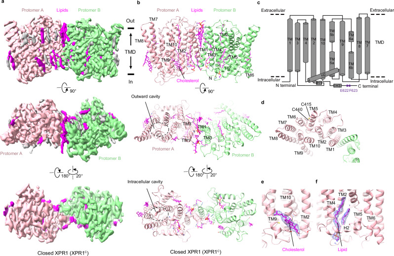
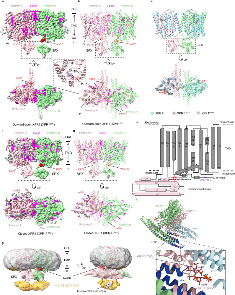
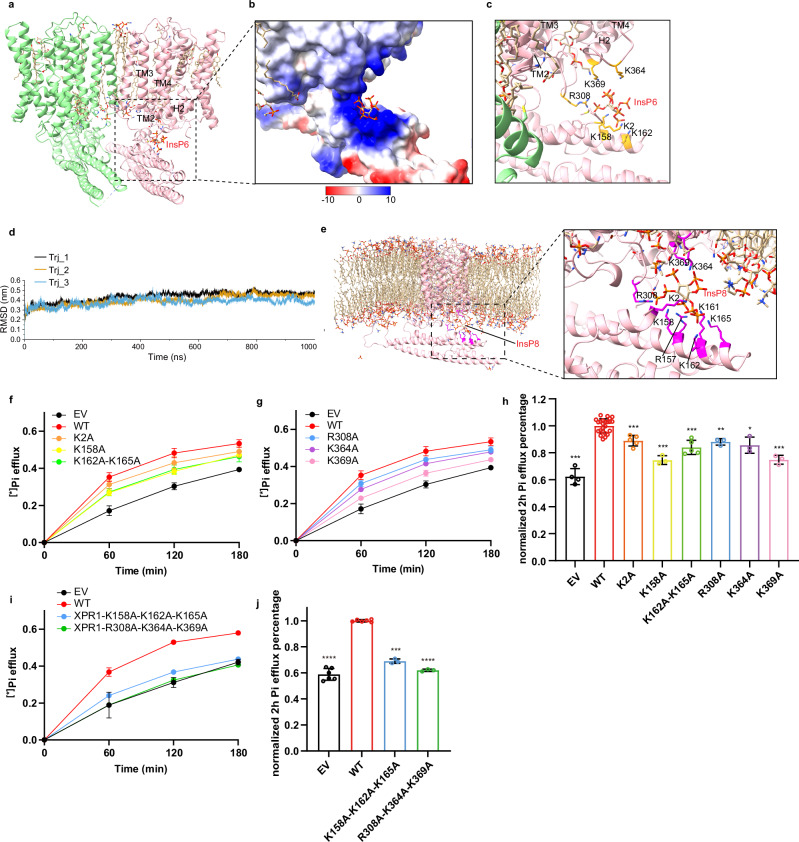
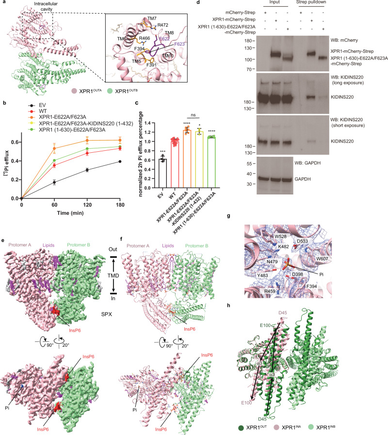
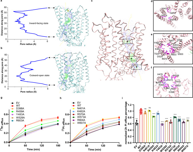
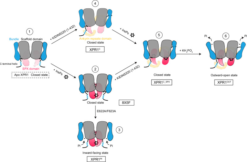
Abstract Inorganic phosphate (Pi) is fundamental to life, and its intracellular concentration must be tightly regulated to prevent toxicity. XPR1, the only known phosphate exporter, plays a crucial role in maintaining this delicate balance. However, the mechanisms underlying the function and regulation of XPR1 remain elusive until now. Here we present cryo-electron microscopy structures of the human XPR1-KIDINS220 complex in both substrate-free closed states and substrate-bound outward-open states, as well as the structure of an XPR1 mutant alone in a substrate-bound inward-facing state. In the presence of inositol hexaphosphate (InsP6) and phosphate, the XPR1-KIDINS220 complex adopts an outward-open conformation. InsP6 binds both the SPX domain and the peripheral juxtamembrane regions of XPR1, indicative of an active phosphate-export state. Conversely, in the absence of either phosphate or InsP6, the complex assumes a closed state, where the extracellular half of transmembrane 9 occupies the outward cavity, and a C-terminal plug-in loop blocks the intracellular cavity. Notably, XPR1 without KIDINS220 adopts a closed state despite the presence of phosphate and InsP6. The functional mutagenesis experiments further demonstrate that InsP6, whose concentrations fluctuate in response to Pi availability, functions synergistically with KIDINS220 to regulate the phosphate export activity of XPR1. These findings not only elucidate the intricate mechanisms of cellular phosphate regulation but also hold promise for the development of targeted therapies for ovarian cancer, where XPR1 plays a significant role.
Overview
The claims and statistics of this paper haven’t been checked yet.
Run the check — free, no account needed. It reads the open-access text and checks the sources it cites.
Integrity signals
Registry
Cites
Statistics
Figures
Verification
Claims the paper makes
The claims and statistics of this paper haven’t been checked yet.
Run the check — free, no account needed. It reads the open-access text and checks the sources it cites.
Self-check
Percentages, sums and reported differences re-computed from the paper’s own numbers.
The self-check needs the paper’s full text.
Citations
Cites
VMD: visual molecular dynamics
Humphrey, W., Dalke, A., & Schulten, K. (1996) · Journal of Molecular Graphics · doi:10.1016/0263-7855(96)00018-5 · Cited by 66,952
Highly accurate protein structure prediction with AlphaFold
Jumper, J., Evans, R., Pritzel, A., Green, T., Figurnov, M., Ronneberger, O., Tunyasuvunakool, K., Bates, R., Žídek, A., Potapenko, A., et al. (2021) · Nature · doi:10.1038/s41586-021-03819-2 · Cited by 43,764
Features and development of Coot
Emsley, P., Lohkamp, B., Scott, W. G., & Cowtan, K. (2010) · Acta Crystallographica Section D Biological Crystallography · doi:10.1107/s0907444910007493 · Cited by 29,915
Abraham, M. J., Murtola, T., Schulz, R., Páll, S., Smith, J. C., Hess, B., & Lindahl, E. (2015) · SoftwareX · doi:10.1016/j.softx.2015.06.001 · Cited by 27,190
cryoSPARC: algorithms for rapid unsupervised cryo-EM structure determination
Punjani, A., Rubinstein, J. L., Fleet, D. J., & Brubaker, M. A. (2017) · Nature Methods · doi:10.1038/nmeth.4169 · Cited by 11,626
CHARMM‐GUI: A web‐based graphical user interface for CHARMM
Jo, S., Kim, T., Iyer, V. G., & Im, W. (2008) · Journal of Computational Chemistry · doi:10.1002/jcc.20945 · Cited by 9,284
Liebschner, D., Afonine, P. V., Baker, M. L., Bunkóczi, G., Chen, V. B., Croll, T. I., Hintze, B., Hung, L.-W., Jain, S., McCoy, A. J., et al. (2019) · Acta Crystallographica Section D Structural Biology · doi:10.1107/s2059798319011471 · Cited by 7,953
MotionCor2: anisotropic correction of beam-induced motion for improved cryo-electron microscopy
Zheng, S. Q., Palovcak, E. J., Armache, J., Verba, K. A., Cheng, Y., & Agard, D. A. (2017) · Nature Methods · doi:10.1038/nmeth.4193 · Cited by 7,559
CHARMM36m: an improved force field for folded and intrinsically disordered proteins
Huang, J., Rauscher, S., Nawrocki, G., Ran, T., Feig, M., de Groot, B. L., Grubmüller, H., & MacKerell, A. D. (2016) · Nature Methods · doi:10.1038/nmeth.4067 · Cited by 7,334
MolProbity: More and better reference data for improved all‐atom structure validation
Williams, C. J., Headd, J. J., Moriarty, N. W., Prisant, M. G., Videau, L. L., Deis, L. N., Verma, V., Keedy, D. A., Hintze, B. J., Chen, V. B., et al. (2018) · Protein Science · doi:10.1002/pro.3330 · Cited by 4,599
UCSF ChimeraX: Tools for structure building and analysis
Meng, E. C., Goddard, T. D., Pettersen, E. F., Couch, G. S., Pearson, Z. J., Morris, J. H., & Ferrin, T. E. (2023) · Protein Science · doi:10.1002/pro.4792 · Cited by 4,442
HOLE: A program for the analysis of the pore dimensions of ion channel structural models
Smart, O. S., Neduvelil, J. G., Wang, X., Wallace, B. A., & Sansom, M. S. P. (1996) · Journal of Molecular Graphics · doi:10.1016/s0263-7855(97)00009-x · Cited by 1,803
Positive-unlabeled convolutional neural networks for particle picking in cryo-electron micrographs
Bepler, T., Morin, A., Rapp, M., Brasch, J., Shapiro, L., Noble, A. J., & Berger, B. (2019) · Nature Methods · doi:10.1038/s41592-019-0575-8 · Cited by 1,606
Casares, D., Escribá, P. V., & Rosselló, C. A. (2019) · International Journal of Molecular Sciences · doi:10.3390/ijms20092167 · Cited by 867
Control of eukaryotic phosphate homeostasis by inositol polyphosphate sensor domains
Wild, R., Gerasimaite, R., Jung, J.-Y., Truffault, V., Pavlovic, I., Schmidt, A., Saiardi, A., Jessen, H. J., Poirier, Y., Hothorn, M., et al. (2016) · Science · doi:10.1126/science.aad9858 · Cited by 627
Kavanaugh, M. P., Miller, D. G., Zhang, W., Law, W., Kozak, S. L., Kabat, D., & Miller, A. D. (1994) · Proceedings of the National Academy of Sciences · doi:10.1073/pnas.91.15.7071 · Cited by 586
Hamburger, D., Rezzonico, E., Petétot, J. M.-C., Somerville, C. R., & Poirier, Y. (2002) · The Plant Cell · doi:10.1105/tpc.000745 · Cited by 555
Mutant of Arabidopsis Deficient in Xylem Loading of Phosphate
Poirier, Y., Thoma, S., Somerville, C., & Schiefelbein, J. (1991) · Plant Physiology · doi:10.1104/pp.97.3.1087 · Cited by 441
Legati, A., Giovannini, D., Nicolas, G., López-Sánchez, U., Quintáns, B., de Oliveira, J. R. M., Sears, R., Ramos, E. M., Spiteri, E., Sobrido, M., et al. (2015) · Nature Genetics · doi:10.1038/ng.3289 · Cited by 322
Phosphate transporters: a tale of two solute carrier families
Virkki, L. V., Biber, J., Murer, H., & Forster, I. C. (2007) · American Journal of Physiology-Renal Physiology · doi:10.1152/ajprenal.00228.2007 · Cited by 264
Battini, J., Rasko, J. E. J., & Miller, A. D. (1999) · Proceedings of the National Academy of Sciences · doi:10.1073/pnas.96.4.1385 · Cited by 236
Inorganic Phosphate Export by the Retrovirus Receptor XPR1 in Metazoans
Giovannini, D., Touhami, J., Charnet, P., Sitbon, M., & Battini, J. (2013) · Cell Reports · doi:10.1016/j.celrep.2013.05.035 · Cited by 205
Tailor, C. S., Nouri, A., Lee, C. G., Kozak, C. A., & Kabat, D. (1999) · Proceedings of the National Academy of Sciences · doi:10.1073/pnas.96.3.927 · Cited by 195
Phosphate as a Signaling Molecule and Its Sensing Mechanism
Michigami, T., Kawai, M., Yamazaki, M., & Ozono, K. (2018) · Physiological Reviews · doi:10.1152/physrev.00022.2017 · Cited by 186
Positive-unlabeled convolutional neural networks for particle picking in cryo-electron micrographs
Bepler, T., Morin, A., Brasch, J., Shapiro, L. S., Noble, A. J., & Berger, B. (2018) · PubMed · doi:10.48550/arxiv.1803.08207 · Cited by 176
Phosphate acquisition and metabolism in plants
Poirier, Y., Jaskolowski, A., & Clúa, J. (2022) · Current Biology · doi:10.1016/j.cub.2022.03.073 · Cited by 164
Wege, S., Khan, G. A., Jung, J., Vogiatzaki, E., Pradervand, S., Aller, I., Meyer, A. J., & Poirier, Y. (2015) · PLANT PHYSIOLOGY · doi:10.1104/pp.15.00975 · Cited by 158
Identification and Cloning of Kidins220, a Novel Neuronal Substrate of Protein Kinase D
Iglesias, T., Cabrera-Poch, N., Mitchell, M., Naven, T. J. P., Rozengurt, E., & Schiavo, G. (2000) · Journal of Biological Chemistry · doi:10.1074/jbc.m005261200 · Cited by 154
Receptors for polytropic and xenotropic mouse leukaemia viruses encoded by a single gene at Rmc1
Yang, Y.-L., Guo, L., Xu, S., Holland, C. A., Kitamura, T., Hunter, K. W., & Cunningham, J. M. (1999) · Nature Genetics · doi:10.1038/6005 · Cited by 140
Wilson, M., Jessen, H. J., & Saiardi, A. (2019) · Journal of Biological Chemistry · doi:10.1074/jbc.ra119.007848 · Cited by 121
Shu, H., Sakisaka, T., Baba, T., Yamada, T., Aoki, K., Matsuda, M., & Takai, Y. (2007) · The Journal of Cell Biology · doi:10.1083/jcb.200610073 · Cited by 115
Li, X., Gu, C.-F., Hostachy, S., Sahu, S., Wittwer, C., Jessen, H., Fiedler, D., Wang, H.-C., & Shears, S. (2020) · Proceedings of the National Academy of Sciences of the United States of America · doi:10.1073/pnas.1908830117 · Cited by 112
Austin, S., & Mayer, A. (2020) · Frontiers in Microbiology · doi:10.3389/fmicb.2020.01367 · Cited by 93
Ansermet, C., Moor, M. B., Centeno, G., Auberson, M., Hu, D. Z., Baron, R., Nikolaeva, S. D., Haenzi, B., Katanaeva, N., Gautschi, I., et al. (2016) · Journal of the American Society of Nephrology · doi:10.1681/asn.2016070726 · Cited by 85
XPR1 mutations are a rare cause of primary familial brain calcification
Anheim, M., López-Sánchez, U., Giovannini, D., Richard, A., Touhami, J., Them, L. N., Rudolf, G., Thibault-Stoll, A., Frébourg, T., Hannequin, D., et al. (2016) · Journal of Neurology · doi:10.1007/s00415-016-8166-4 · Cited by 77
Bondeson, D. P., Paolella, B. R., Asfaw, A., Rothberg, M. V., Skipper, T. A., Langan, C. R., Mesa, G., Gonzalez, A. E., Surface, L. E., Ito, K., et al. (2022) · Nature Cancer · doi:10.1038/s43018-022-00360-7 · Cited by 76
The French PFBC study group, Ramos, E. M., Carecchio, M., de Lemos, R. R., Ferreira, J., Legati, A., Sears, R., Hsu, S. C., Panteghini, C., Magistrelli, L., et al. (2018) · European Journal of Human Genetics · doi:10.1038/s41431-018-0185-4 · Cited by 75
López-Sánchez, U., Tury, S., Nicolas, G., Wilson, M., Jurici, S., Ayrignac, X., Courgnaud, V., Saiardi, A., Sitbon, M., & Battini, J. (2020) · Journal of Biological Chemistry · doi:10.1074/jbc.ra119.011376 · Cited by 66
Guo, X., Zou, X., Wang, C., Yao, X., Su, H., Lai, L., Chen, H., Lai, J., Liu, Y., Chen, D., et al. (2019) · Human Mutation · doi:10.1002/humu.23703 · Cited by 47
Chen, W.-C., Li, Q., Pan, Q., Zhang, H., Fu, X.-Y., Fan, Y., Wang, J.-N., & Yang, A. (2019) · Journal of Experimental & Clinical Cancer Research · doi:10.1186/s13046-019-1155-6 · Cited by 42
Xenotropic and polytropic retrovirus receptor 1 regulates procoagulant platelet polyphosphate
Mailer, R. K., Allende, M., Heestermans, M., Schweizer, M., Deppermann, C., Frye, M., Pula, G., Odeberg, J., Gelderblom, M., Rose‐John, S., et al. (2020) · Blood · doi:10.1182/blood.2019004617 · Cited by 40
Role of transporters in regulating mammalian intracellular inorganic phosphate
Jennings, M. L. (2023) · Frontiers in Pharmacology · doi:10.3389/fphar.2023.1163442 · Cited by 37
Structural basis for inositol pyrophosphate gating of the phosphate channel XPR1
Lu, Y., Yue, C.-X., Zhang, L., Yao, D., Xia, Y., Zhang, Q., Zhang, X., Li, S., Shen, Y., Mi, C., et al. (2024) · Science · doi:10.1126/science.adp3252 · Cited by 37
Human XPR1 structures reveal phosphate export mechanism
Yan, R., Chen, H., Liu, C., et al. (2024) · Nature · doi:10.1038/s41586-024-07852-9 · Cited by 35
Wege, S., & Poirier, Y. (2013) · FEBS Letters · doi:10.1016/j.febslet.2013.12.013 · Cited by 33
López-Sánchez, U., Nicolas, G., Richard, A., Maltête, D., Charif, M., Ayrignac, X., Goizet, C., Touhami, J., Labesse, G., Battini, J., et al. (2019) · Scientific Reports · doi:10.1038/s41598-019-43255-x · Cited by 33
Pi sensing and signalling: from prokaryotic to eukaryotic cells
Qi, W., Baldwin, S. A., Muench, S. P., & Baker, A. (2016) · Biochemical Society Transactions · doi:10.1042/bst20160026 · Cited by 27
PHOSPHATE exporter XPR1/SLC53A1 is required for the tumorigenicity of epithelial ovarian cancer
Akasu‐Nagayoshi, Y., Hayashi, T., Kawabata, A., Shimizu, N., Yamada, A. Y., Yokota, N., Nakato, R., Shirahige, K., Okamoto, A., & Akiyama, T. (2022) · Cancer Science · doi:10.1111/cas.15358 · Cited by 27
Li, X., Kirkpatrick, R. B., Wang, X., Tucker, C. J., Shukla, A., Jessen, H. J., Wang, H., Shears, S. B., & Gu, C. (2024) · Cell Reports · doi:10.1016/j.celrep.2024.114316 · Cited by 25
Xu, X., Li, X., Sun, H., Cao, Z., Gao, R., Niu, T., Wang, Y., Ma, T., Chen, R., Wang, C., et al. (2019) · Journal of Bone and Mineral Research · doi:10.1002/jbmr.3866 · Cited by 24
Tang, L., Hou, B., Zhang, X., Xi, Z.-Y., Li, C., & Xu, L. (2020) · Brain and Development · doi:10.1016/j.braindev.2020.09.014 · Cited by 14
Arase, H., Yamada, S., Torisu, K., Tokumoto, M., Taniguchi, M., Tsuruya, K., Nakano, T., & Kitazono, T. (2022) · Calcified Tissue International · doi:10.1007/s00223-022-00947-3 · Cited by 10
Physiological regulation of phosphate homeostasis
Marks, J., & Unwin, R. J. (2022) · Vitamins and hormones · doi:10.1016/bs.vh.2022.04.007 · Cited by 10
Jiang, Y., Li, X., Feng, J., Li, K., Wang, O., Xing, X., & Xia, W.-B. (2020) · Journal of Endocrinological Investigation · doi:10.1007/s40618-020-01371-w · Cited by 8
XPR1 Mediates the Pancreatic β-Cell Phosphate Flush
Barker, C. J., Tessaro, F. H. G., Ferreira, S. D. S., Simas, R., Ayala, T. S., Köhler, M. G., Rajasekaran, S. S., Martins, J. O., Daré, E., & Berggren, P. (2020) · Diabetes · doi:10.2337/db19-0633 · Cited by 8
Primary Familial Brain Calcification With XPR1 Mutation Presenting With Cognitive Dysfunction
Yoon, S., Chung, S. J., & Kim, Y. J. (2024) · Journal of Clinical Neurology · doi:10.3988/jcn.2023.0284 · Cited by 6
A Japanese family with idiopathic basal ganglia calcification carrying a novel XPR1 variant
Orimo, K., Kakumoto, T., Hara, R., et al. (2023) · Journal of the Neurological Sciences · doi:10.1016/j.jns.2023.120732 · Cited by 5
Huawei, Z. O. U., & Xiang, C. (2021) · Journal of Zhejiang University (Medical Sciences) · doi:10.3724/zdxbyxb-2021-0052 · Cited by 2
Cited by
Metadata
- Authors
- Zuo, P., Wang, W., Dai, Z., Zheng, J., Yu, S., Wang, G., Yin, Y., Liang, L., & Yin, Y.
- Affiliations
- Peking University (CN)
- Corresponding author
- Yin, Yue
- Liang, Ling
- Venue
- Nature Communications
- Published
- 2025-03-24
- Volume
- 16
- Issue
- 1
- Pages
- 2879-2879
- ISSN
- 2041-1723 (ISSN-L)
- PMC
- PMC11933459
- OpenAlex
- W4408788073
- Cited by
- 13
- References
- 58
- Citations by year
- 2026: 7 · 2025: 6
- FWCI
- 3.91
- Type
- journal article
- Language
- en
- Open access
- gold
- doi.org
- Licence
- cc-by-nc-nd
- creativecommons.org/licenses/by-nc-nd/4.0 (tdm)
- creativecommons.org/licenses/by-nc-nd/4.0 (vor)
- Locations
- Nature Communications (publishedVersion, cc-by-nc-nd)
- bioRxiv (Cold Spring Harbor Laboratory) (acceptedVersion)
- PubMed Central (publishedVersion, cc-by-nc-nd)
- PubMed (publishedVersion)
- RePEc: Research Papers in Economics (submittedVersion)
- DOAJ (DOAJ: Directory of Open Access Journals) (submittedVersion, cc-by-sa)
- Funders
- National Natural Science Foundation of China (82030081, 32171224)
- Topics
- Life Sciences › Biochemistry, Genetics and Molecular Biology › Molecular Biology › Ion Transport and Channel Regulation
- Life Sciences › Biochemistry, Genetics and Molecular Biology › Molecular Biology › Ion channel regulation and function
- Health Sciences › Medicine › Oncology › Drug Transport and Resistance Mechanisms
- Keywords
- XPR1, Kidins220, phytic acid, inositol pyrophosphates, SPX domain, juxtamembrane region, inorganic phosphate homeostasis, cryo-EM structure
- MeSH terms
- Protein Domains, Xenotropic and Polytropic Retrovirus Receptor, Humans, Inositol Phosphates, Membrane Proteins, Models, Molecular, Phosphates, Phytic Acid, Protein Binding, Protein Conformation, Cryoelectron Microscopy, Receptors, G-Protein-Coupled, HEK293 Cells
About these checks
Registry, retraction, Cites and statistics results are computed, not judged, with one exception: a printed reference that no index lists is read once by a model before it counts as a citation error. Claims are extracted by a model and kept only when their quote appears verbatim in the text; each cited source is then checked, with a model’s help, for whether it says what the claim relies on. Those verdicts can be wrong — read the claim’s quote and the cited source before relying on one.
Statistics and arithmetic are recomputed, not judged.
Confidence bands are shown only for fields calibrated on our annotated test set. None is calibrated yet, so no band is shown.
What counts as a claim ›How we measure confidence ›How we check this ›
Are you an author, or do you see a problem with this page? Request a review
Data sources
Bibliographic data: Semantic Scholar, OpenAlex, Crossref.
Contains information from the Semantic Scholar Academic Graph Datasets, made available under the ODC Attribution License (ODC-BY 1.0).
Semantic Scholar’s data is described in: Kinney, R., et al. (2023). The Semantic Scholar Open Data Platform. arXiv:2301.10140.
