

DIS3 mutations enhance AID-driven translocations during B-cell activation, promoting transformation to multiple myeloma
Open accessThe DOI matches its Crossref record; 117 of the 117 references it cites were found in a registry or an index; none of the 97 sources its claims cite is checked for support yet.
Abstract
DIS3, a key nuclear RNA-degrading enzyme, is essential for immunoglobulin class switch recombination (CSR), promoting activation-induced cytidine deaminase (AID) activity on both DNA strands to induce double-strand DNA breaks. During somatic hypermutation, AID-dependent lesions predominantly occur on the non-template DNA strand. Dominant mutations impairing DIS3 exoribonucleolytic activity are common in multiple myeloma (MM), but their role in carcinogenesis remains unclear. Here we show, using a knock-in mouse model, that the clinically relevant DIS3 G766R variant causes chromosomal translocations in B-cells, characterized by aberrant AID activity signatures. The mice develop pristane-induced plasmacytomas, modeling early-stage MM. In clinical MM samples, DIS3 mutations correlate with IGH translocations and AID-driven lesions in driver genes. Mechanistically, mutated DIS3 accumulates on chromatin-bound RNA, particularly at aberrant AID target sites, promoting mutations on both DNA strands. This results in increased AID-dependent double-strand DNA breaks, fostering microhomology-mediated oncogenic rearrangements. Translocations occur specifically during CSR, which remains functionally intact. The DIS3 G766R mutation does not disrupt chromatin architecture in activated B cells but exploits spatial proximity to permanently juxtapose enhancers and proto-oncogenes, facilitating transformation. Thus, gain-of-function DIS3 mutations enhance AID promiscuity, driving IGH translocations and MM development without broadly affecting B-cell physiology.
Overview
CoChat Summary
TL;DR
In a Dis3 G766R knock-in mouse and clinical MM data, MM-associated DIS3 mutations act as gain-of-function alleles that stall on chromatin-bound RNA, increase AID-dependent double-strand breaks and IGH translocations, and promote plasmacytoma/MM transformation without disrupting CSR or Igh chromatin architecture. The mutant DIS3 does not broadly deregulate the transcriptome/proteome but enhances AID promiscuity at off-target loci.
Key findings
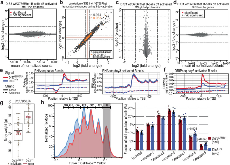
- Heterozygous Dis3G766R/+ mice are viable with subtle non-coding transcriptome/R-loop changes; homozygous Dis3G766R is embryonically lethal. They show 16 DEGs in naïve B cells, 3 DEGs after 3 days of activation, no DEGs after 5 days, no transcript upregulated >2-fold, and no significant proteome changes, but are smaller and show reduced proliferation.
- CSR and Igh chromatin architecture remain intact: ~20% of cells switch to IgG1 at day 3 in both genotypes; CoMMpass patients with somatic or germline DIS3 mutations show no CSR disruption; MicroC detects 20,603 WT and 20,279 mutant loops, with no statistically significant difference in 3′RR–Eµ interactions.
- Pristane-treated Dis3G766R/+ mice develop plasmacytomas at higher incidence than WT (risk ratio 7.19; p = 0.0073) with more Igh translocations and chimeric reads. In CoMMpass, IGH translocations occur in 64.3% of non-recurrent DIS3-mutated patients, 64.5% of recurrent DIS3-mutated patients, and 33.2% of others; AID/DNA-repair signatures are enriched in MM driver genes, with SBS26 at 35% in recurrent, 23% in non-recurrent, and 15% in the rest.
- Mechanistically, mutant DIS3 stalls on chromatin-bound RNA at AID target sites: DamID libraries from DIS3G766R cells yield 2.5-fold more DNA; DIS3–AID PLA spots increase upon activation; γ-H2AX foci increase in activated Dis3G766R/+ B cells and in CH12F3-2A cells in an AID-dependent manner; AID strand bias is reduced; microhomology at breakpoints is modestly longer (7 bp vs 5 bp in WT); translocations localize near enhancers, promoters, CTCF, and Rad21.
Methods
- Knock-in Dis3 G766R mice were generated on C57BL/6N and backcrossed to BALB/c for plasmacytoma experiments; primary splenic B cells, MEFs, and CH12F3-2A cells (WT and AID−/−) were used, with in vitro B-cell activation and pristane-induced plasmacytoma as the in vivo model.
- Assays included RNA-seq, global proteomics, DRIPc-seq, MicroC, DamID-seq, proximity ligation assay, γ-H2AX imaging, flow cytometry, mate-pair and whole-genome DNA sequencing, and analysis of CoMMpass clinical samples.
- CoMMpass groups comprised 22 patients with recurrent DIS3 mutations, 48 with other DIS3 variants, and 993 with WT DIS3; analyses used DESeq2, SVDetect, Manta, SigProfilerMatrixGenerator, and mmsig.
Limitations
- Stated in the paper: recurrent MM DIS3 alleles could not be modeled because they cause strong dominant-negative effects and early embryonic lethality, so the study uses the milder heterozygous G766R variant; DIS3 chromatin immunoprecipitation was extremely challenging because its chromatin interaction is indirect and transient; C57BL/6 is naturally resistant to plasmacytoma, requiring BALB/c backcrossing for sufficient WT control material.
- Evident from the text: the clinical CoMMpass findings are correlative associations between DIS3 mutations, IGH translocations, and mutational signatures, not direct causal proof in human MM; the mouse model is heterozygous-only and may not fully recapitulate human MM progression.
Model-written summary. Check it against the paper before relying on it.
Integrity signals
Registry
Cites
Statistics
Figures


Verification
Claims the paper makes
0 Valid · 0 Not valid · 47 Not checked · 134 Own results and interpretations
A result the paper reports from its own data — it cites no other source for it, so there is none to check. The quote shows it is in the paper.
In the paper
“This reduced size corresponds to a negative impact on cell proliferation both in activated B-cells and MEFs (Fig. 1h, Supplementary Fig. S3e, f), aligning with a conserved phenotype of DIS3 dysfunction, which includes reduced mitotic progression and increased cell death5,44–47.”
§ DIS3 mutations enhance AID-driven translocations during B-cell activation, promoting transformation to multiple myeloma > Results > Heterozygous MM-associated Dis3G766R/+ mutation leads to subtle deregulation of non-coding regions of the genome
plainstates a cause- Asserted: passes — The authors put it forward as true.
- Checkable: passes — It can be checked against evidence.
- Load-bearing: unclear — It is unclear whether the paper’s conclusions depend on it.
- Based: passes — The paper gives a reason why it holds.
A result the paper reports from its own data — it cites no other source for it, so there is none to check. The quote shows it is in the paper.
In the paper
“However, global analysis of the RNA-seq signal centered over active transcriptional start sites (TSS) revealed focal enrichments in regions corresponding to RNA Pol II promoter-proximal pausing and upstream regions from which PROMPTs originate (Fig. 1e and Supplementary Fig. S2d, e).”
§ DIS3 mutations enhance AID-driven translocations during B-cell activation, promoting transformation to multiple myeloma > Results > Heterozygous MM-associated Dis3G766R/+ mutation leads to subtle deregulation of non-coding regions of the genome
plainstates an association- Asserted: passes — The authors put it forward as true.
- Checkable: passes — It can be checked against evidence.
- Load-bearing: unclear — It is unclear whether the paper’s conclusions depend on it.
- Based: passes — The paper gives a reason why it holds.
A result the paper reports from its own data — it cites no other source for it, so there is none to check. The quote shows it is in the paper.
In the paper
“DIS3G766R is predicted to be highly pathogenic (Supplementary Fig. S1a)”
§ DIS3 mutations enhance AID-driven translocations during B-cell activation, promoting transformation to multiple myeloma > Introduction
plain- Asserted: passes — The authors put it forward as true.
- Checkable: passes — It can be checked against evidence.
- Load-bearing: passes — The paper’s conclusions depend on it.
- Based: passes — The paper gives a reason why it holds.
A result the paper reports from its own data — it cites no other source for it, so there is none to check. The quote shows it is in the paper.
In the paper
“In the case of Dis3G766R/+ B-cells compared to WT, no accumulation was detected when looking at individual ncRNA species.”
§ DIS3 mutations enhance AID-driven translocations during B-cell activation, promoting transformation to multiple myeloma > Results > Heterozygous MM-associated Dis3G766R/+ mutation leads to subtle deregulation of non-coding regions of the genome
plain- Asserted: passes — The authors put it forward as true.
- Checkable: passes — It can be checked against evidence.
- Load-bearing: unclear — It is unclear whether the paper’s conclusions depend on it.
- Based: passes — The paper gives a reason why it holds.
A result the paper reports from its own data — it cites no other source for it, so there is none to check. The quote shows it is in the paper.
In the paper
“Notably, in Dis3G766R/+ B-cells, DRIP-seq analysis revealed a much more prominent accumulation of R-loops in the promoter-proximal pausing regions and over PROMPTs (Fig. 1f).”
§ DIS3 mutations enhance AID-driven translocations during B-cell activation, promoting transformation to multiple myeloma > Results > Heterozygous MM-associated Dis3G766R/+ mutation leads to subtle deregulation of non-coding regions of the genome
plainstates an association- Asserted: passes — The authors put it forward as true.
- Checkable: passes — It can be checked against evidence.
- Load-bearing: passes — The paper’s conclusions depend on it.
- Based: passes — The paper gives a reason why it holds.
88 more claims
Create a free account to see all 181 claims and to verify the 97 references still open.
- It may state two separate claims as one. 55
- Probably not a substantial claim. 43
Claims other papers make about this one
Each is checked against this paper’s own text: does it say what the citing paper says it does?
In the citing paper
“Proposed mechanisms underlying DIS3-related mutagenesis include alterations in the pattern of CTCF and cohesin binding, and the establishment of abnormally open chromatin states that favor promiscuous editing by the activation-induced cytidine deaminase AID.14,16,49”
From RNA degradation by DIS3 is a necessary step in the resolution of backtracked transcription complexes (2026) · cites this paper as [16]
Self-check
Percentages, sums and reported differences re-computed from the paper’s own numbers.
Each contradiction between two passages listed here was confirmed by an independent check.
Rated minor: it would not touch a result — a value no result rests on, a p-value that stays on the same side of the significance level, or a conflict that is clerical, resolved elsewhere in the paper, or outside its own findings.
In the paper
§ DIS3 mutations enhance AID-driven translocations during B-cell activation, promoting transformation to multiple myeloma > Methods > Microscopic analysis
“For Fig. 5f,g cells were imaged using Olympus IX81 wide-field fluorescence microscope”
§ DIS3 mutations enhance AID-driven translocations during B-cell activation, promoting transformation to multiple myeloma > Results > Mutational profiles revealed signatures of promiscuous AID activity and a prevalence of translocations in patients with MM DIS3 alleles
“c–g Contribution of COSMIC mutational signatures in all somatic single-nucleotide variants (SNVs) acquired by CoMMpass patients with WT DIS3, a mutant DIS3, or a recurrent MM DIS3 variant in whole exome sequencing (WES): exome-wide (c), in all somatic DIS3 mutations (d), expressed PROMPTs (e), in genes known to be AID off-targets (f) and in MM driver genes (g).”
Rated minor: it would not touch a result — a value no result rests on, a p-value that stays on the same side of the significance level, or a conflict that is clerical, resolved elsewhere in the paper, or outside its own findings.
In the paper
§ DIS3 mutations enhance AID-driven translocations during B-cell activation, promoting transformation to multiple myeloma > Results > MM DIS3 variants do not affect CSR and genome architecture in B-cells
“n = 8 DIS3G766R/+ n = 7 DIS3+/+ individuals in immunized and n = 3 / genotype for naïve mice”
§ DIS3 mutations enhance AID-driven translocations during B-cell activation, promoting transformation to multiple myeloma > Methods > Splenic Germinal Center Phenotyping
“three biological replicates per genotype for naïve animals and eight individuals per genotype at day 14 post-immunization with the Moderna mRNA vaccine”
Rated minor: it would not touch a result — a value no result rests on, a p-value that stays on the same side of the significance level, or a conflict that is clerical, resolved elsewhere in the paper, or outside its own findings.
In the paper
§ DIS3 mutations enhance AID-driven translocations during B-cell activation, promoting transformation to multiple myeloma > Results > Mutant DIS3 stalling on chromatin-bound substrates, resulting in increased AID-dependent dsDNA breaks in activated B-cells
“Analysis was restricted to cells with at least one PLA spot identified (PLA-positive cells).”
§ DIS3 mutations enhance AID-driven translocations during B-cell activation, promoting transformation to multiple myeloma > Methods > Microscopic analysis
“Cells containing at least one PLA spot were filtered out and for each experimental condition, a total number of spots in a cell and its mean were calculated.”
Citations
Cites
STAR: ultrafast universal RNA-seq aligner
Dobin, A., Davis, C. A., Schlesinger, F., Drenkow, J., Zaleski, C., Jha, S., Batut, P., Chaisson, M., & Gingeras, T. R. (2013) · Bioinformatics · doi:10.1093/bioinformatics/bts635 · Cited by 51,709
Cutadapt removes adapter sequences from high-throughput sequencing reads
Martin, M. (2011) · EMBnet.journal · doi:10.14806/ej.17.1.200 · Cited by 33,889
featureCounts: an efficient general purpose program for assigning sequence reads to genomic features
Liao, Y., Smyth, G. K., & Shi, W. (2014) · Bioinformatics · doi:10.1093/bioinformatics/btt656 · Cited by 27,219
The Perseus computational platform for comprehensive analysis of (prote)omics data
Tyanova, S., Temu, T., Sinitcyn, P., Carlson, A., Hein, M. Y., Geiger, T., Mann, M., & Cox, J. (2016) · Nature Methods · doi:10.1038/nmeth.3901 · Cited by 8,803
Li, H. (2011) · Bioinformatics · doi:10.1093/bioinformatics/btr509 · Cited by 6,486
Poewe, W., Seppi, K., Tanner, C. M., Halliday, G. M., Brundin, P., Volkmann, J., Schrag, A.-E., & Lang, A. E. (2017) · Nature Reviews Disease Primers · doi:10.1038/nrdp.2017.13 · Cited by 5,415
The MaxQuant computational platform for mass spectrometry-based shotgun proteomics
Tyanova, S., Temu, T., & Cox, J. (2016) · Nature Protocols · doi:10.1038/nprot.2016.136 · Cited by 4,797
Juicer Provides a One-Click System for Analyzing Loop-Resolution Hi-C Experiments
Durand, N. C., Shamim, M. S., Machol, I., Rao, S. S. P., Huntley, M. H., Lander, E. S., & Aiden, E. L. (2016) · Cell Systems · doi:10.1016/j.cels.2016.07.002 · Cited by 3,836
Muramatsu, M., Kinoshita, K., Fagarasan, S., Yamada, S., Shinkai, Y., & Honjo, T. (2000) · Cell · doi:10.1016/s0092-8674(00)00078-7 · Cited by 3,474
(2015) · doi:10.1093/bioinformatics/btv710 · Cited by 2,234
Research works printed but not linked: 1
- Laffleur, B. et al. RNA exosome drives early B cell development via noncoding RNA processing mechanisms. Sci. Immunol. 7, 1–14 (2022). PMC9357289 35658015 · DOI · Paper page
Cited by
AID in non-Hodgkin B-cell lymphomas: The consequences of on- and off-target activity
Leeman‐Neill, R. J., Bhagat, G., & Basu, U. (2024) · Advances in immunology · doi:10.1016/bs.ai.2024.03.005 · Cited by 6
Brouze, M., Szpila, M., Czerwińska, A. M., Antczak, W., Mroczek, S., Kuliński, T., Hojka-Osińska, A. M., Cysewski, D., Gewartowska, O., Adamska, D., et al. (2025) · RNA · doi:10.1261/rna.080350.124 · Cited by 6
Self-citationKuliński, T. M., Gewartowska, O., Mroczek, S., Szpila, M., Sałas, K., Liudkovska, V., & Dziembowski, A. (2023) · doi:10.1101/2023.07.27.550471 · Cited by 4
Self-citationComparative Molecular Insights and Computational Modeling of Multiple Myeloma and Osteosarcoma
Ardelean, A. I., Silberschmidt, V. V., & Ioniţă, M. (2026) · International Journal of Molecular Sciences · doi:10.3390/ijms27083611 · Cited by 1
RNA degradation by DIS3 is a necessary step in the resolution of backtracked transcription complexes
Enervald, E., Jain, S., Tarbier, M., Sinha, I., Jain, A., Banerjee, P., Planells, J., McHugh, T., & Visa, N. (2026) · Cell Reports · doi:10.1016/j.celrep.2026.117886
Metadata
- Authors
- Kuliński, T. M., Gewartowska, O., Mahé, M., Kasztelan, K., Durys, N., Stroynowska-Czerwińska, A., Jedynak-Slyvka, M., Owczarek, E. P., Chaudhury, D., Nowotny, M., Pękowska, A., Séraphin, B., & Dziembowski, A.
- Affiliations
- International Institute of Molecular and Cell Biology (PL)
- Centre National de la Recherche Scientifique (FR)
- Institute of Biochemistry and Biophysics, Polish Academy of Sciences (PL)
- Instytut Biologii Doświadczalnej im. Marcelego Nenckiego (PL)
- Corresponding author
- Kuliński, Tomasz M.
- Dziembowski, Andrzej
- ORCID
- Venue
- Nature Communications
- Published
- 2026-03-14
- Volume
- 17
- Issue
- 1
- Pages
- 3976
- ISSN
- 2041-1723 (ISSN-L)
- PMC
- PMC13133341
- OpenAlex
- W7135404926
- Cited by
- 7
- References
- 116
- Citations by year
- 2026: 4
- FWCI
- 12.31
- Type
- journal article
- Language
- en
- Open access
- gold
- doi.org
- Licence
- cc-by
- creativecommons.org/licenses/by/4.0 (tdm)
- creativecommons.org/licenses/by/4.0 (vor, 48 d)
- Locations
- Nature Communications (publishedVersion, cc-by)
- bioRxiv (Cold Spring Harbor Laboratory) (submittedVersion, cc-by-nd)
- PubMed Central (publishedVersion, cc-by)
- PubMed (publishedVersion)
- HAL (submittedVersion)
- HAL (submittedVersion, cc-by)
- DOAJ (DOAJ: Directory of Open Access Journals) (submittedVersion, cc-by-sa)
- Funders
- Narodowe Centrum Nauki (UMO-2016/22/A/NZ4/00380, UMO-2013/10/M/NZ4/00299, UMO-2019/33/B/NZ2/02437, UMO-2021/43/B/NZ2/02934, UMO-2021/42/E/NZ2/00392)
- EC | Horizon 2020 Framework Programme (810425)
- Topics
- Health Sciences › Medicine › Hematology › Multiple Myeloma Research and Treatments
- Life Sciences › Biochemistry, Genetics and Molecular Biology › Molecular Biology › DNA Repair Mechanisms
- Health Sciences › Medicine › Pathology and Forensic Medicine › Lymphoma Diagnosis and Treatment
- Keywords
- DIS3, multiple myeloma, activation-induced cytidine deaminase, immunoglobulin class switch recombination, somatic hypermutation, chromosomal translocation, IGH translocation, B cell activation, DNA double-strand breaks, gain-of-function mutations
- MeSH terms
- AICDA (Activation-Induced Cytidine Deaminase), Animals, B-Lymphocytes, Cell Transformation, Neoplastic, Cytidine Deaminase, Humans, Lymphocyte Activation, Multiple Myeloma, Mutation, Translocation, Genetic, Immunoglobulin Class Switching, Mice, DNA Breaks, Double-Stranded, Exosome Multienzyme Ribonuclease Complex
About these checks
Registry, retraction, Cites and statistics results are computed, not judged, with one exception: a printed reference that no index lists is read once by a model before it counts as a citation error. Claims are extracted by a model and kept only when their quote appears verbatim in the text; each cited source is then checked, with a model’s help, for whether it says what the claim relies on. Those verdicts can be wrong — read the claim’s quote and the cited source before relying on one.
A language model drew the claims from the paper on 2026-11-22, under version 3 of the claim definition (model deepseek-v4.1-flash, prompt extract-v3).
A second model labelled each claim on 2026-11-22 (model jev-1.13-20260917, definition revision gold-guideline-v0.2).
Statistics and arithmetic are recomputed, not judged.
Confidence bands are shown only for fields calibrated on our annotated test set. None is calibrated yet, so no band is shown.
What counts as a claim ›How we measure confidence ›How we check this ›
Are you an author, or do you see a problem with this page? Request a review
How to cite
Cite CoChat for the checks on this page, and the paper for its own findings.
- This page
- CoChat. (2026). Citation check: DIS3 mutations enhance AID-driven translocations during B-cell activation, promoting transformation to multiple myeloma. https://app.cochat.ai/papers/10.1038/s41467-026-70386-3
- The paper
- Kuliński, T. M., Gewartowska, O., Mahé, M., Kasztelan, K., Durys, N., Stroynowska-Czerwińska, A., Jedynak-Slyvka, M., Owczarek, E. P., Chaudhury, D., Nowotny, M., Pękowska, A., Séraphin, B., & Dziembowski, A. (2026). DIS3 mutations enhance AID-driven translocations during B-cell activation, promoting transformation to multiple myeloma. Nature Communications. https://doi.org/10.1038/s41467-026-70386-3
Data sources
Bibliographic data: Semantic Scholar, OpenAlex, Crossref.
Contains information from the Semantic Scholar Academic Graph Datasets, made available under the ODC Attribution License (ODC-BY 1.0).
Semantic Scholar’s data is described in: Kinney, R., et al. (2023). The Semantic Scholar Open Data Platform. arXiv:2301.10140.
