

The DOI matches its Crossref record; 72 of the 72 references it cites were found in a registry or an index; 27 of the 44 sources its claims cite were checked for support.
Connectome-constrained networks predict neural activity across the fly visual system
Open accessAbstract
With experimental measurements of only the connectivity of a biological neural network, we can predict the neural activity underlying a specified neural computation. We constructed a model neural network with the experimentally determined connectivity for 64 cell types in the motion pathways of the fruit fly optic lobe but with unknown parameters for the single-neuron and single-synapse properties, and task-optimized it on visual motion detection.
Overview
CoChat Summary
## TL;DR
The paper shows that a deep mechanistic network constrained only by the fly optic-lobe connectome and optimized for optic-flow computation can predict neural responses across the visual system, matching measurements from 26 prior studies. It also argues that sparse connectivity and task optimization are key to making single-neuron predictions from connectome data.
## Key findings
- The model includes 45,669 neurons, 1,513,231 connections, 64 cell types, and only 734 free parameters because cell-type parameters and synapse-count-based constraints are shared across the tiled hexagonal lattice.
- In validation against prior physiology, the ensemble predicted the correct ON/OFF contrast selectivity for all 32 cell types with experimentally established contrast preferences; the best single model got 30 of 32 correct.
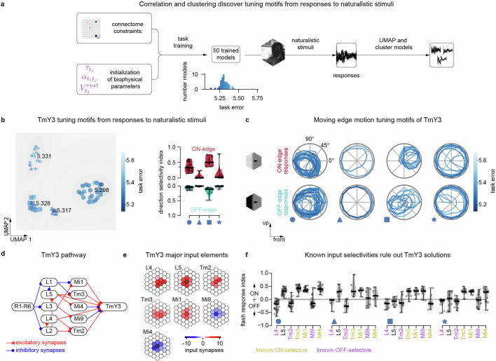
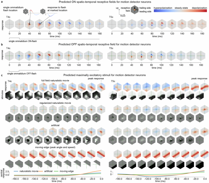
- For motion tuning, the ensemble correctly predicted that T4 neurons are ON-motion selective and T5 neurons are OFF-motion selective, and also predicted the lack of motion tuning in the listed input neurons to T4/T5.
- In synthetic-connectome experiments, sparse networks improved prediction from connectivity alone: median Pearson correlation was 0.85 for 10% connectivity versus 0.38 for 80% connectivity; with connection-strength estimates, correlations were >0.9 across connectivities.
## Methods
- Built a connectome-constrained, task-optimized deep mechanistic network (DMN) for the fly visual system using local reconstructions from FlyEM/FIB datasets, tiled into a spatially invariant hexagonal lattice.
- Trained the model end-to-end on optic-flow prediction from 23 Sintel video sequences (with augmentations), using a recurrent DMN plus a feedforward decoder and backpropagation through time.
- Evaluated model predictions by simulating responses to flash stimuli, moving edges, and naturalistic video, then comparing predicted tuning to reported experimental data from 26 studies.
## Limitations
- The paper explicitly notes that the model is reductionist and cannot account for electrical synapses, nonlinear chemical synapses, neuromodulation, or glia.
- The connectome was approximated as perfectly periodic / translation invariant across the visual field; this is a modeling simplification stated in the paper.
- The text makes evident that the model is limited to the motion pathways and the optic-flow task only, so its scope does not cover all computations of the fly visual system.
Model-written summary. Check it against the paper before relying on it.
Integrity signals
Registry
Cites
Statistics
Figures




Verification
Claims the paper makes
16 Valid · 1 Not valid · 5 Not checked · 67 Own results and interpretations
Every source the paper cites for this claim was checked and supports it.
In the paper
“We found that our connectome-constrained and task-optimized DMN accurately predicts the separation of the visual system into light-increment (ON) and light-decrement (OFF) channels, as well as the generation of direction selectivity in the well-known T4 and T5 motion detector neurons24.”
§ Connectome-constrained networks predict neural activity across the fly visual system > Main Electrical signals propagating through networks of neurons form the basis of computations such as visual motion detection. The propagation of neural activity is shaped by both the functional properties of individual neurons and their synaptic connectivity. Additional factors10,13, including electrical synapses, neuromodulation and glia, are known to further influence neural activity on multiple timescales. Volume electron microscopy can now be used to comprehensively measure the connectivity of each neuron in a neural circuit, and even entire nervous systems1–9. However, we do not yet have the means to also comprehensively measure all other biological details, including the dynamical properties of every neuron and synapse in the same circuit13. For these reasons, there has been considerable debate about the utility of connectome measurements for understanding brain function14. It is unclear whether it is possible to use only measurements of connectivity to generate accurate predictions about how the neural circuit functions, especially in the absence of direct measurements of neural activity from a living brain. There is considerable evidence from computer science and neuroscience that there is not necessarily a strong link between the connectivity of a neural network and its computational function. Universal function approximation theorems for artificial neural networks15 imply that the same computational task can be performed by many different networks with very different neural connectivity. Empirically, there exist many classes of general-purpose artificial neural network architectures that can be trained to perform the same computational task11. Such differences in connectivity can correspond to qualitatively different computational mechanisms16. Similarly, in neuroscience there have been competing proposals for the same computation (for instance, the computation of visual motion)17,18. Furthermore, even circuits with the same connectivity can function differently19. Thus, neither the connectivity of a circuit alone, nor its computational task alone, can uniquely determine the mechanism of circuit function20. Here we show that the connectivity of a neural circuit, together with knowledge of its computational task, enables accurate predictions of the role played by individual neurons in the circuit in the computational task. We constructed a differentiable21 model neural network with a close correspondence to the brain, whose connectivity was given by connectome measurements and with unknown single-neuron and single-synapse parameters. We optimized the unknown parameters of the model using techniques from deep learning11, to enable the model to accomplish the computational task22. We call such models connectome-constrained and task-optimized deep mechanistic networks (DMNs) (Fig. 1a). We applied this approach to model the motion pathways in the optic lobe of the Drosophila visual system. We constructed a DMN with experimentally measured connectivity1–5, and unknown parameters for the single-neuron dynamics and the strength of a unitary synapse. We optimized the model parameters on the computer vision task of detecting motion in dynamic visual stimuli12. Visual motion computation in the fly and its mechanistic underpinnings have been extensively studied23. Thus, we were able to compare the detailed predictions of our model with experimental measurements of neural activity in response to visual stimuli, on a neuron-by-neuron basis. We found that our connectome-constrained and task-optimized DMN accurately predicts the separation of the visual system into light-increment (ON) and light-decrement (OFF) channels, as well as the generation of direction selectivity in the well-known T4 and T5 motion detector neurons24. We release our model as a resource for the community (https://github.com/TuragaLab/flyvis).
What the sources say
- Supportedcites [24] Currier, T. A., Pang, M. M. & Clandinin, T. R. Visual processing in the fly, from photoreceptors to behavior. Genetics 224, iyad064 (2023). source
In the section ‘The lobula complex performs directional motion computations’ (page unavailable), the review identifies T4 and T5 as motion detectors and says direction selectivity first emerges in their dendrites. This supports the biological point for which the citation is used; the DMN’s predictive accuracy is the citing authors’ own finding, not a result attributed to this review. (LLM, conf=0.90)
boostedstates a cause- Asserted: passes — The authors put it forward as true.
- Checkable: passes — It can be checked against evidence.
- Load-bearing: passes — The paper’s conclusions depend on it.
- Based: passes — The paper gives a reason why it holds.
- Rests on 4 assumptions not shown here.
- Supported
Every source the paper cites for this claim was checked and supports it.
In the paper
“We found that our connectome-constrained and task-optimized DMN accurately predicts the separation of the visual system into light-increment (ON) and light-decrement (OFF) channels, as well as the generation of direction selectivity in the well-known T4 and T5 motion detector neurons24.”
§ Connectome-constrained networks predict neural activity across the fly visual system > Main Electrical signals propagating through networks of neurons form the basis of computations such as visual motion detection. The propagation of neural activity is shaped by both the functional properties of individual neurons and their synaptic connectivity. Additional factors10,13, including electrical synapses, neuromodulation and glia, are known to further influence neural activity on multiple timescales. Volume electron microscopy can now be used to comprehensively measure the connectivity of each neuron in a neural circuit, and even entire nervous systems1–9. However, we do not yet have the means to also comprehensively measure all other biological details, including the dynamical properties of every neuron and synapse in the same circuit13. For these reasons, there has been considerable debate about the utility of connectome measurements for understanding brain function14. It is unclear whether it is possible to use only measurements of connectivity to generate accurate predictions about how the neural circuit functions, especially in the absence of direct measurements of neural activity from a living brain. There is considerable evidence from computer science and neuroscience that there is not necessarily a strong link between the connectivity of a neural network and its computational function. Universal function approximation theorems for artificial neural networks15 imply that the same computational task can be performed by many different networks with very different neural connectivity. Empirically, there exist many classes of general-purpose artificial neural network architectures that can be trained to perform the same computational task11. Such differences in connectivity can correspond to qualitatively different computational mechanisms16. Similarly, in neuroscience there have been competing proposals for the same computation (for instance, the computation of visual motion)17,18. Furthermore, even circuits with the same connectivity can function differently19. Thus, neither the connectivity of a circuit alone, nor its computational task alone, can uniquely determine the mechanism of circuit function20. Here we show that the connectivity of a neural circuit, together with knowledge of its computational task, enables accurate predictions of the role played by individual neurons in the circuit in the computational task. We constructed a differentiable21 model neural network with a close correspondence to the brain, whose connectivity was given by connectome measurements and with unknown single-neuron and single-synapse parameters. We optimized the unknown parameters of the model using techniques from deep learning11, to enable the model to accomplish the computational task22. We call such models connectome-constrained and task-optimized deep mechanistic networks (DMNs) (Fig. 1a). We applied this approach to model the motion pathways in the optic lobe of the Drosophila visual system. We constructed a DMN with experimentally measured connectivity1–5, and unknown parameters for the single-neuron dynamics and the strength of a unitary synapse. We optimized the model parameters on the computer vision task of detecting motion in dynamic visual stimuli12. Visual motion computation in the fly and its mechanistic underpinnings have been extensively studied23. Thus, we were able to compare the detailed predictions of our model with experimental measurements of neural activity in response to visual stimuli, on a neuron-by-neuron basis. We found that our connectome-constrained and task-optimized DMN accurately predicts the separation of the visual system into light-increment (ON) and light-decrement (OFF) channels, as well as the generation of direction selectivity in the well-known T4 and T5 motion detector neurons24. We release our model as a resource for the community (https://github.com/TuragaLab/flyvis).
What the sources say
- Supportedcites [24] Currier, T. A., Pang, M. M. & Clandinin, T. R. Visual processing in the fly, from photoreceptors to behavior. Genetics 224, iyad064 (2023). source
The Optic lobes section (Fig. 5 caption; page unavailable) describes distinct ON and OFF motion pathways leading to T4 and T5. The cited paper supports the visual-system organization used as a comparison; the DMN’s predictive accuracy is presented as the citing authors’ own finding, not a finding attributed to this source. (LLM, conf=0.90)
boostedstates a cause- Asserted: passes — The authors put it forward as true.
- Checkable: passes — It can be checked against evidence.
- Load-bearing: passes — The paper’s conclusions depend on it.
- Based: passes — The paper gives a reason why it holds.
- Rests on 4 assumptions not shown here.
- Supported
The sources with a verdict support this claim; the rest weren’t checked yet, have no full text we could read, or their verdicts aren’t rated yet.
In the paper
“Our reductionist model cannot, for example, account for the role played in this circuit by electrical synapses46, nonlinear chemical synapses47 and neuromodulation48.”
§ Connectome-constrained networks predict neural activity across the fly visual system > Discussion
What the sources say
- Not checked — no full text of the sourcecites [46] Ammer, G., Leonhardt, A., Bahl, A., Dickson, B. J. & Borst, A. Functional specialization of neural input elements to the Drosophila ON motion detector. Curr. Biol. 25, 2247–2253 (2015). source
- Supportedcites [47] Groschner, L. N., Malis, J. G., Zuidinga, B. & Borst, A. A biophysical account of multiplication by a single neuron. Nature 603, 119–123 (2022). source
Citation 47 is attached to “nonlinear chemical synapses,” not to the citing authors’ statement about their model’s limitations. The source paper’s Main section describes a supralinear interaction between excitatory and inhibitory synaptic signals in the fruit-fly motion circuit; the Discussion calls it “multiplicative disinhibition.” Page numbers were not provided. (LLM, conf=0.90)
- Supportedcites [48] Strother, J. A. et al. Behavioral state modulates the ON visual motion pathway of Drosophila. Proc. Natl Acad. Sci. USA 115, E102–E111 (2018). source
Citation 48 is attached to neuromodulation, not to the other synapse types or the citing authors’ assessment of their model. The Discussion describes behavioral-state modulation in the ON motion pathway, and the Results report evidence that octopamine neurons mediate it, with especially substantial effects on Mi4 neurons. Page numbers are not provided in our copy. (LLM, conf=0.92)
plain- Asserted: passes — The authors put it forward as true.
- Checkable: passes — It can be checked against evidence.
- Load-bearing: unclear — It is unclear whether the paper’s conclusions depend on it.
- Based: passes — The paper gives a reason why it holds.
The sources with a verdict support this claim; the rest weren’t checked yet, have no full text we could read, or their verdicts aren’t rated yet.
In the paper
“We built a computational model of the fly visual system that is consistent with available connectome data1–5, has biophysically plausible neural dynamics, and can be computationally trained to solve an ethologically relevant behavioural task, namely the estimation of optic flow.”
§ Connectome-constrained networks predict neural activity across the fly visual system > Methods > Construction of spatially invariant connectome from local reconstructions
What the sources say
- Not checked — no full text of the sourcecites [1] Rivera-Alba, M. et al. Wiring economy and volume exclusion determine neuronal placement in the Drosophila brain. Curr. Biol. 21, 2000–2005 (2011). source
- Supportedcites [2] Takemura, S. et al. Synaptic circuits and their variations within different columns in the visual system of Drosophila. Proc. Natl Acad. Sci. USA 112, 13711–13716 (2015). source
The citation identifies a source of available connectome data, rather than attributing the citing authors’ model to this paper. The Results describe an EM reconstruction of seven fly medulla columns and a dataset of connections among 20 cell types, supporting its use as connectome data. (LLM, conf=0.87)
- Supportedcites [3] Takemura, S. et al. The comprehensive connectome of a neural substrate for ‘ON’ motion detection in Drosophila. eLife 6, e24394 (2017). source
The citation is attached to “available connectome data,” not to the citing authors’ assessment of their model. The source’s Materials and methods (Connectome reconstruction; page unavailable) describes reconstructed neurons, mapped synapses, and connectivity matrices, confirming that it supplies such data. It does not itself evaluate the citing authors’ model. (LLM, conf=0.88)
- Supportedcites [4] Shinomiya, K. et al. Comparisons between the ON- and OFF-edge motion pathways in the Drosophila brain. eLife 8, e40025 (2019). source
The citation identifies a source of fly visual-system connectome data. The Abstract reports a connectome with detailed connectivity information for inputs to both the T4 and T5 motion pathways in an optic-lobe EM dataset; the Introduction likewise describes its analysis of those pathways. This supports citing the paper as available connectome data used to constrain the model. (LLM, conf=0.90)
- Not checked — no full text of the sourcecites [5] Shinomiya, K., Nern, A., Meinertzhagen, I. A., Plaza, S. M. & Reiser, M. B. Neuronal circuits integrating visual motion information in Drosophila melanogaster. Curr. Biol. 32, 3529–3544 (2022). source
plainstates an association- Asserted: passes — The authors put it forward as true.
- Checkable: passes — It can be checked against evidence.
- Load-bearing: passes — The paper’s conclusions depend on it.
- Based: passes — The paper gives a reason why it holds.
The cited sources support only part of this claim — read each source’s verdict below.
In the paper
“By contrast, every neuron and synapse in our connectome-constrained model49–53 has a direct correspondence to neurons and synapses in the brain.”
§ Connectome-constrained networks predict neural activity across the fly visual system > Discussion
What the sources say
- Partly supportedcites [49] Tschopp, F. D., Reiser, M. B. & Turaga, S. C. A connectome based hexagonal lattice convolutional network model of the Drosophila visual system. Preprint at https://arxiv.org/abs/1806.04793 (2018). sourceExperimental — accuracy not yet measured
The source does ground its model's neurons and edges in real connectome data: Section 2 (p. 2) states the model is built 'Based on publications of lamina [12-14] and medulla [9-11] connectomes... 43 neuron types', and Section 2.1 (p. 3) that 'we replaced all synapses counted between two connected distinct neurons in the connectome by a single synapse', weighted by the synapse count. But the absolute 'every neuron and synapse has a direct correspondence' overstates it: the locally described connectome is spatially repeated across the lattice (p. 2), morphology is deliberately ignored (p. 2), biases carry no connectome information and delays/decoder layers are assumed (Sections 2.1, 2.2, p. 3), weights are subsequently fine-tuned by backpropagation, and the Discussion (p. 11) concedes 'incomplete filters due to missing connectomic information', an assumed T5 configuration, and tangential neurons (Mt8, Mi11, Tm28) not included at all. (LLM, conf=0.72)
- Partly supportedcites [50] Mano, O., Creamer, M. S., Badwan, B. A. & Clark, D. A. Predicting individual neuron responses with anatomically constrained task optimization. Curr. Biol. 31, 4062–4075 (2021). sourceExperimental — accuracy not yet measured
The Introduction says connectomic constraints create an a priori correspondence between specific model nodes and individual biological neurons. But the Discussion’s “Circuit features neglected in these models” describes simplified inputs and omitted lateral and feedback connections; the paper does not establish a direct correspondence for every model synapse. (LLM, conf=0.86)
- Not matchedcites [51] Mi, L. et al. Connectome-constrained latent variable model of whole-brain neural activity. In Proc. International Conference on Learning Representations https://openreview.net/forum?id=CJzi3dRlJE- (2022).
Couldn’t be matched to a paper in Crossref, so it can’t be checked.
- Supportedcites [52] Shiu, P. K. et al. A leaky integrate-and-fire computational model based on the connectome of the entire adult Drosophila brain reveals insights into sensorimotor processing. Preprint at bioRxiv https://doi.org/10.1101/2023.05.02.539144 (2023). source
The Methods (Computational Model; page unavailable) state that all 127,400 proofread Flywire neurons are included and that connection weights come from Flywire synaptic connectivity. This supports direct correspondence between the model’s neurons and connections and the reconstructed brain connectome; it does not imply that the model reproduces every physiological property of those neurons or synapses. (LLM, conf=0.88)
- Partly supportedcites [53] Beiran, M. & Litwin-Kumar, A. Prediction of neural activity in connectome-constrained recurrent networks. Preprint at bioRxiv https://doi.org/10.1101/2024.02.22.581667 (2024). sourceExperimental — accuracy not yet measured
The Introduction says connectome-constrained models use structural connectivity, and the Discussion’s ‘Assessment of connectome-constrained solutions’ says they address the contribution of each specific neuron. But this paper studies simulated networks (Discussion, ‘Challenges for connectome-constrained neural networks’); it does not establish that every neuron and synapse in the citing authors’ model directly corresponds to one in a brain. Page numbers are unavailable in our copy. (LLM, conf=0.84)
plainstates an association- Asserted: passes — The authors put it forward as true.
- Checkable: passes — It can be checked against evidence.
- Load-bearing: passes — The paper’s conclusions depend on it.
- Based: passes — The paper gives a reason why it holds.
- Partly supported
46 more claims
Create a free account to see all 89 claims and every source verdict.
- It may state two separate claims as one. 26
- Probably not a substantial claim. 18
Claims other papers make about this one
Each is checked against this paper’s own text: does it say what the citing paper says it does?
The source backs part of the claim, not all of it — or backs it for a different population, condition or comparison.
In the citing paper
“In previous studies, an absence of information across columns has necessitated treating each column as identical in simulations of the optic lobe85.”
From Neuronal wiring diagram of an adult brain (2024) · cites this paper as [85]
Experimental — accuracy not yet measuredThe source backs part of the claim, not all of it — or backs it for a different population, condition or comparison.
In the citing paper
“The Drosophila melanogaster central brain connectome, comprising more than 125,000 neurons and 50 million synaptic connections, allows brainwide analyses of how the fly processes sensory information1,2,11–16.”
From A Drosophila computational brain model reveals sensorimotor processing (2024) · cites this paper as [15]
Experimental — accuracy not yet measuredThe cited source says what the claim attributes to it.
In the citing paper
“Finally, the connectome now enables brain simulations—partial connectomes of the early visual system of the fly3 had already inspired simulations of visual processing85.”
From Neuronal wiring diagram of an adult brain (2024) · cites this paper as [85]
The check wasn’t confident either way, so it gives no verdict — never disputed.
In the citing paper
“Using this small number of neurons, flies can nevertheless generate complex behaviors including walking over complex terrain 30 , rapid corrective maneuvers during flight 31 , courtship sequences 32 , aggression towards competitors 33 , and learning 34 .”
From NeuroMechFly v2: simulating embodied sensorimotor control in adult Drosophila (2024) · cites this paper as [31]
Experimental — accuracy not yet measured
Self-check
Percentages, sums and reported differences re-computed from the paper’s own numbers.
Each contradiction between two passages listed here was confirmed by an independent check.
Rated minor: it would not touch a result — a value no result rests on, a p-value that stays on the same side of the significance level, or a conflict that is clerical, resolved elsewhere in the paper, or outside its own findings.
In the paper
§ Connectome-constrained networks predict neural activity across the fly visual system > DMN ensemble predicts known activity
“This is also the case for the model with the best task performance (task-optimal model), which correctly predicts the preferred contrast of 30 of 32 cells (Fig. 2b).”
§ Connectome-constrained networks predict neural activity across the fly visual system > DMN ensemble predicts known activity
“the median flash response index (FRI) across the ensemble predicts the correct ON- and OFF-preferred contrast selectivity for all 32 cell types for which contrast selectivity has been experimentally established”
Citations
Cites
Scikit-learn: Machine Learning in Python
Pedregosa, F., Varoquaux, G., Gramfort, A., Michel, V., Thirion, B., Grisel, O., Blondel, M., Müller, A., Nothman, J., Louppe, G., et al. (2012) · arXiv · doi:10.48550/arxiv.1201.0490 · Cited by 71,016
Learning representations by back-propagating errors
Rumelhart, D. E., Hinton, G. E., & Williams, R. J. (1986) · Nature · doi:10.1038/323533a0 · Cited by 31,892 · cited 1×
“…differentiable 61 DMN model of the fly visual system can be interpreted as a continuous-time neural ordinary differential equation 62 with a deep convolutional recurrent neural network 63 architecture that is trained to perform a computer vision task using backpropagation through time 64,65 .”
PyTorch: An Imperative Style, High-Performance Deep Learning Library
Paszke, A., Gross, S., Massa, F., Lerer, A., Bradbury, J., Chanan, G., Killeen, T., Lin, Z., Gimelshein, N., Antiga, L., et al. (2019) · arXiv · doi:10.48550/arxiv.1912.01703 · Cited by 31,473 · cited 5×
“The decoder uses PyTorch-native implementations for 2D convolutions, batch normalization, softplus activation and dropout.”
MethodInfluentialMultilayer feedforward networks are universal approximators
Hornik, K., Stinchcombe, M., & White, H. (1989) · Neural Networks · doi:10.1016/0893-6080(89)90020-8 · Cited by 21,728
Backpropagation Applied to Handwritten Zip Code Recognition
LeCun, Y., Boser, B., Denker, J. S., Henderson, D., Howard, R. E., Hubbard, W., & Jackel, L. D. (1989) · Neural Computation · doi:10.1162/neco.1989.1.4.541 · Cited by 11,891
Convolutional LSTM Network: A Machine Learning Approach for Precipitation Nowcasting
Shi, X., Chen, Z., Wang, H., Yeung, D.-Y., Wong, W.-K., & Woo, W.-C. (2015) · arXiv · doi:10.48550/arxiv.1506.04214 · Cited by 7,709 · cited 1×
“…differentiable 61 DMN model of the fly visual system can be interpreted as a continuous-time neural ordinary differential equation 62 with a deep convolutional recurrent neural network 63 architecture that is trained to perform a computer vision task using backpropagation through time 64,65 .”
Dimensionality reduction for visualizing single-cell data using UMAP
Becht, E., McInnes, L., Healy, J., Dutertre, C.-A., Kwok, I. W. H., Ng, L. G., Ginhoux, F., & Newell, E. W. (2019) · Nature Biotechnology · doi:10.1038/nbt.4314 · Cited by 5,358 · cited 1×
“…reduction to two dimensions using the UMAP (uniform manifold approximation and projection) algorithm, and fitted Gaussian mixtures of 2 to 5 components, with the number of components that minimize the Bayesian information criterion, using the Python libraries umap-learn and scikit-learn 38,74 .”
MethodBackpropagation through time: what it does and how to do it
Werbos, P. J. (1990) · Proceedings of the IEEE · doi:10.1109/5.58337 · Cited by 4,879 · cited 1×
“…differentiable 61 DMN model of the fly visual system can be interpreted as a continuous-time neural ordinary differential equation 62 with a deep convolutional recurrent neural network 63 architecture that is trained to perform a computer vision task using backpropagation through time 64,65 .”
Flownet: Learning optical flow with convolutional networks
Dosovitskiy, A., Fischer, P., Ilg, E., Hausser, P., Hazirbas, C., Golkov, V., Smagt, P. V. D., Cremers, D., & Brox, T. (2015) · 2015 IEEE International Conference on Computer Vision (ICCV) · doi:10.1109/iccv.2015.316 · Cited by 4,313 · cited 2×
“To rank models on the basis of their task performance, we computed the standard optic flow metric of average end-to-end point error (EPE) 72 , which calculates the average over all time steps and pixels (that is, here columns) of the error between predicted optic flow and ground-truth optic flow and averaged across th…”
MethodHierarchical models of object recognition in cortex
Riesenhuber, M., & Poggio, T. (1999) · Nature Neuroscience · doi:10.1038/14819 · Cited by 3,309
Research works printed but not linked: 7
- Goodfellow, I., Bengio, Y. & Courville, A. Deep Learning (MIT Press, 2016).
- Raghu, M., Unterthiner, T., Kornblith, S., Zhang, C. & Dosovitskiy, A. Do vision transformers see like convolutional neural networks? Adv. Neural Inf. Process. Syst. 34, 12116–12128 (2021).
- Reichardt, W. in Sensory Communication (ed. Rosenblith, W. A.) Ch. 17 (MIT Press, 1961).
- Mi, L. et al. Connectome-constrained latent variable model of whole-brain neural activity. In Proc. International Conference on Learning Representations https://openreview.net/forum?id=CJzi3dRlJE- (2022).
- Chen, R. T. Q., Rubanova, Y., Bettencourt, J. & Duvenaud, D. K. Neural ordinary differential equations. In Proc. Advances in Neural Information Processing Systems Vol. 31 (eds Bengio, S. et al.) 6571–6583 (Curran Associates, 2018).
- Krizhevsky, A., Sutskever, I. & Hinton, G. E. ImageNet classification with deep convolutional neural networks. In Proc. Advances in Neural Information Processing Systems (eds Pereira, F. et al.) 1097–1105 (Curran Associates, 2012).
- Frankle, J. & Carbin, M. The lottery ticket hypothesis: finding sparse, trainable neural networks. In Proc. International Conference on Learning Representations https://openreview.net/forum?id=rJl-b3RcF7 (2018).
Cited by
Neuronal wiring diagram of an adult brain
Dorkenwald, S., Matsliah, A., Sterling, A. R., Schlegel, P., Yu, S.-C., McKellar, C. E., Lin, A., Costa, M., Eichler, K., Yin, Y., et al. (2024) · Nature · doi:10.1038/s41586-024-07558-y · Cited by 570
“Finally, the connectome now enables brain simulations—partial con-nectomes of the early visual system of the fly 3 had already inspired simulations of visual processing 85 .”
BackgroundMethodA Drosophila computational brain model reveals sensorimotor processing
Shiu, P. K., Sterne, G. R., Spiller, N., Franconville, R., Sandoval, A., Zhou, J., Simha, N., Kang, C. H., Yu, S., Kim, J. S., et al. (2024) · Nature · doi:10.1038/s41586-024-07763-9 · Cited by 97
“15), and need a clearly defined behavioural task.”
BackgroundNeuronal parts list and wiring diagram for a visual system
Matsliah, A., Yu, S.-C., Kruk, K., Bland, D., Burke, A. T., Gager, J., Hebditch, J., Silverman, B., Willie, K. P., Willie, R., et al. (2024) · Nature · doi:10.1038/s41586-024-07981-1 · Cited by 85
“The columns of the optic lobe form a hexagonal lattice, rather than the square lattice used in computer vision, but it is a highly regular lattice nonetheless, and the activities of the neurons in each cell type are analogous to a feature map in a convolutional net 67 .”
MethodDecoding the brain: From neural representations to mechanistic models
Mathis, M. W., Rotondo, A. P., Chang, E. F., Tolias, A. S., & Mathis, A. (2024) · Cell · doi:10.1016/j.cell.2024.08.051 · Cited by 128
Connectome-driven neural inventory of a complete visual system
Nern, A., Loesche, F., Takemura, S.-Y., Burnett, L. E., Dreher, M., Gruntman, E., Hoeller, J., Huang, G. B., Januszewski, M., Klapoetke, N. C., et al. (2025) · Nature · doi:10.1038/s41586-025-08746-0 · Cited by 115
Metadata
- Authors
- Lappalainen, J. K., Tschopp, F. D., Prakhya, S., McGill, M., Nern, A., Shinomiya, K., Takemura, S.-Y., Gruntman, E., Macke, J. H., & Turaga, S. C.
- Affiliations
- Howard Hughes Medical Institute (US)
- California Institute of Technology (US)
- Max Planck Institute for Intelligent Systems (DE)
- Corresponding author
- Turaga, Srinivas C.
- ORCID
- Venue
- Nature
- Published
- 2024-09-11
- Volume
- 634
- Issue
- 8036
- Pages
- 1132-1140
- ISSN
- 0028-0836 (ISSN-L)
- 1476-4687
- PMC
- PMC11525180
- OpenAlex
- W4402437891
- Cited by
- 128
- References
- 69
- Citations by year
- 2026: 64 · 2025: 44 · 2024: 17
- FWCI
- 30.28
- Type
- journal article
- Language
- en
- Open access
- hybrid
- doi.org
- Licence
- cc-by
- creativecommons.org/licenses/by/4.0 (tdm)
- creativecommons.org/licenses/by/4.0 (vor)
- Locations
- Nature (publishedVersion, cc-by)
- PubMed Central (publishedVersion, cc-by)
- PubMed (publishedVersion)
- RePEc: Research Papers in Economics (submittedVersion)
- Funders
- Howard Hughes Medical Institute
- International Max Planck Research School for Environmental, Cellular and Molecular Microbiology
- European Commission (101089288)
- Deutsche Forschungsgemeinschaft (206/4-1, CC BY 4.0, 390727645, SFB1233)
- Bundesministerium für Bildung und Forschung (01IS18039A, CC BY 4.0, FKZ: 01IS18039A)
- Topics
- Life Sciences › Neuroscience › Cognitive Neuroscience › Neural dynamics and brain function
- Life Sciences › Neuroscience › Cellular and Molecular Neuroscience › Neurobiology and Insect Physiology Research
- Life Sciences › Neuroscience › Cognitive Neuroscience › Visual perception and processing mechanisms
- Keywords
- connectome, neural network model, neural activity prediction, visual motion detection, sparse connectivity, neural circuit function, parameter optimization, deep learning, Drosophila
- MeSH terms
- Deep Learning, Animals, Drosophila melanogaster, Models, Neurological, Motion Perception, Nerve Net, Neurons, Optic Lobe, Nonmammalian, Synapses, Visual Pathways, Neural Networks, Computer, Connectome
About these checks
Registry, retraction, Cites and statistics results are computed, not judged, with one exception: a printed reference that no index lists is read once by a model before it counts as a citation error. Claims are extracted by a model and kept only when their quote appears verbatim in the text; each cited source is then checked, with a model’s help, for whether it says what the claim relies on. Those verdicts can be wrong — read the claim’s quote and the cited source before relying on one.
The claim verdicts this report counts are experimental: their accuracy is not yet measured.
A language model drew the claims from the paper on 2026-10-10, under version 3 of the claim definition (model deepseek-v4.1-flash, prompt extract-v3).
A second model labelled each claim on 2026-10-10 (model jev-1.13-20260917, definition revision gold-guideline-v0.2).
Cited sources are checked with a language model’s help (gpt-6-sol and qwen3.8-omni-flash).
Statistics and arithmetic are recomputed, not judged.
Confidence bands are shown only for fields calibrated on our annotated test set. None is calibrated yet, so no band is shown.
What counts as a claim ›How we measure confidence ›How we check this ›
Are you an author, or do you see a problem with this page? Request a review
How to cite
Cite CoChat for the checks on this page, and the paper for its own findings.
- This page
- CoChat. (2026). Citation check: Connectome-constrained networks predict neural activity across the fly visual system. https://app.cochat.ai/papers/10.1038/s41586-024-07939-3
- The paper
- Lappalainen, J. K., Tschopp, F. D., Prakhya, S., McGill, M., Nern, A., Shinomiya, K., Takemura, S.-Y., Gruntman, E., Macke, J. H., & Turaga, S. C. (2024). Connectome-constrained networks predict neural activity across the fly visual system. Nature. https://doi.org/10.1038/s41586-024-07939-3
Data sources
Bibliographic data: Semantic Scholar, OpenAlex, Crossref.
Contains information from the Semantic Scholar Academic Graph Datasets, made available under the ODC Attribution License (ODC-BY 1.0).
Semantic Scholar’s data is described in: Kinney, R., et al. (2023). The Semantic Scholar Open Data Platform. arXiv:2301.10140.
