

The DOI matches its Crossref record; 102 of the 102 references it cites were found in a registry or an index; 79 of the 94 sources its claims cite were checked for support.
A Drosophila computational brain model reveals sensorimotor processing
Open accessPublished version of a preprint
Abstract
We create a leaky integrate-and-fire computational model of the entire Drosophila brain, on the basis of neural connectivity and neurotransmitter identity, to study circuit properties of feeding and grooming behaviours.
Overview
CoChat Summary
## TL;DR
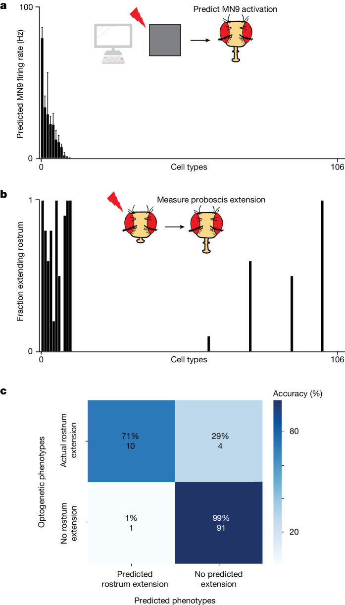
The paper builds a whole-brain leaky integrate-and-fire model of the adult Drosophila central brain using FlyWire connectivity and neurotransmitter predictions, and shows it can predict sensorimotor transformations in feeding and grooming circuits. Across 164 experimentally testable predictions, 91% matched empirical results, with especially strong performance on taste pathways and the antennal grooming circuit.
## Key findings
- The model used all FlyWire neurons: 127,400 proofread neurons from connectome materialization v.630, with more than 125,000 neurons and 50 million synaptic connections in the underlying adult central brain connectome.
- Across 164 predictions that could be tested empirically, 91% were consistent with experimental results; excluding the optogenetic split-GAL4 experiments, accuracy was 84%.
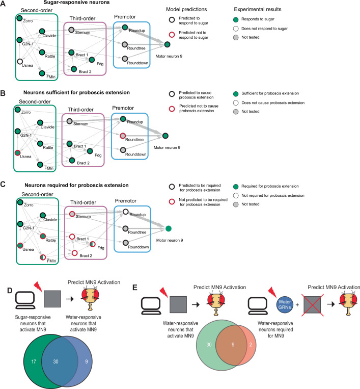
- For feeding initiation, the model predicted that activating sugar GRNs activates proboscis motor neurons including MN6, MN8, MN9, and MN11; in total, it identified 47 neurons predicted to be sugar-responsive and sufficient for feeding initiation, with 14 also predicted to be required for MN9 activity.
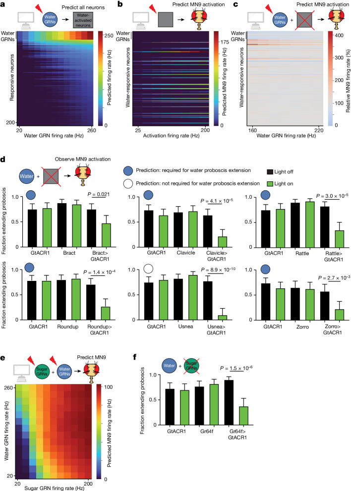
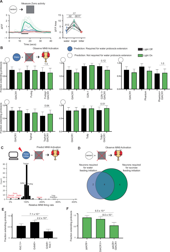
- In the water pathway, the model identified 39 water-responsive neurons sufficient for MN9 activation and 9 neurons predicted to be both necessary and sufficient for water feeding initiation; experiments confirmed water responses in Fudog and Zorro and showed 5 of 6 predicted water-silencing neurons decreased proboscis extension.
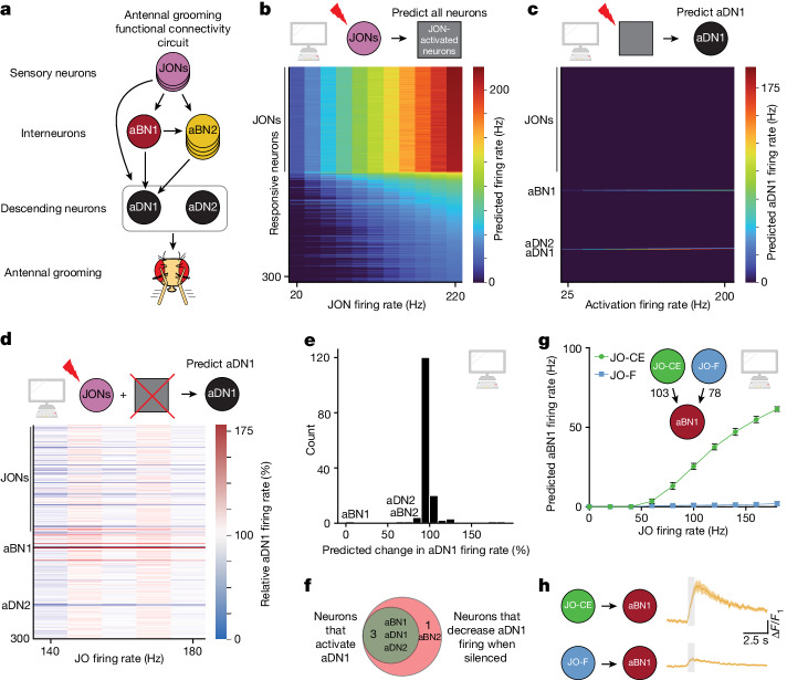
- For antennal grooming, the model identified aBN1, aBN2, aDN1, and aDN2 as JON-responsive; it predicted that JO-CE activates aBN1 robustly while JO-F does not, which was confirmed by calcium imaging.
## Methods
- Built a whole-brain leaky integrate-and-fire spiking model in Brian2 using FlyWire synaptic weights and neurotransmitter predictions, with Wsyn = 0.275 mV as the single free parameter; baseline firing was assumed to be 0 Hz.
- Simulated sensory input by driving specific neurons with Poisson-distributed firing and ran 30 simulations of 1,000 ms per experiment; for taste circuits, activation frequencies ranged from 10–200 Hz for sugar and 20–260 Hz for water.
- Validated predictions with optogenetics, silencing, calcium imaging, and behavioural PER assays in flies, including screens of SEZ split-GAL4 lines and experiments on taste and antennal grooming circuits.
## Limitations
- Stated by the paper: the model treats neurons as only excitatory or inhibitory, ignores neural morphology, receptor dynamics, gap junctions, neuropeptides, neuromodulation, and non-spiking neurons, and assumes basal firing of 0 Hz; therefore, absolute firing rates are unlikely to be accurate.
- Stated by the paper: the model may perform poorly in circuits with extensive basal inhibition or extensive neuromodulation/extrasynaptic signalling, and its accuracy is limited by the accuracy of the underlying synapse and neurotransmitter predictions.
- Evident from the text: many validations are based on a relatively small number of specifically chosen circuits and genetic lines, so the results may not generalize to all brain regions or behaviours.
Model-written summary. Check it against the paper before relying on it.
Integrity signals
Registry
Cites
Statistics
Figures


Verification
Claims the paper makes
42 Valid · 4 Not valid · 2 Not checked · 111 Own results and interpretations
At least one cited source doesn’t support this claim or points to a different paper, and none confirmed it — see the verdicts below.
In the paper
“Our calcium imaging experiments (Extended Data Figs. 1c and 3a) combined with previous experiments4 confirm that five neurons predicted to respond to sugar and water do respond to both sugar and water in vivo: Clavicle, Fudog, Phantom, Rattle and Zorro.”
§ A Drosophila computational brain model reveals sensorimotor processing > Main
What the sources say
- Disputedcites [4] Shiu, P. K., Sterne, G. R., Engert, S., Dickson, B. J. & Scott, K. Taste quality and hunger interactions in a feeding sensorimotor circuit. eLife 11, e79887 (2022). sourceExperimental — accuracy not yet measured
The Results section, “Feeding initiation neurons respond to sugar taste detection” (page unavailable), reports that Phantom responded to both water and sugar, but Clavicle did so only in a thirsty-like state. It says the other sugar-responsive neurons did not respond to water in food-deprived flies, contrary to the claim that all five named neurons were confirmed to respond to both. (LLM, conf=0.88)
Checked by a reasoning model — may be wrong.
plainstates an association- Asserted: passes — The authors put it forward as true.
- Checkable: passes — It can be checked against evidence.
- Load-bearing: passes — The paper’s conclusions depend on it.
- Based: passes — The paper gives a reason why it holds.
- Disputed
At least one cited source doesn’t support this claim or points to a different paper, and none confirmed it — see the verdicts below.
In the paper
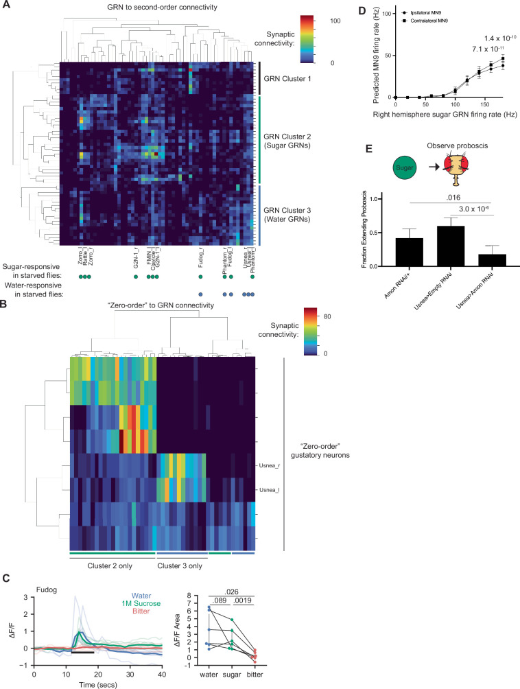
“Thus, we conclude that cluster 2 corresponds to sugar GRNs, consistent with their previous assignment23, and cluster 3 corresponds to water GRNs.”
§ A Drosophila computational brain model reveals sensorimotor processing > Methods > Classification of GRNs Bitter and Ir94e GRNs were identified previously by their distinctive morphology23. Water and sugar GRNs were identified previously by morphology; to confirm these assignments and to incorporate new GRNs identified in Flywire, we performed hierarchical clustering using the connectivity of GRNs onto second-order neurons, using cosine distance as a measure of similarity70. This identified three broad clusters of GRNs (Extended Data Fig. 1a). We compared this clustering with the response properties of second-order neurons in starved flies, the nutritional state for which we have the most complete data4. Cluster 2 GRNs synapse onto, among others, G2N-1 and FMIN, which respond exclusively to sugar. Cluster 3 neurons synapse comparatively strongly onto Fudog and Phantom, which respond to both water and sugar in starved flies; cluster 3 neurons also synapse more strongly, on average, onto Usnea than cluster 2 neurons. Usnea responds exclusively to water. Thus, we conclude that cluster 2 corresponds to sugar GRNs, consistent with their previous assignment23, and cluster 3 corresponds to water GRNs. We speculate that cluster 1 GRNs are Ppk23-glut GRNs31 that may respond to salt; however, little is known about this pathway, making testing this hypothesis challenging. We also examined the connectivity of ‘zero-order’ gustatory neurons onto cluster 2 and 3 GRNs. This clustering corresponds strongly with the GRN to second-order clustering (Extended Data Fig. 1), verifying that these clusters reflect distinct taste modalities. As a conservative measure, we chose to examine only those sugar GRNs that fully correspond between the GRN to second-order clustering and the zero-order to GRN clustering. Recently, the FlyWire brain (FAFB) dataset (on the basis of which the Flywire dataset was prepared) was found to be left–right inverted1,2. In this paper, we refer to the true biological side. Thus, we note that ‘right hemisphere GRNs’ in this paper correspond to ‘left hemisphere GRNs’ described previously4,23.
What the sources say
- Disputedcites [23] Engert, S., Sterne, G. R., Bock, D. D. & Scott, K. Drosophila gustatory projections are segregated by taste modality and connectivity. eLife 11, e78110 (2022). sourceExperimental — accuracy not yet measured
The Results section (‘Different GRN classes can be identified by morphology and connectivity’) assigns group 2 to bitter GRNs, not sugar GRNs. It identifies group 4 as the hypothesized sugar-GRN group. The paper calls these clusters ‘groups,’ so its numbering does not support the cited assignment of cluster 2. (LLM, conf=0.88)
Checked by a reasoning model — may be wrong.
plainstates an association- Asserted: passes — The authors put it forward as true.
- Checkable: passes — It can be checked against evidence.
- Load-bearing: passes — The paper’s conclusions depend on it.
- Based: passes — The paper gives a reason why it holds.
- Disputed
At least one cited source doesn’t support this claim or points to a different paper, and none confirmed it — see the verdicts below.
In the paper
“Because GRN reconstructions on the left hemisphere are more complete23, probably due to challenges imaging and reconstructing neurons at the brain’s periphery, we performed unilateral left hemisphere activation for all simulations, except for Extended Data Fig. 1d.”
§ A Drosophila computational brain model reveals sensorimotor processing > Methods > Computational modelling of water and sugar GRN activity
What the sources say
- Disputedcites [23] Engert, S., Sterne, G. R., Bock, D. D. & Scott, K. Drosophila gustatory projections are segregated by taste modality and connectivity. eLife 11, e78110 (2022). sourceExperimental — accuracy not yet measured
The Introduction and Discussion report that 83–96% of expected GRNs were reconstructed on the right hemisphere, compared with 54–63% on the left. The Results section (“GRN axons contain presynaptic and postsynaptic sites”) also says EM-volume misalignments prevented identification of additional left-hemisphere GRNs. The source therefore says the right, not the left, is more complete. (LLM, conf=0.90)
Checked by a reasoning model — may be wrong.
hedged- Asserted: passes — The authors put it forward as true.
- Checkable: passes — It can be checked against evidence.
- Load-bearing: passes — The paper’s conclusions depend on it.
- Based: passes — The paper gives a reason why it holds.
- Disputed
At least one cited source doesn’t support this claim or points to a different paper, and none confirmed it — see the verdicts below.
In the paper
“Thus, we conclude that cluster 2 corresponds to sugar GRNs, consistent with their previous assignment23, and cluster 3 corresponds to water GRNs.”
§ A Drosophila computational brain model reveals sensorimotor processing > Methods > Classification of GRNs Bitter and Ir94e GRNs were identified previously by their distinctive morphology23. Water and sugar GRNs were identified previously by morphology; to confirm these assignments and to incorporate new GRNs identified in Flywire, we performed hierarchical clustering using the connectivity of GRNs onto second-order neurons, using cosine distance as a measure of similarity70. This identified three broad clusters of GRNs (Extended Data Fig. 1a). We compared this clustering with the response properties of second-order neurons in starved flies, the nutritional state for which we have the most complete data4. Cluster 2 GRNs synapse onto, among others, G2N-1 and FMIN, which respond exclusively to sugar. Cluster 3 neurons synapse comparatively strongly onto Fudog and Phantom, which respond to both water and sugar in starved flies; cluster 3 neurons also synapse more strongly, on average, onto Usnea than cluster 2 neurons. Usnea responds exclusively to water. Thus, we conclude that cluster 2 corresponds to sugar GRNs, consistent with their previous assignment23, and cluster 3 corresponds to water GRNs. We speculate that cluster 1 GRNs are Ppk23-glut GRNs31 that may respond to salt; however, little is known about this pathway, making testing this hypothesis challenging. We also examined the connectivity of ‘zero-order’ gustatory neurons onto cluster 2 and 3 GRNs. This clustering corresponds strongly with the GRN to second-order clustering (Extended Data Fig. 1), verifying that these clusters reflect distinct taste modalities. As a conservative measure, we chose to examine only those sugar GRNs that fully correspond between the GRN to second-order clustering and the zero-order to GRN clustering. Recently, the FlyWire brain (FAFB) dataset (on the basis of which the Flywire dataset was prepared) was found to be left–right inverted1,2. In this paper, we refer to the true biological side. Thus, we note that ‘right hemisphere GRNs’ in this paper correspond to ‘left hemisphere GRNs’ described previously4,23.
What the sources say
- Disputedcites [23] Engert, S., Sterne, G. R., Bock, D. D. & Scott, K. Drosophila gustatory projections are segregated by taste modality and connectivity. eLife 11, e78110 (2022). sourceExperimental — accuracy not yet measured
In Results (‘Different GRN classes can be identified by morphology and connectivity’), the source assigns group 3 to candidate low-salt GRNs, not water GRNs. It identifies group 6 as the best match for water GRNs. These are the source’s own group numbers; the passage does not establish how they relate to clusters numbered in the citing paper. (LLM, conf=0.88)
Checked by a reasoning model — may be wrong.
plainstates an association- Asserted: passes — The authors put it forward as true.
- Checkable: passes — It can be checked against evidence.
- Load-bearing: passes — The paper’s conclusions depend on it.
- Based: passes — The paper gives a reason why it holds.
- Disputed
Every source the paper cites for this claim was checked and supports it.
In the paper
“Here we create a leaky integrate-and-fire computational model of the entire Drosophila brain, on the basis of neural connectivity and neurotransmitter identity3, to study circuit properties of feeding and grooming behaviours.”
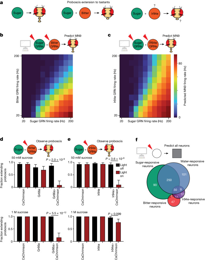
§ A Drosophila computational brain model reveals sensorimotor processing > Abstract The recent assembly of the adult Drosophila melanogaster central brain connectome, containing more than 125,000 neurons and 50 million synaptic connections, provides a template for examining sensory processing throughout the brain1,2. Here we create a leaky integrate-and-fire computational model of the entire Drosophila brain, on the basis of neural connectivity and neurotransmitter identity3, to study circuit properties of feeding and grooming behaviours. We show that activation of sugar-sensing or water-sensing gustatory neurons in the computational model accurately predicts neurons that respond to tastes and are required for feeding initiation4. In addition, using the model to activate neurons in the feeding region of the Drosophila brain predicts those that elicit motor neuron firing5—a testable hypothesis that we validate by optogenetic activation and behavioural studies. Activating different classes of gustatory neurons in the model makes accurate predictions of how several taste modalities interact, providing circuit-level insight into aversive and appetitive taste processing. Additionally, we applied this model to mechanosensory circuits and found that computational activation of mechanosensory neurons predicts activation of a small set of neurons comprising the antennal grooming circuit, and accurately describes the circuit response upon activation of different mechanosensory subtypes6–10. Our results demonstrate that modelling brain circuits using only synapse-level connectivity and predicted neurotransmitter identity generates experimentally testable hypotheses and can describe complete sensorimotor transformations.
What the sources say
- Supportedcites [3] Eckstein, N. et al. Neurotransmitter classification from electron microscopy images at synaptic sites in Drosophila melanogaster. Cell 187, 2574–2594.e23 (2024). source
The citation is attached to “neurotransmitter identity,” not to the citing authors’ statement that they created a leaky integrate-and-fire model. The source’s Abstract reports transmitter predictions across a Drosophila whole brain, and its Results (“An overview of neurotransmitter usage in the nervous system”) report neuron-level predictions for the FAFB-FlyWire dataset. (LLM, conf=0.93)
plain- Asserted: passes — The authors put it forward as true.
- Checkable: passes — It can be checked against evidence.
- Load-bearing: passes — The paper’s conclusions depend on it.
- Based: passes — The paper gives a reason why it holds.
- Supported
88 more claims · 16 with their cited sources checked
Create a free account to see all 159 claims and to verify the 2 references still open.
- It may state two separate claims as one. 51
- Probably not a substantial claim. 20
Claims other papers make about this one
Each is checked against this paper’s own text: does it say what the citing paper says it does?
The cited source says what the claim attributes to it.
In the citing paper
“Shiu et al.34 and Pospisil et al.39 use the connectome to model Drosophila brain dynamics and include connection weights (number of synapses) and putative connection signs (excitatory or inhibitory).”
From Neuronal wiring diagram of an adult brain (2024) · cites this paper as [34]
The cited source says what the claim attributes to it.
In the citing paper
“This effort has now been extended to leverage the full connectome34,39 and to—for example—predict taste responses of neurons34.”
From Neuronal wiring diagram of an adult brain (2024) · cites this paper as [34]
In the citing paper
“Here, using connectome-informed models7–9 and functional studies, we explain two fundamental mechanisms by which Drosophila implement context-appropriate halting.”
From Neural circuit mechanisms underlying context-specific halting in Drosophila (2024) · cites this paper as [7]
In the citing paper
“Notably, connectomics revealed that this halting pathway diverges from the previously described proboscis extension pathway7,41 (Fig. 5a).”
From Neural circuit mechanisms underlying context-specific halting in Drosophila (2024) · cites this paper as [7]
Citations
Cites
Fiji: an open-source platform for biological-image analysis
Schindelin, J., Arganda-Carreras, I., Frise, E., Kaynig, V., Longair, M., Pietzsch, T., Preibisch, S., Rueden, C., Saalfeld, S., Schmid, B., et al. (2012) · Nature Methods · doi:10.1038/nmeth.2019 · Cited by 66,890 · cited 2×
“First, in Fiji, z stacks for each timepoint were maximum intensity projected and then movement corrected using the StackReg plugin with ‘Rigid Body’ or ‘Translation’ transformation 79 .”
MethodUltrasensitive fluorescent proteins for imaging neuronal activity
Chen, T.-W., Wardill, T. J., Sun, Y., Pulver, S. R., Renninger, S. L., Baohan, A., Schreiter, E. R., Kerr, R. A., Orger, M. B., Jayaraman, V., et al. (2013) · Nature · doi:10.1038/nature12354 · Cited by 6,474
A pyramid approach to subpixel registration based on intensity
Thevenaz, P., Ruttimann, U. E., & Unser, M. (1998) · IEEE Transactions on Image Processing · doi:10.1109/83.650848 · Cited by 3,078
Independent optical excitation of distinct neural populations
Klapoetke, N. C., Murata, Y., Kim, S. S., Pulver, S. R., Birdsey-Benson, A., Cho, Y. K., Morimoto, T. K., Chuong, A. S., Carpenter, E. J., Tian, Z., et al. (2014) · Nature Methods · doi:10.1038/nmeth.2836 · Cited by 2,344 · cited 1×
“Experiments were performed as in the GtAcr1 experiments, except that flies were exposed to red light rather than green light 77 .”
MethodNeuronal Dynamics: From Single Neurons to Networks and Models of Cognition
Gerstner, W., Kistler, W. M., Naud, R., & Paninski, L. (2014) · doi:10.1017/cbo9781107447615 · Cited by 1,515 · cited 4×
“…that generate behaviour, we implement a simple leaky integrate-and-fire model using the connection weights derived from the entire adult Drosophila central brain connectome of reconstructed electron microscopy neurons 1,17–20 , as well as neurotransmitter predictions for each neuron 3,21 .”
MethodInfluentialA connectome and analysis of the adult Drosophila central brain
Scheffer, L. K., Xu, C. S., Januszewski, M., Lu, Z., Takemura, S.-Y., Hayworth, K. J., Huang, G. B., Shinomiya, K., Maitlin-Shepard, J., Berg, S., et al. (2020) · eLife · doi:10.7554/elife.57443 · Cited by 1,128 · cited 3×
“Two different approaches have been used thus far to analyze the recent Drosophila hemibrain and larval connectomes 13,16 .”
BackgroundMethodSelf-citationA Complete Electron Microscopy Volume of the Brain of Adult Drosophila melanogaster
Zheng, Z., Lauritzen, J. S., Perlman, E., Robinson, C. G., Nichols, M., Milkie, D., Torrens, O., Price, J., Fisher, C. B., Sharifi, N., et al. (2018) · Cell · doi:10.1016/j.cell.2018.06.019 · Cited by 1,050 · cited 3×
“We find that computational activation of labellar sugar-sensing GRNs activates several proboscis MNs involved in feeding, including MNs 6, 8, 9 and 11 (Fig.”
BackgroundSelf-citationThe organization of the chemosensory system in Drosophila melanogaster: a rewiew
Stocker, R. F. (1994) · Cell and Tissue Research · doi:10.1007/bf00305372 · Cited by 950 · cited 1×
“GRNs respond to specific taste categories, such as appetitive sugar or aversive bitter compounds, resulting in acceptance (that is, proboscis extension and feeding) or avoidance, respectively 24,33,35,38 .”
BackgroundNeuromodulation of neuronal circuits: back to the future
Marder, E. (2012) · Neuron · doi:10.1016/j.neuron.2012.09.010 · Cited by 904
Brian 2, an intuitive and efficient neural simulator
Stimberg, M., Brette, R., & Goodman, D. F. (2019) · eLife · doi:10.7554/elife.47314 · Cited by 749
Research works printed but not linked: 1
- Dethier, V. G. The Hungry Fly: a Physiological Study of the Behavior Associated With Feeding (Harvard Univ. Press, 1976).
Cited by
Neuronal wiring diagram of an adult brain
Dorkenwald, S., Matsliah, A., Sterling, A. R., Schlegel, P., Yu, S.-C., McKellar, C. E., Lin, A., Costa, M., Eichler, K., Yin, Y., et al. (2024) · Nature · doi:10.1038/s41586-024-07558-y · Cited by 570
“Also included are neurons that extend into the brain through the nerves and neck connective, which are essential for tracing sensorimotor pathways, as illustrated here and in the accompanying studies 11,12,34,36–45 .”
BackgroundInfluentialSelf-citationWhole-brain annotation and multi-connectome cell typing of Drosophila
Schlegel, P., Yin, Y., Bates, A. S., Dorkenwald, S., Eichler, K., Brooks, P., Han, D. S., Gkantia, M., dos Santos, M., Munnelly, E. J., et al. (2024) · Nature · doi:10.1038/s41586-024-07686-5 · Cited by 413
“It will also make numerous network analyses possible as well as substantially reduce the degrees of freedom in brain scale modelling 65,66 .”
BackgroundNeuronal wiring diagram of an adult brain
Dorkenwald, S., Matsliah, A., Sterling, A. R., Schlegel, P., Yu, S.-C., McKellar, C. E., Lin, A., Costa, M., Eichler, K., Yin, Y., et al. (2023) · doi:10.1101/2023.06.27.546656 · Cited by 149
Self-citationConnectome-constrained networks predict neural activity across the fly visual system
Lappalainen, J. K., Tschopp, F. D., Prakhya, S., McGill, M., Nern, A., Shinomiya, K., Takemura, S.-Y., Gruntman, E., Macke, J. H., & Turaga, S. C. (2024) · Nature · doi:10.1038/s41586-024-07939-3 · Cited by 128
Whole-brain annotation and multi-connectome cell typing quantifies circuit stereotypy in Drosophila
Schlegel, P., Yin, Y., Bates, A. S., Dorkenwald, S., Eichler, K., Brooks, P., Han, D. S., Gkantia, M., dos Santos, M., Munnelly, E. J., et al. (2023) · doi:10.1101/2023.06.27.546055 · Cited by 102
Self-citation
Metadata
- Authors
- Shiu, P. K., Sterne, G. R., Spiller, N., Franconville, R., Sandoval, A., Zhou, J., Simha, N., Kang, C. H., Yu, S., Kim, J. S., Dorkenwald, S., Matsliah, A., Schlegel, P., Yu, S.-C., McKellar, C. E., Sterling, A., Costa, M., Eichler, K., Bates, A. S., Eckstein, N., Funke, J., Jefferis, G. S. X. E., Murthy, M., Bidaye, S. S., Hampel, S., Seeds, A. M., & Scott, K.
- Affiliations
- University of California, Berkeley (US)
- University of Rochester Medicine (US)
- Max Planck Florida Institute for Neuroscience (US)
- Howard Hughes Medical Institute (US)
- Sungkyunkwan University (KR)
- Princeton University (US)
- MRC Laboratory of Molecular Biology (GB)
- University of Cambridge (GB)
- University of Puerto Rico, Medical Sciences Campus (PR)
- Corresponding author
- Shiu, Philip K.
- ORCID
- Shiu, Philip K.
- Sterne, Gabriella R.
- Spiller, Nico
- Franconville, Romain
- Sandoval, Andrea
- Simha, Neha
- Yu, Seongbong
- Kim, Jinseop S.
- Dorkenwald, Sven
- Matsliah, Arie
- Schlegel, Philipp
- Yu, Szi-chieh
- McKellar, Claire E.
- Sterling, Amy
- Costa, Marta
- Eichler, Katharina
- Bates, Alexander Shakeel
- Eckstein, Nils
- Funke, Jan
- Jefferis, Gregory S. X. E.
- Murthy, Mala
- Bidaye, Salil S.
- Hampel, Stefanie
- Seeds, Andrew M.
- Scott, Kristin
- Venue
- Nature
- Published
- 2024-10-02
- Volume
- 634
- Issue
- 8032
- Pages
- 210-219
- ISSN
- 0028-0836 (ISSN-L)
- 1476-4687
- PMC
- PMC11446845
- OpenAlex
- W4403062868
- Semantic Scholar
- 1c747c588fa46133a6e0146d1eeb81c5be67150a
- Cited by
- 97
- References
- 84
- Citations by year
- 2026: 39 · 2025: 35 · 2024: 20
- FWCI
- 16.38
- Type
- journal article
- Language
- en
- Open access
- hybrid
- doi.org
- Licence
- cc-by
- creativecommons.org/licenses/by/4.0 (tdm)
- creativecommons.org/licenses/by/4.0 (vor)
- Locations
- Nature (publishedVersion, cc-by)
- bioRxiv (Cold Spring Harbor Laboratory) (acceptedVersion, cc-by-nc-nd)
- PubMed Central (publishedVersion, cc-by)
- PubMed (publishedVersion)
- PubMed Central (submittedVersion, cc-by-nc-nd)
- Apollo (University of Cambridge) (submittedVersion, cc-by)
- RePEc: Research Papers in Economics (submittedVersion)
- Funders
- National Science Foundation (2014862)
- Wellcome Trust (220343/Z/20/Z, 203261/Z/16/Z)
- National Institutes of Health (1rf1ns121911-01)
- National Institute of Mental Health (RF1MH117815, RF1MH129268, RF1MH120679)
- National Institute on Deafness and Other Communication Disorders (F32DC018225, R01DC013280)
- National Institute of Diabetes and Digestive and Kidney Diseases (F32DK117671)
- National Institute of Neurological Disorders and Stroke (RF1 NS121911, U24NS126935, R01NS121911, R35NS111580)
- Topics
- Life Sciences › Neuroscience › Cellular and Molecular Neuroscience › Neurobiology and Insect Physiology Research
- Life Sciences › Neuroscience › Cognitive Neuroscience › Neural dynamics and brain function
- Life Sciences › Neuroscience › Cellular and Molecular Neuroscience › Neuroscience and Neuropharmacology Research
- Keywords
- Drosophila melanogaster, computational models of the brain, leaky integrate-and-fire neurons, brain connectome, sensorimotor processing, gustatory neurons, taste processing, feeding behaviour, grooming, mechanosensory neurons, neurotransmitter identity
About these checks
Registry, retraction, Cites and statistics results are computed, not judged, with one exception: a printed reference that no index lists is read once by a model before it counts as a citation error. Claims are extracted by a model and kept only when their quote appears verbatim in the text; each cited source is then checked, with a model’s help, for whether it says what the claim relies on. Those verdicts can be wrong — read the claim’s quote and the cited source before relying on one.
The claim verdicts this report counts are experimental: their accuracy is not yet measured.
A language model drew the claims from the paper on 2026-10-10, under version 3 of the claim definition (model deepseek-v4.1-flash, prompt extract-v3).
A second model labelled each claim on 2026-10-10 (model jev-1.13-20260917, definition revision gold-guideline-v0.2).
Cited sources are checked with a language model’s help (gpt-6-sol).
Statistics and arithmetic are recomputed, not judged.
Confidence bands are shown only for fields calibrated on our annotated test set. None is calibrated yet, so no band is shown.
What counts as a claim ›How we measure confidence ›How we check this ›
Are you an author, or do you see a problem with this page? Request a review
How to cite
Cite CoChat for the checks on this page, and the paper for its own findings.
- This page
- CoChat. (2026). Citation check: A Drosophila computational brain model reveals sensorimotor processing. https://app.cochat.ai/papers/10.1038/s41586-024-07763-9
- The paper
- Shiu, P. K., Sterne, G. R., Spiller, N., Franconville, R., Sandoval, A., Zhou, J., Simha, N., Kang, C. H., Yu, S., Kim, J. S., Dorkenwald, S., Matsliah, A., Schlegel, P., Yu, S.-C., McKellar, C. E., Sterling, A., Costa, M., Eichler, K., Bates, A. S., . . . Scott, K. (2024). A Drosophila computational brain model reveals sensorimotor processing. Nature. https://doi.org/10.1038/s41586-024-07763-9
Data sources
Bibliographic data: Semantic Scholar, OpenAlex, Crossref.
Contains information from the Semantic Scholar Academic Graph Datasets, made available under the ODC Attribution License (ODC-BY 1.0).
Semantic Scholar’s data is described in: Kinney, R., et al. (2023). The Semantic Scholar Open Data Platform. arXiv:2301.10140.
