

The DOI matches its Crossref record; 56 of the 56 references it cites were found in a registry or an index.
High-definition spatial transcriptomic profiling of immune cell populations in colorectal cancer
Open accessAbstract
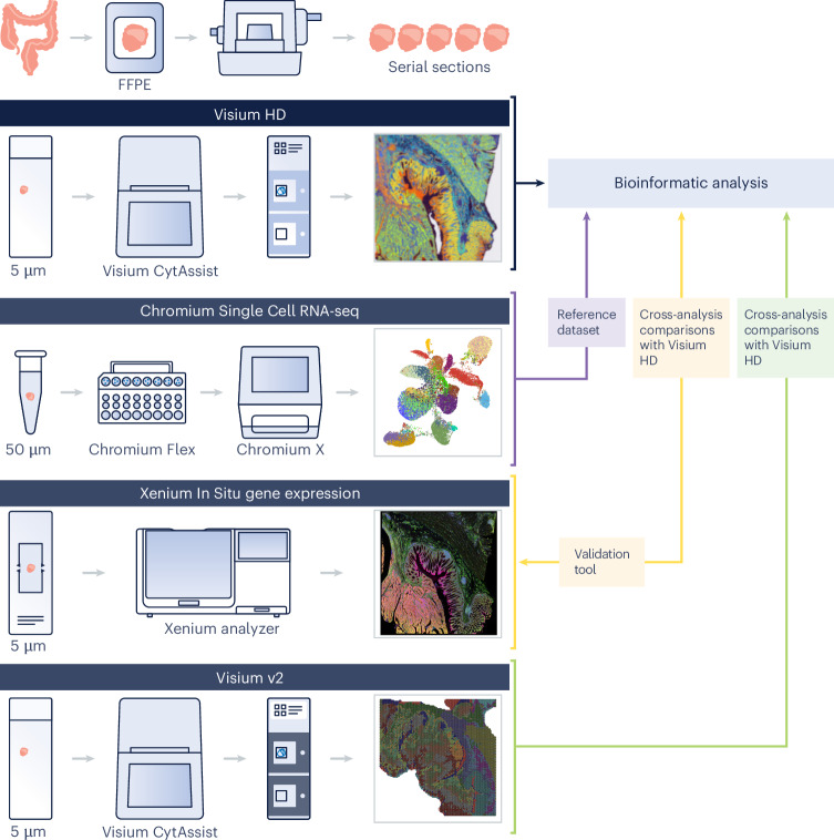
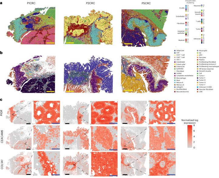
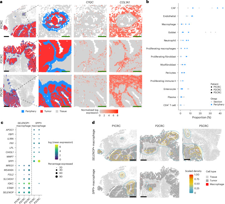
A comprehensive understanding of cellular behavior and response to the tumor microenvironment (TME) in colorectal cancer (CRC) remains elusive. Here, we introduce the high-definition Visium spatial transcriptomic technology (Visium HD) and investigate formalin-fixed paraffin-embedded human CRC samples (n = 5). We demonstrate the high sensitivity, single-cell-scale resolution and spatial accuracy of Visium HD, generating a highly refined whole-transcriptome spatial profile of CRC samples. We identify transcriptomically distinct macrophage subpopulations in different spatial niches with potential pro-tumor and anti-tumor functions via interactions with tumor and T cells. In situ gene expression analysis validates our findings and localizes a clonally expanded T cell population close to macrophages with anti-tumor features. Our study demonstrates the power of high-resolution spatial technologies to understand cellular interactions in the TME and paves the way for larger studies that will unravel mechanisms and biomarkers of CRC biology, improving diagnosis and disease management strategies.
Overview
The claims and statistics of this paper haven’t been checked yet.
Run the check — free, no account needed. It reads the open-access text and checks the sources it cites.
Integrity signals
Registry
Cites
Statistics
Figures
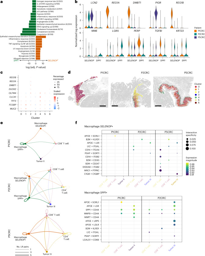
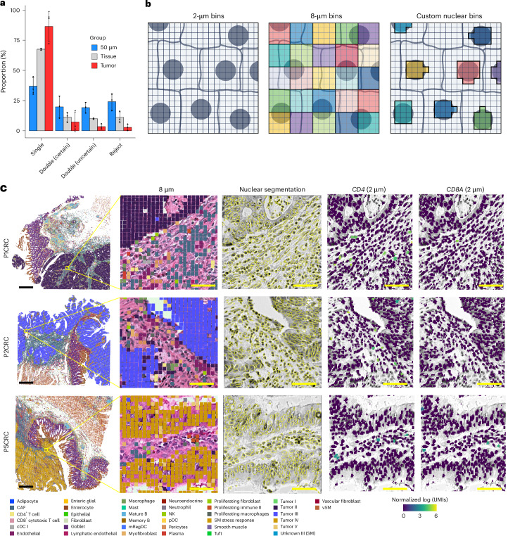
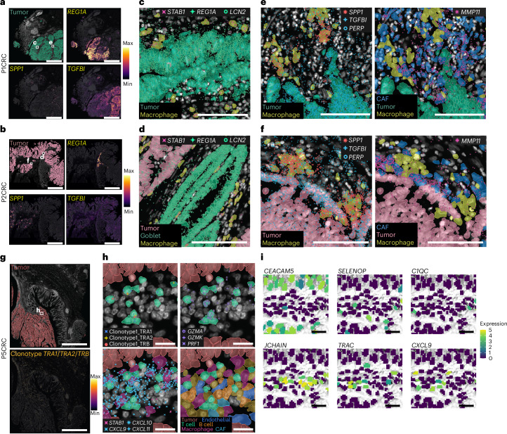
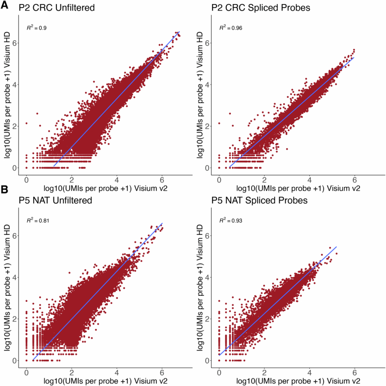
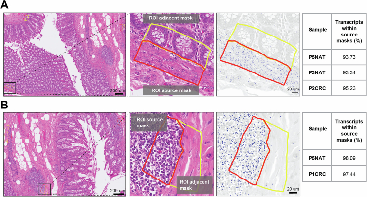
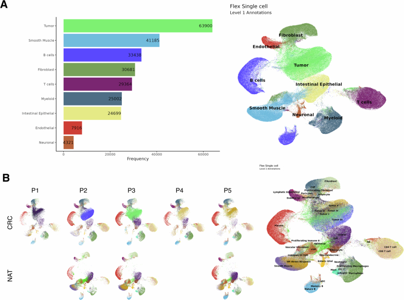
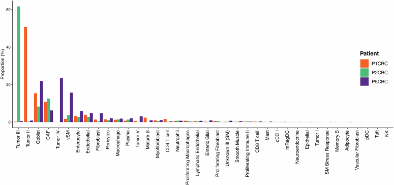
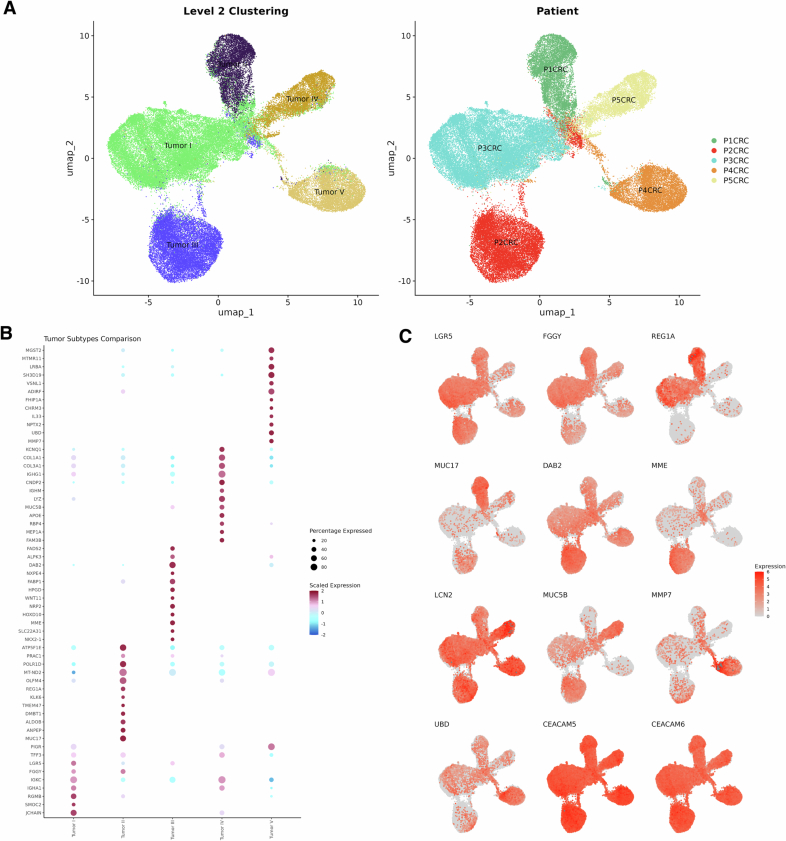
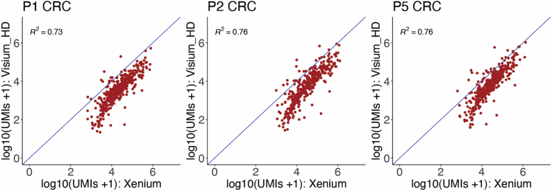
Verification
Claims the paper makes
The claims and statistics of this paper haven’t been checked yet.
Run the check — free, no account needed. It reads the open-access text and checks the sources it cites.
Claims other papers make about this one
Each is checked against this paper’s own text: does it say what the citing paper says it does?
The source backs part of the claim, not all of it — or backs it for a different population, condition or comparison.
In the citing paper
“Moreover, vascular-immune interactions were primarily inferred from histological proximity, flow cytometry and gene-expression patterns; spatial transcriptomics and single-cell multiomics approaches will be valuable for elucidating cell-type-specific crosstalk at single-cell resolution 54,55 .”
From Vascularized human skin assembloids model compartmental skin organization and immune responsiveness in vitro (2026) · cites this paper as [54]
Experimental — accuracy not yet measured
Self-check
Percentages, sums and reported differences re-computed from the paper’s own numbers.
The self-check needs the paper’s full text.
Citations
Cites
The Molecular Signatures Database Hallmark Gene Set Collection
Liberzon, A., Birger, C., Thorvaldsdóttir, H., Ghandi, M., Mesirov, J. P., & Tamayo, P. (2015) · Cell Systems · doi:10.1016/j.cels.2015.12.004 · Cited by 13,807
QuPath: Open source software for digital pathology image analysis
Bankhead, P., Loughrey, M. B., Fernández, J. A., Dombrowski, Y., McArt, D. G., Dunne, P. D., McQuaid, S., Gray, R. T., Murray, L. J., Coleman, H. G., et al. (2017) · Scientific Reports · doi:10.1038/s41598-017-17204-5 · Cited by 8,908
Dictionary learning for integrative, multimodal and scalable single-cell analysis
Hao, Y., Stuart, T., Kowalski, M. H., Choudhary, S., Hoffman, P., Hartman, A., Srivastava, A., Molla, G., Madad, S., Fernandez-Granda, C., et al. (2024) · Nature Biotechnology · doi:10.1038/s41587-023-01767-y · Cited by 5,631
The consensus molecular subtypes of colorectal cancer
Guinney, J., Dienstmann, R., Wang, X., de Reyniès, A., Schlicker, A., Soneson, C., Marisa, L., Roepman, P., Nyamundanda, G., Angelino, P., et al. (2015) · Nature Medicine · doi:10.1038/nm.3967 · Cited by 5,170
Global colorectal cancer burden in 2020 and projections to 2040
Xi, Y., & Xu, P. (2021) · Translational Oncology · doi:10.1016/j.tranon.2021.101174 · Cited by 2,633
Immunotherapy in colorectal cancer: Rationale, challenges and potential
Ganesh, K., Stadler, Z. K., Cercek, A., Mendelsohn, R. B., Shia, J., Segal, N. H., & Diaz, L. A. (2019) · Nature Reviews Gastroenterology & Hepatology · doi:10.1038/s41575-019-0126-x · Cited by 1,954 · cited 1×
“Figure 7D has been updated to show SELENOP +/ STAB1 + macrophages near REG1A + cells.”
CXCL9, CXCL10, CXCL11/CXCR3 axis for immune activation - a target for novel cancer therapy
Tokunaga, R., Zhang, W., Naseem, M., Puccini, A., Berger, M. D., Soni, S., McSkane, M., Baba, H., & Lenz, H.-J. (2018) · Cancer Treatment Reviews · doi:10.1016/j.ctrv.2017.11.007 · Cited by 1,627 · cited 2×
“Figure 7D has been updated to show SELENOP +/ STAB1 + macrophages near REG1A + cells.”
Single-Cell Analyses Inform Mechanisms of Myeloid-Targeted Therapies in Colon Cancer
Zhang, L., Li, Z., Skrzypczynska, K. M., Fang, Q., Zhang, W., O’Brien, S. A., He, Y., Wang, L., Zhang, Q., Kim, A., et al. (2020) · Cell · doi:10.1016/j.cell.2020.03.048 · Cited by 1,620 · cited 1×
“These cells were localized in microenvironments with a higher density of SELENOP +/ STAB1 + macrophages (Figure 4D) .”
Colon Cancer Survival Rates With the New American Joint Committee on Cancer Sixth Edition Staging
O'Connell, J. B., Maggard, M. A., & Ko, C. Y. (2004) · JNCI Journal of the National Cancer Institute · doi:10.1093/jnci/djh275 · Cited by 1,598 · cited 1×
“Additionally, comparing these metrics with other high-resolution methods like Stereo-seq or Seq-scope could enhance the manuscript's appeal and provide a clearer benchmark for readers.”
Robust decomposition of cell type mixtures in spatial transcriptomics
Cable, D. M., Murray, E., Zou, L. S., Goeva, A., Macosko, E. Z., Chen, F., & Irizarry, R. A. (2022) · Nature Biotechnology · doi:10.1038/s41587-021-00830-w · Cited by 1,487
Research works printed but not linked: 1
- Avraham-Davidi, I. et al. Spatially defined multicellular functional units in colorectal cancer revealed from single cell and spatial transcriptomics. eLife 14, RP104815 (2025).
Cited by
Sorted by citations
Items 101–200 of 316
Frank, M., Ghirardello, G., Howie, J. M., Braun, N., Zirnbauer, R., Stecher, C., Gruener, S., Regele, H. M., Barozzi, I., Bergmann, M. M., et al. (2025) · Cancer Letters · doi:10.1016/j.canlet.2025.218228 · Cited by 2
Park, S. Y., & Kim, Y. H. (2025) · Journal of Digestive Cancer Research · doi:10.52927/jdcr.2025.13.3.269 · Cited by 1
Zhu, Y., Yao, Y., Wang, Y., Qiu, Z., Zhou, D., Huang, H., Chen, K., Sun, Q., Chen, J., Li, Y., et al. (2026) · Biomarker Research · doi:10.1186/s40364-025-00888-7 · Cited by 2
“GO enrichment analysis of differentially expressed genes in neutrophils from the673-KO group compared to the WT group exclusion, highlighting their role in sustaining an immunosuppressive microenvironment [48].”
Noh, M., Lee, S. J., Kim, S., & Lim, S. (2026) · Briefings in Bioinformatics · doi:10.1093/bib/bbag029 · Cited by 1
“…spatial transcriptomics (S T) datasets: HER2-Amplified Invasive Ductal Carcinoma (IDC) [25], HER2-Positive Breast Tumor (Her2ST) [26], Human Breast Cancer Ductal Carcinoma (BCDC) [27], 10x Visium HD Human Colorectal Cancer (HCRC) [28], and Human Dorsolateral Pre-frontal Cortex (DLPFC) [29].”
Spatial Transcriptomics Meets Histochemistry: Insights from Glioblastoma as a Model System
Tomioka, S., Kitazawa, R., Haraguchi, R., Takaoka, Y., Inoue, A., & Kitazawa, S. (2026) · ACTA HISTOCHEMICA ET CYTOCHEMICA · doi:10.1267/ahc.25-00065 · Cited by 1
Advances in spatial omics for the analysis of prostate cancer
Xu, J., Wang, D., Di, X., & Liu, Y. (2026) · Analytical and Bioanalytical Chemistry · doi:10.1007/s00216-026-06396-3 · Cited by 1
HRCHY-CytoCommunity identifies hierarchical tissue organization in cell-type spatial maps
Xie, R., Wang, Z., Liu, J., Xu, H., Xu, Y., Lin, J., Hu, Y., & Gao, L. (2026) · Nature Communications · doi:10.1038/s41467-026-70069-z · Cited by 1
Qian, J., Bao, H., Yang, H., Zhang, J., Shao, X., & Fan, X. (2026) · Targetome · doi:10.48130/targetome-0026-0009 · Cited by 1
Decoding the immune–tumor synapse for data-driven design of next-generation immunotherapies
van Rossum, I., van Spriel, A. B., & van Mierlo, G. (2026) · Trends in Immunology · doi:10.1016/j.it.2025.12.009 · Cited by 2
Single-Cell Sequencing Plus Spatial Transcriptomics
Wang, Y., Wu, X., Cheng, X., Fang, R., Zhang, P., Wang, H., Yu, S., Zhang, H., & Zhao, M. (2026) · American Journal Of Pathology · doi:10.1016/j.ajpath.2026.04.001 · Cited by 1
The Hodgkin Lymphoma Microenvironment: Insights from Spatial Transcriptomics
Alonso‐Alonso, R., Menendez, V., Vázquez, E. M., Solórzano, J. L., & García, J. F. (2026) · International Journal of Molecular Sciences · doi:10.3390/ijms27083689 · Cited by 2
Organoids glimpse: the nexus for diverse tumor heterogeneity
Xu, J., Feng, Q., Xia, Y., Liao, L., Dai, Z., Cai, Z., & Xie, S. M. (2026) · Frontiers in Cell and Developmental Biology · doi:10.3389/fcell.2026.1817560 · Cited by 1
“Spatial transcriptomics and multiplex imaging studies have demonstrated that distinct anatomical compartments are often enriched for markedly different immune cell subsets and effector programs (Pelka et al., 2021; Oliveira et al., 2025).”
Hu, J., He, H., Xu, J., MacDonald, W. A., He, Y., Liu, T., Chen, W. C. W., & Li, G. (2026) · Communications Biology · doi:10.1038/s42003-026-10259-z · Cited by 1
Switchable and biodegradable nanostructures for safe and precise tumor diagnostics
Reszegi, R. R., Zhen, W., Chen, X. S., & Zhang, J. (2026) · Chemical Society Reviews · doi:10.1039/d5cs00925a · Cited by 1
Wu, F., Danzeng, Q., Wu, R., Shen, Y., & Shi, G. (2026) · Frontiers in Immunology · doi:10.3389/fimmu.2026.1835325 · Cited by 1
Chang, L., Yan, Y., 徐翠香, & Wang, J. (2026) · Journal of Nanobiotechnology · doi:10.1186/s12951-026-04629-2 · Cited by 1
“These cellular components can be broadly classified into anti-tumor and pro-tumor populations [5].”
Lin, K., Zhang, N., Zhang, Z., Zhou, X., Qiao, B., Yang, D., Guo, J., Zeng, Z. Q., Xie, X., & Zhuang, B. (2026) · Hepatology · doi:10.1097/hep.0000000000001714 · Cited by 1
Zhang, F., Kong, Y., Fu, X., Zuo, J., Guo, Q., Wang, J., Xu, M., Zeng, Q., Zhang, Y., ling, J., et al. (2026) · International Endodontic Journal · doi:10.1111/iej.70200 · Cited by 1
Hincapie, R., Mantovani, O. M., McFaline‐Figueroa, J. L., & Correa, S. (2026) · Med-X · doi:10.1007/s44258-026-00087-5 · Cited by 3
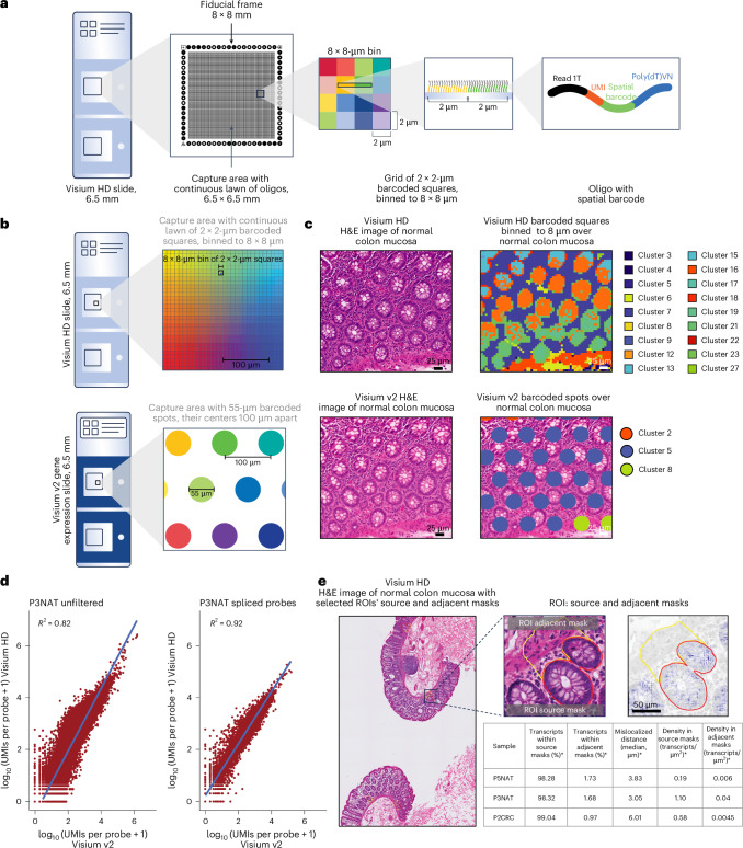
“The 10x Genomics Visium HD platform is among the few that achieve single-cell-scale spatial resolution using 2 x 2 µm barcoded squares while maintaining a large tissue coverage area of 6.5 x 6.5 mm [147].”
InfluentialMapping safety in space: the emerging role of spatial transcriptomics in safe drug development
Golfinos, A. E., Vleet, T. V., Kohnken, R. A., Mahalingaiah, P. K. S., Fossey, S., Buck, W., Liang, W., Mayana, S. A. K., Tuladhar, A., Trairatphisan, P., et al. (2026) · Frontiers in Toxicology · doi:10.3389/ftox.2026.1817521 · Cited by 1
Zilio, S., Rouault, A., Seradj, M., Marigo, I., Prévost‐Blondel, A., Peranzoni, E., & Bercovici, N. (2026) · Frontiers in Immunology · doi:10.3389/fimmu.2026.1851873 · Cited by 1
Spatial Transcriptomics in Breast Cancer: Advances and Applications
Cao, Y., Xu, K., Li, X. Y., Zhang, J., Jin, W., & Liu, Y.-X. (2026) · Biology · doi:10.3390/biology15131061 · Cited by 1
Dissecting human organ development using spatial technologies
Zhang, X., Vandana, J. J., Greenspun, B. C., & Chen, S. (2026) · Developmental Cell · doi:10.1016/j.devcel.2026.05.004 · Cited by 1
Next-generation kidney tissue analysis — spatial omics and digital pathology
Yoshikawa, T., Bülow, R. D., Boor, P., & Yanagita, M. (2026) · Nature Reviews Nephrology · doi:10.1038/s41581-026-01102-z · Cited by 3
“The spot diameter of Visium v1 and v2 (55 μm) lacks true single-cell resolution 15,19 ; however, VisiumHD offers higher resolution (2 μm) and may benefit kidney research 28 .”
Bonilha, C. S. (2026) · Tissue and Cell · doi:10.1016/j.tice.2026.103813 · Cited by 1
Ji, X., Fang, P., Wan, Y., Zhao, X., Xiong, C., Guo, F., Yang, Q., Yan‐Do, R., Qi, L., Xu, C., et al. (2026) · Nature Biomedical Engineering · doi:10.1038/s41551-026-01745-0 · Cited by 2
Zhan, X., Huang, M., Zhang, Y., Liu, Z., Liu, C., Guo, Y., Liu, Y., Zhou, W., Yan, Y., Zeng, H., et al. (2026) · Nature Communications · doi:10.1038/s41467-026-75405-x · Cited by 1
Computational analysis in spatial transcriptomics: methods and perspectives
Zhou, Q., Jiang, Y., Ruan, P., Xiao, G., & Xie, Y. (2026) · Briefings in Bioinformatics · doi:10.1093/bib/bbag417 · Cited by 1
“Recent advancements, such as 10 × Genomics Visium HD [ 13] and Stereo-seq [ 14], have pushed the resolution to the submicron level (2 μ m and 0.5 μ m, respectively), enabling spatially resolved transcriptomic analysis at near-cellular or subcellular levels.”
InfluentialChen, Y., Liu, Y., Wang, Z., Zeng, Y., Chao, Z., Jiang, P., Chen, H., Wang, J., Xiao, J., & Yang, C. (2026) · Nature Computational Science · doi:10.1038/s43588-026-01054-1 · Cited by 1
Transcriptomic profiling of shed cells enables spatial mapping of cellular turnover in human organs
Barkai, T., Yakubovsky, O., Kohanim, Y. K., Halpern, K. B., Shir, S., Oren, N., Fine, M., Kelmer, P., Talmon, A., Israeli, A., et al. (2025) · Molecular Systems Biology · doi:10.1038/s44320-025-00154-w · Cited by 1
“We used two independent datasets – the pan-GI cell atlas (Oliver et al, 2024) and the colon Visium HD (Oliveira et al, 2025).”
InfluentialSpacGPA: annotating spatial transcriptomes through de novo interpretable gene programsPreprint
Xu, Y., Chen, L., & Ma, S. (2025) · bioRxiv (Cold Spring Harbor Laboratory) · doi:10.1101/2025.10.01.679918 · Cited by 1
Annotation-Free Prediction of Cancer Cells and Glands and Spatial Analysis of Immune CellsPreprint
Jung, K. J., Ghose, S., Cho, S., et al. (2025) · bioRxiv (Cold Spring Harbor Laboratory) · doi:10.1101/2025.11.09.687528 · Cited by 1
Joint imputation and deconvolution of gene expression across spatial transcriptomics platformsPreprint
Zheng, H., Sarkar, H., & Raphael, B. J. (2025) · Genome Research · doi:10.1101/gr.280555.125 · Cited by 1
MisTIC: Missegmented Transcript Inference Correction for Improved Spatial Transcriptomics AnalysisPreprint
Yang, Y., DePasquale, E. A. K., Adeleke, D., Xie, X., Lu, Z., Nelson, M. D., Xiao, G., Wang, X., & Wang, Y. (2025) · bioRxiv (Cold Spring Harbor Laboratory) · doi:10.64898/2025.12.11.693759 · Cited by 5
Wang, X., Que, N., Liu, H., Tao, C., Jiang, Y., Jiang, Y., Zhu, P., Xu, J., Yan, H., Ye, B., et al. (2025) · bioRxiv (Cold Spring Harbor Laboratory) · doi:10.64898/2025.12.23.696267 · Cited by 1
“For example, smaller capture units increase spatial resolution but typically result in fewer transcripts captured per unit, reducing sensitivity to low-abundance genes 5–7 .”
Wang, T., Li, L., & Huang, Y. (2026) · bioRxiv (Cold Spring Harbor Laboratory) · doi:10.64898/2026.01.22.701193 · Cited by 1
Brunn, N., Guitart, L. C., Farhadyar, K., et al. (2026) · bioRxiv (Cold Spring Harbor Laboratory) · doi:10.64898/2026.03.18.712664 · Cited by 1
“The framework operates on spatial transcriptomics data generated using diverse technologies [10, 11, 12].”
Solano, A. N., Yip, R. K. H., Wang, C., Amann‐Zalcenstein, D., Rajasekhar, P., Zaman, I., Motyer, A., Cmero, M., Xu, Y., Pan, Y., et al. (2026) · bioRxiv (Cold Spring Harbor Laboratory) · doi:10.64898/2026.04.29.721531 · Cited by 2
“The recent introduction of Visium HD, with a 2 µm pitch and an effective bin size of 8 µm, now delivers sequencing-based profiling at near-cellular resolution in a commercially accessible format (24) , narrowing the gap with iST.”
Pan-cancer virtual spatial transcriptomics from routine histology with PhoenixPreprint
Tran, M., Gindra, R. H., Putze, P., Senbai, 康. K., Palla, G., Šket, T., Falcomatà, C., Wang, C., Guo, R., Boxberg, M., et al. (2026) · bioRxiv (Cold Spring Harbor Laboratory) · doi:10.64898/2026.04.25.720812 · Cited by 4
Draganić, K., Isaev, S., Pototschnig, I., Valcanover, D., Pfneissl, J., Stadler, M., Hocevar, M., Lotz, V., Wasinger, G., Pisibon, C., et al. (2026) · bioRxiv (Cold Spring Harbor Laboratory) · doi:10.64898/2026.05.28.728363 · Cited by 1
Co-delivered PD-L1 rescues the protective efficacy mediated by an AAV-expressed HIV-1 bNAbPreprint
Kuipa, M., Koroma, A. A., Leguizamo, I., Dhole, P., Barot, Y., Lee, M. Y.-H., Tharp, G. K., Liang, S., Chouinard, M., Ehnert, S., et al. (2026) · bioRxiv (Cold Spring Harbor Laboratory) · doi:10.64898/2026.05.29.728706 · Cited by 1
A multimodal foundation model to integrate histology with spatial molecular programsPreprint
Zhang, Z., Qin, B., Zhao, Y., Qi, Z., Xu, H., Wang, Y., Zheng, W., Dai, J., Chen, A., Wang, H., et al. (2026) · bioRxiv (Cold Spring Harbor Laboratory) · doi:10.64898/2026.06.01.729028 · Cited by 1
Cross-Modality Alignment of Spatial Transcriptomics, Multiplexed Imaging, and Histology with PHARAOHPreprint
Li, M., Yao, S., Jing, Z., Jiang, S., Li, W., Yamamoto, Y., Dumoulin, B., Sachs, N., Mäegdefessel, L., Suszták, K., et al. (2026) · Research Square · doi:10.21203/rs.3.rs-9954002/v1 · Cited by 1
Handa, D., Martin-Linares, C. P., Stein-O’Brien, G., Ling, J., & Chitra, U. (2026) · bioRxiv (Cold Spring Harbor Laboratory) · doi:10.64898/2026.08.01.742217 · Cited by 1
Kim, M.-J., Lee, S., & Kang, K.-S. (2026) · Experimental & Molecular Medicine · doi:10.1038/s12276-026-01840-x
Elbialy, A. (2025) · bioRxiv (Cold Spring Harbor Laboratory) · doi:10.1101/2025.07.03.663047
“By using matched Xenium single-cell data as ground truth 16 , we provide the first definitive evidence that computational enhancement can accurately reconstruct single-cell signals from multi-cellular measurements.”
InfluentialAbdelkareem, A. O., Gill, G. S., Manoharan, V. T., Verhey, T. B., & Morrissy, A. S. (2026) · Briefings in Bioinformatics · doi:10.1101/2025.08.02.668292 · Cited by 1
Srikakulam, N., Sukrithan, V., & Miles, W. O. (2025) · Research Square · doi:10.21203/rs.3.rs-7245242/v1
Bridging histology and spatial gene expression across scales
Ma, Y. (2025) · Nature Methods · doi:10.1038/s41592-025-02806-z
Single cell-scale spatial transcriptome profiling of the adult cycling mouse uterusPreprint
Ung, E., Gibson, T. J., Barker, L. J., et al. (2025) · bioRxiv (Cold Spring Harbor Laboratory) · doi:10.1101/2025.09.22.677812
“To overcome these knowledge gaps, we performed unbiased single-cell-scale spatial transcriptomics on the wild type cycling mouse uterus at each stage of the estrous cycle with 10X Visium HD technology 31 .”
A gene program dictionary of human cellsPreprint
Xu, Y., Wang, Y., Geng, Z., Qin, Y., & Ma, S. (2025) · bioRxiv (Cold Spring Harbor Laboratory) · doi:10.1101/2025.10.29.685322
Du, Z., Wang, D., Chen, Q., Ou, Y., Huang, X., Zhou, X., & Zheng, X. (2025) · Bioinformatics · doi:10.1093/bioinformatics/btaf619
“We applied the STAHD method to analyze human tonsil ST data generated by the 10x Visium-HD platform.”
InfluentialSathe, A., Zhang, M., Bai, X., Kang, J. I., Meka, R. S., Sun, H., Grimes, S. M., Khan, A., Liu, M., Luksik, A. S., et al. (2025) · bioRxiv (Cold Spring Harbor Laboratory) · doi:10.1101/2025.11.14.688538 · Cited by 1
Montierth, M. D., Yan, H., Xie, L., Németh, K., Pan, X., Li, R., Ercan, C., Peng, Y., Sinjab, A. F., Zhou, T., et al. (2025) · bioRxiv (Cold Spring Harbor Laboratory) · doi:10.1101/2025.11.21.689822
Panwar, P., Guo, B., Zhou, H., Hicks, S. C., & Ghazanfar, S. (2025) · bioRxiv (Cold Spring Harbor Laboratory) · doi:10.64898/2025.11.30.691081 · Cited by 1
“…approach (e.g., 10x Visium [10], Slide-seq [11], Slide-seqv2 [12]), and spatial resolution, from low-resolution spots containing multiple cells (e.g., 10x Visium, Nanostring GeoMx DSP [13]) to high-resolution subcellular information capture (e.g., 10x Visium HD [14], STOmics Stereo-seq [15]).”
geneSCOPE: gene spatial co-occurrence of pairwise expression
Zhang, S., Saeki, K., & Haeno, H. (2026) · Briefings in Bioinformatics · doi:10.1093/bib/bbag302
“New high-definition in situ assays further increase molecule density, even in human tissues [ 16– 18].”
Morphological Landscape Mapping Decodes Pathological Heterogeneity and Proteomic Programs in HCCPreprint
Wang, X., Cheng, Y., Peng, X., Li, J., Han, J., Yang, Z., Ding, G., Zhao, H., Qu, P., Sun, Q., et al. (2025) · medRxiv · doi:10.64898/2025.11.27.25341163
Strope, B., Varghese, D., Bowie, W., Wang, S., & Zhu, Q. (2025) · bioRxiv (Cold Spring Harbor Laboratory) · doi:10.64898/2025.12.22.696102
“We also evaluated two retrieval methods made using single-cell spatial transcriptomic data, namely Xenium-5K 50,93,94 and Visium HD 93,95 .”
HEDeST: An Integrative Approach to Enhance Spatial Transcriptomic Deconvolution with HistologyPreprint
Gortana, L., Chadoutaud, L., Bourgade, R., Barillot, E., & Walter, T. (2026) · bioRxiv (Cold Spring Harbor Laboratory) · doi:10.64898/2026.01.06.697922 · Cited by 1
“More recently, technologies such as Visium HD have aimed to increase resolution to the subcellular level [13].”
Fishing with Two Lines: A Hybrid Approach to Spatial Transcriptomic DiscoveryPreprint
Williams-Katek, A. L., Mallapragada, S., Mee, E. D., et al. (2026) · bioRxiv (Cold Spring Harbor Laboratory) · doi:10.64898/2026.01.07.698201
Dissecting and steering cell dynamics using spatially-informed RNA velocity with veloAgentPreprint
Yoon, B., Raghavan, V., & Ding, J. (2026) · bioRxiv (Cold Spring Harbor Laboratory) · doi:10.64898/2026.01.09.698589
Piao, H., Chen, D., Feng, T., Wang, Y., Pei, S., & Wang, Q. (2026) · Research Square · doi:10.21203/rs.3.rs-8197716/v1
Bergheim, I., Cadranel, J. F. D., Chen, J., Ding, W., Eferl, R., García‐Ruiz, C., Jaeschke, H., Kazerouni, F., Lonardo, A., Mann, D. A., et al. (2026) · Exploration of Digestive Diseases · doi:10.37349/edd.2026.1005110
Spatial immune hubs defined by conserved activated dendritic cells are remodeled by immunotherapyPreprint
Minowa, T., Keshari, S., He, S., Saha, A., Shavkunov, A. S., Pineda, J. E., Yeo, Y. Y., Zhu, B., Mohanty, V., Jindal, S., et al. (2026) · bioRxiv (Cold Spring Harbor Laboratory) · doi:10.64898/2026.02.10.703136
Sekulovski, N., Kabir, M., Rengarajan, A., Carleton, A. E., Schmidt, J. K., Lin, C., & Taniguchi, K. (2026) · bioRxiv (Cold Spring Harbor Laboratory) · doi:10.64898/2026.02.10.705168
Análisis de célula única en cáncer: aplicaciones clínicas y hoja de ruta para su implementación
Garzón-Dangond, J. M., Lopez, M. N., & Cardona, A. F. (2026) · Revista Colombiana de Hematología y Oncología · doi:10.51643/22562915.841
An Australian brain bank and the future of alcohol and major neuropsychiatric disorders research
Stevens, J., Smith, C. C., Maskey, D., Novelli, M. G., Bronfenbrener, J., Hofer, M. J., & Sutherland, G. T. (2026) · Frontiers in Neurology · doi:10.3389/fneur.2026.1746346
“However, new sequencing-based spatial transcriptomics platforms are designed for FFPE tissue (33).”
Reconstructing multi-scale tissue spatial architecture from single-cell RNA-seq with REMAPPreprint
Jiang, S., Coleman, K., Chen, Z., Jin, K., Liu, Y., Lee, D. H., Hwang, T. H., Xiao, R., Jin, J., Walsh, C. A., et al. (2026) · bioRxiv (Cold Spring Harbor Laboratory) · doi:10.64898/2026.02.21.707167 · Cited by 1
Nomura, R., Sakai, S. A., Kageyama, S., Tsuchihara, K., & Yamashita, R. (2026) · bioRxiv (Cold Spring Harbor Laboratory) · doi:10.64898/2026.02.26.707418
Spatial organization of myofibroblastic and complement-secreting CAFs in neuroendocrine tumorsPreprint
Niedra, H., Spriņģe, M. L., Tiltina, K., Pečulis, R., Saksis, R., Nazarovs, J., Ozoliņš, A., Vilisova, S., Senterjakova, N., Geriņa-Bērziņa, A., et al. (2026) · bioRxiv (Cold Spring Harbor Laboratory) · doi:10.64898/2026.02.26.708188
“Recent advances in spatial transcriptomics enable high-resolution mapping of gene expression in intact tissue, allowing simultaneous interrogation of tumor cells and stromal and immune compartments within preserved architecture (Oliveira et al , 2025).”
Synora: vector-based boundary detection for spatial omicsPreprint
Li, J.-T., Liang, Z., Fu, Z., Chen, H., Liang, Y., Liu, N., Wu, Q., Liu, Z., Zheng, Y., Huo, J., et al. (2026) · bioRxiv (Cold Spring Harbor Laboratory) · doi:10.64898/2026.02.26.708395
“Recent advances in spatial omics technologies, including spatial transcriptomics (Visium HD, Xenium, CosMx) and multiplexed protein imaging (CODEX, MIBI-TOF), now enable single-cell resolution mapping of these interfaces 11-16 .”
Kuang, Z., Su, Q., Liang, W., et al. (2026) · Oncogene · doi:10.1038/s41388-026-03729-5
Stage-resolved geography of mouse skeletal stem cells
Liu, Y. L., Tang, X. T., Chen, L. V., & Zhou, B. O. (2026) · Journal of Bone and Mineral Metabolism · doi:10.1007/s00774-026-01714-2
Plying potency assays for immunotherapy of solid tumors
Burns, J. S., Gjeta, R., Lesný, P., & Vives, J. (2026) · Frontiers in Immunology · doi:10.3389/fimmu.2026.1770222
Metastasis-Specific Biomarker SPP1 and its Characterization in Colorectal Cancer with HIV Infection
Gao, Y., Guan, Y., Yang, P., Ning, Q., Chang, J., Chen, D., Zhang, Y., & Zhang, Y. (2026) · Current HIV Research · doi:10.2174/011570162x424254251230043415
Scalable discovery of spatial multicellular patterns via neighborhood-to-sequence transformation
Zhao, F., Wang, S., Wang, Z., Cui, Y., Zhao, Y., & Yuan, Z. (2026) · Communications Biology · doi:10.1038/s42003-026-09923-1
A tissue multiplexing approach to maximize spatial omics output from pathology specimens
Kao, Y.-C., Lee, K., Zhou, C., et al. (2026) · Frontiers in Medicine · doi:10.3389/fmed.2026.1749401
The Rayleigh Quotient and Contrastive Principal Component Analysis IIPreprint
Jackson, K. C., Carilli, M., & Pachter, L. S. (2026) · bioRxiv (Cold Spring Harbor Laboratory) · doi:10.64898/2026.04.08.717236
“1A) to a dataset containing colorectal cancer (CRC) and nearby non-tumor tissue samples profiled with Xenium, Visium V2, Visium HD, and Chromium Single Cell Gene Expression Flex (Oliveira et al., 2025).”
Ein halbes Jahrhundert Genexpressionsanalyse
Redmer, T., Radke, J., & Radbruch, H. (2026) · Die Pathologie · doi:10.1007/s00292-026-01551-0
Liu, S., Cui, C., Kong, L., Xu, Q., Cheng, H., Zhou, L., & Sun, Y. (2026) · MedComm – Oncology · doi:10.1002/mog2.70067
Spatiotemporal profiling reveals the role of inflammatory niche in driving prostate cancerPreprint
Nazir, A., Wang, H., Lu, Z., Lau, J., Peale, F., Jesudason, R., Connolly, K. A., Andrusivová, Ž., Lau, J., Gierke, S., et al. (2026) · bioRxiv (Cold Spring Harbor Laboratory) · doi:10.64898/2026.04.19.719485
Magoulopoulou, A., Chatzinikolaou, M. P., Metousis, A., Hu, T., Yu, H., Zerdes, I., Wang, K., Foukakis, T., Long, M., Leandersson, K., et al. (2026) · EBioMedicine · doi:10.1016/j.ebiom.2026.106264
Benchmarking cell-type-specific spatially variable gene detection methods
Yao, H., Mu, S., He, F., & Fang, Z. (2026) · Briefings in Bioinformatics · doi:10.1093/bib/bbag190
“In colon cancer tissues, macrophages near REG1A + LCN2 + cancer cells and goblet cells were marked by SELENOP and STAB1 overexpression, whereas those near TGFBI + PERP + cancer cells or FAP + fibroblasts were marked by SPP1 [18, 19].”
Wanchai, V., Bustamante-Gomez, N. C., Kurilung, A., Beenken, K. E., Cortes, S., Smeltzer, M. S., Leung, Y., Xiong, J., Almeida, M., O’Brien, C. A., et al. (2026) · bioRxiv (Cold Spring Harbor Laboratory) · doi:10.64898/2026.04.24.720489
Uzun, S., Haefliger, S., Zinner, C., Pant, A., Beenen, A. C., Bendik, N., Heusler, H., Stalder, A. K., Whipman, J., Mertz, K. D., et al. (2026) · medRxiv · doi:10.64898/2026.04.27.26351449 · Cited by 1
Comprehensive characterization of primary ciliated cells in inflammatory skin diseases
Ike, M., & Toriyama, M. (2026) · Folia Pharmacologica Japonica · doi:10.1254/fpj.25073
STAT: A multi-agent framework for integrated and interactive spatial transcriptomics analysisPreprint
Chen, Y. H., Han, S., Chao, Z., Liu, Y., Zhang, F., Chen, H., Wang, J., Xiao, J., & Yang, C. (2026) · bioRxiv (Cold Spring Harbor Laboratory) · doi:10.64898/2026.05.01.722244 · Cited by 3
“…breast cancer cohort [33] on which the agent runs a diverse suite of spatial analyses through natural language dialogue and drawn-on-tissue ROIs, and a Visium HD colorectal cancer (CRC) cohort [34] on which we reproduce the central analysis of the original study from natural language prompts alone.”
InfluentialAn optimized workflow for spatial transcriptomics across early development in XenopusPreprint
Zhou, C., Das, S., Defard, T., Borgman, K. J. E., Seal, S., Kappès, V., Walter, T., Simeonova, I., Almouzni, G., & Monsoro-Burq, A. H. (2026) · bioRxiv (Cold Spring Harbor Laboratory) · doi:10.64898/2026.05.07.723548
Shree, A., Aditya, V., Kumar, T., & Zafar, H. (2026) · bioRxiv (Cold Spring Harbor Laboratory) · doi:10.64898/2026.05.11.724248
Emerging Technologies and Innovations
Lucchetti, D., Colella, F., Toma, F., Cellini, B., & Sgambato, A. (2026) · Current clinical pathology · doi:10.1007/978-3-032-18790-1_18
Gu, Y., Dong, B., Teng, X., Chen, L., Yang, Y., Sun, X. Y., Song, S., Qian, B., Sun, Y., Zhang, J., et al. (2026) · Molecular Cancer · doi:10.1186/s12943-026-02686-7
“Recent studies had demonstrated that the TNF signaling pathway and NF-κB pathway were significantly enriched in Selenop ⁺ macrophages, which could secrete pro-inflammatory cytokines such as TNF-α and IL-1β to recruit and activate CD8 T cells [43].”
Shi, Y., Hai, L., 侯国庆, Wang, Z., Wang, L., Lv, Z., Hao, X., Chen, S., Xia, R., Cao, J., et al. (2026) · Frontiers in Immunology · doi:10.3389/fimmu.2026.1827555 · Cited by 1
Hermans, D., Fussell, S. C., Ramirez-Valdez, A., Shepard, S., Poulard, R., Zumalave, S., Sievers, B. L., Garliss, C. M., Coble, V. L., Lynn, G. M., et al. (2026) · bioRxiv (Cold Spring Harbor Laboratory) · doi:10.64898/2026.05.12.724323
Wang, Q. Y., Zheng, H., Zheng, Z., et al. (2026) · Cancer Control · doi:10.1177/10732748261453119
Decoding spatial transcriptomics across multicellular and subcellular resolutions
Zhao, C., Liu, T., Miller, L. M., Li, H., Zhao, H., Barnett, D., Yuan, R., Zhang, H., Wang, Q., Eddens, T. J., et al. (2026) · Nature Communications · doi:10.1038/s41467-026-72872-0
Liu, Y., Xiong, C., Huang, G., Xu, M., Li, J., Zhou, M., Liang, F., & Chen, R. (2026) · Journal of Translational Medicine · doi:10.1186/s12967-026-08160-8
“This typically shows a gradual decrease in CXCL16 from the tumor margin toward the tumor core [111–113].”
Deciphering interaction syntax via decoupling intrinsic lineages and niche pressurePreprint
Guo, Q., Zhong, W., Zeng, Z. Q., Nie, Q. H., Zhou, P., & Zhang, L. (2026) · bioRxiv (Cold Spring Harbor Laboratory) · doi:10.64898/2026.05.18.725889
Cui, Y., Cui, Y., Wang, R., Zeng, X., Zhu, Z., Yi, D., Guo, R., Sakurai, T., Nakai, K., Ye, X., et al. (2026) · Advanced Intelligent Systems · doi:10.1002/aisy.70429
Huang, M., Hu, H., Li, M. F., Zhang, J., Zhang, H., Ye, X., Ma, T., Sun, H., Ye, L., & Hou, T. (2026) · Journal of Chemical Information and Modeling · doi:10.1021/acs.jcim.6c00494
Decoding cardiac homeostasis and injury: the evolving landscape of spatial transcriptomics
Hanna, A. (2026) · Frontiers in Cell and Developmental Biology · doi:10.3389/fcell.2026.1772507
“The Visium HD platform (Oliveira et al., 2025) employs 2 µm bins to provide whole transcriptome profiling on a single-cell resolution.”
Metadata
- Authors
- Oliveira, M. F. D., Romero, J. P., Chung, M., Williams, S. R., Gottscho, A. D., Gupta, A., Pilipauskas, S. E., Mohabbat, S., Raman, N., Sukovich, D. J., Patterson, D. M., Visium HD Development Team, de Oliveira, M. F., Romero, J. P., Williams, S. R., Patterson, D. M., Taylor, S. E. B., & Taylor, S. E. B.
- Affiliations
- 10X Genomics (United States) (US)
- Venue
- Nature Genetics
- Published
- 2025-06
- Volume
- 57
- Issue
- 6
- Pages
- 1512-1523
- ISSN
- 1061-4036 (ISSN-L)
- 1546-1718
- PMC
- PMC12165841
- OpenAlex
- W4411069266
- Cited by
- 321
- References
- 56
- Citations by year
- 2026: 253 · 2025: 68
- FWCI
- 95.95
- Type
- journal article
- Language
- en
- Open access
- hybrid
- doi.org
- Licence
- Locations
- Nature Genetics (publishedVersion, cc-by-nc-nd)
- Europe PMC (PubMed Central) (publishedVersion, cc-by-nc-nd)
- PubMed Central (publishedVersion, cc-by-nc-nd)
- PubMed (publishedVersion)
- Topics
- Life Sciences › Biochemistry, Genetics and Molecular Biology › Molecular Biology › Single-cell and spatial transcriptomics
- Health Sciences › Medicine › Oncology › Cancer Immunotherapy and Biomarkers
- Life Sciences › Immunology and Microbiology › Immunology › Immune cells in cancer
- Keywords
- colorectal cancer, tumor microenvironment, spatial transcriptomics, Visium HD, macrophage subpopulations, T cell clonal expansion, immune cell interactions, formalin-fixed paraffin-embedded tissue, in situ gene expression, antitumor immunity
- MeSH terms
- Female, Humans, Macrophages, Male, T-Lymphocytes, Biomarkers, Tumor, Colorectal Neoplasms, Gene Expression Regulation, Neoplastic, Gene Expression Profiling, Single-Cell Analysis, Tumor Microenvironment, Transcriptome
About these checks
Registry, retraction, Cites and statistics results are computed, not judged, with one exception: a printed reference that no index lists is read once by a model before it counts as a citation error. Claims are extracted by a model and kept only when their quote appears verbatim in the text; each cited source is then checked, with a model’s help, for whether it says what the claim relies on. Those verdicts can be wrong — read the claim’s quote and the cited source before relying on one.
Statistics and arithmetic are recomputed, not judged.
Confidence bands are shown only for fields calibrated on our annotated test set. None is calibrated yet, so no band is shown.
What counts as a claim ›How we measure confidence ›How we check this ›
Are you an author, or do you see a problem with this page? Request a review
