

The DOI matches its Crossref record; 54 of the 54 references it cites were found in a registry or an index.
SARS-CoV-2 501Y.V2 escapes neutralization by South African COVID-19 donor plasma
Open accessPublished version of a preprint
Abstract
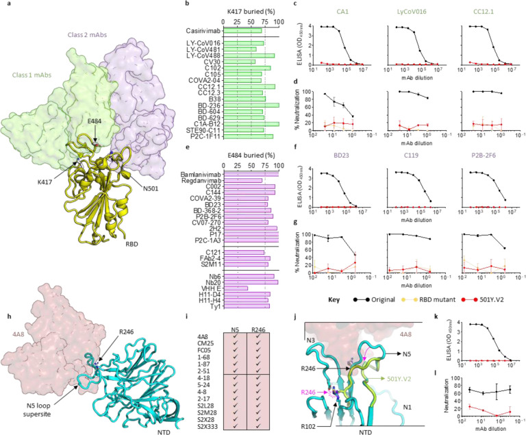
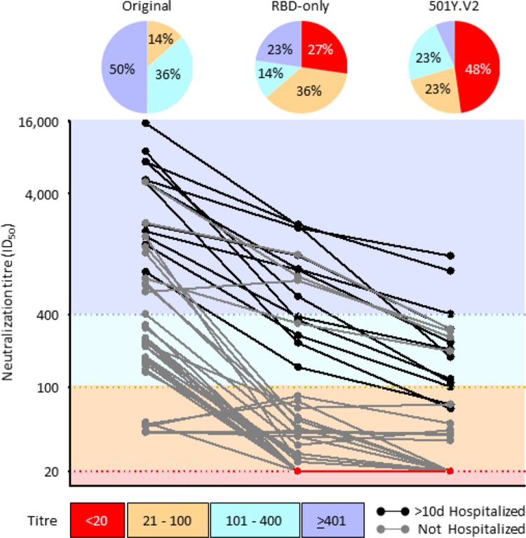
SARS-CoV-2 501Y.V2 (B.1.351), a novel lineage of coronavirus causing COVID-19, contains substitutions in two immunodominant domains of the spike protein. Here, we show that pseudovirus expressing 501Y.V2 spike protein completely escapes three classes of therapeutically relevant antibodies. This pseudovirus also exhibits substantial to complete escape from neutralization, but not binding, by convalescent plasma. These data highlight the prospect of reinfection with antigenically distinct variants and foreshadows reduced efficacy of spike-based vaccines.
Overview
The claims and statistics of this paper haven’t been checked yet.
Run the check — free, no account needed. It reads the open-access text and checks the sources it cites.
Integrity signals
Registry
Cites
Statistics
Figures
Verification
Claims the paper makes
The claims and statistics of this paper haven’t been checked yet.
Run the check — free, no account needed. It reads the open-access text and checks the sources it cites.
Claims other papers make about this one
Each is checked against this paper’s own text: does it say what the citing paper says it does?
The source backs part of the claim, not all of it — or backs it for a different population, condition or comparison.
In the citing paper
“Similarly, sera from PIDs who experienced symptomatic SARS-CoV-2 infection and who had detectable anti-RBD IgG titers before vaccination displayed generally weak nAb titers against Wuhan-Hu-1 at 1 to 9 months after infection and lower or nonexistent titers against the B.1.351 variants, in agreement with another study (69).”
From mRNA vaccination boosts cross-variant neutralizing antibodies elicited by SARS-CoV-2 infection (2021) · cites this paper as [69]
Experimental — accuracy not yet measuredThe source backs part of the claim, not all of it — or backs it for a different population, condition or comparison.
In the citing paper
“In multiple studies, B.1.351 was the most resistant to neutralization by convalescent or vaccinee sera, with 6- to 15-fold less neutralization activity for sera from individuals immunized with vaccines based on the virus strain first described in January 2020 (Wuhan-Hu-1, spike also called WA1) (2–9).”
From Durability of mRNA-1273 vaccine–induced antibodies against SARS-CoV-2 variants (2021) · cites this paper as [6]
Experimental — accuracy not yet measuredThe source backs part of the claim, not all of it — or backs it for a different population, condition or comparison.
In the citing paper
“As previously reported for other vaccines29–33, the neutralizing antibody titres against certain SARS-CoV-2 variants, such as the B.1.351 (Beta) and B.1.617.2 (Delta) variants, were lower than those against the parental strain WA1/2020.”
From Optimization of non-coding regions for a non-modified mRNA COVID-19 vaccine (2022) · cites this paper as [32]
Experimental — accuracy not yet measuredIn the citing paper
“recent reports suggest that these variants are partially resistant to neutralization by monoclonal antibodies against spike protein, by convalescent plasma and, notably, by sera from participants who have received severe acute respiratory syndrome coronavirus 2 (SARS-CoV-2) vaccines91,94,95.”
From Prospects for durable immune control of SARS-CoV-2 and prevention of reinfection (2021) · cites this paper as [94]
In the citing paper
“The sensitivity of B.1.351 to neutralizing antibodies from convalescent donors infected with the prototype lineage virus, assessed with a spike-pseudovirus neutralization assay, indicated that 48% of serum samples were unable to neutralize B.1.351, with the rest showing a reduction in neutralization titers by a factor of 3 to 86.21”
From Efficacy of the ChAdOx1 nCoV-19 Covid-19 Vaccine against the B.1.351 Variant (2021) · cites this paper as [21]
In the citing paper
“In contrast, mutations in B.1.351 and P.1 variants are associated with the more pronounced loss of neutralizing capacity.15,16,22,23,25”
From Impact of SARS-CoV-2 variants on the total CD4+ and CD8+ T cell reactivity in infected or vaccinated individuals (2021) · cites this paper as [23]
In the citing paper
“Mutations in the B.1.351and P.1 strains, including E484K and K417N/T, are of high concern, since they partly compromise neutralization generated by previous infection or vaccination [23][24][25] or may increase inherent viral fitness.”
From Sensitivity of infectious SARS-CoV-2 B.1.1.7 and B.1.351 variants to neutralizing antibodies (2021) · cites this paper as [23]
In the citing paper
“The E484K mutation, which can mediate antibody escape of B.1.351and P.1 variants, has been recently detected in the B.1.1.7 lineage in the United Kingdom, and thus represents a major potential threat for previously immune populations.”
From Sensitivity of infectious SARS-CoV-2 B.1.1.7 and B.1.351 variants to neutralizing antibodies (2021) · cites this paper as [23]
In the citing paper
“Most of these prior studies have evaluated sera from vaccinated individuals at time points soon after the first or second dose and have had limited data on the durability of such responses.”
From Durability of mRNA-1273 vaccine–induced antibodies against SARS-CoV-2 variants (2021) · cites this paper as [6]
In the citing paper
“Both the Beta and Delta variants are associated with decreased neutralization by some anti-receptor binding-domain (anti-RBD) and anti-N-terminal domain (anti-NTD) monoclonal antibodies though both Beta and Delta each remain responsive to at least one anti-RBD ( 8 , 9 , 21 ).”
From Increased risk of SARS-CoV-2 reinfection associated with emergence of Omicron in South Africa (2021) · cites this paper as [8]
In the citing paper
“In addition, Beta and Delta are relatively poorly neutralized by convalescent sera obtained from unvaccinated individuals infected with non-VOC virus ( 7 – 9 , 21 ).”
From Increased risk of SARS-CoV-2 reinfection associated with emergence of Omicron in South Africa (2021) · cites this paper as [8]
Self-check
Percentages, sums and reported differences re-computed from the paper’s own numbers.
The self-check needs the paper’s full text.
Citations
Cites
Berman, H. M. (2000) · Nucleic Acids Research · doi:10.1093/nar/28.1.235 · Cited by 40,196
A pneumonia outbreak associated with a new coronavirus of probable bat origin
Zhou, P., Yang, X.-L., Wang, X.-G., Hu, B., Zhang, L., Zhang, W., Si, H.-R., Zhu, Y., Li, B., Huang, C.-L., et al. (2020) · Nature · doi:10.1038/s41586-020-2012-7 · Cited by 21,969
The PyMOL Molecular Graphics System
DeLano, W. L. (2002) · Medical Entomology and Zoology · Cited by 18,147
Structure, function and antigenicity of the SARS-CoV-2 spike glycoprotein
Walls, A. C., Park, Y.-J., Tortorici, M. A., Wall, A., McGuire, A. T., & Veesler, D. (2020) · doi:10.1101/2020.02.19.956581 · Cited by 8,586
Korber, B., Fischer, W. M., Gnanakaran, S., Yoon, H., Theiler, J., Abfalterer, W., Hengartner, N., Giorgi, E. E., Bhattacharya, T., Foley, B., et al. (2020) · Cell · doi:10.1016/j.cell.2020.06.043 · Cited by 4,281
Nextstrain: real-time tracking of pathogen evolutionPreprint
Hadfield, J., Megill, C., Bell, S. M., et al. (2017) · bioRxiv · doi:10.1101/224048 · Cited by 3,172
Nextstrain: real-time tracking of pathogen evolution
Hadfield, J., Megill, C., Bell, S. M., Huddleston, J., Potter, B., Callender, C., Sagulenko, P., Bedford, T., & Neher, R. A. (2018) · Bioinformatics · doi:10.1093/bioinformatics/bty407 · Cited by 3,154
Structural and Functional Basis of SARS-CoV-2 Entry by Using Human ACE2
Wang, Q., Zhang, Y., Wu, L., Niu, S., Song, C., Zhang, Z., Lu, G., Qiao, C., Hu, Y., Yuen, K.-Y., et al. (2020) · Cell · doi:10.1016/j.cell.2020.03.045 · Cited by 2,892
Tegally, H., Wilkinson, E., Giovanetti, M., Iranzadeh, A., Fonseca, V., Giandhari, J., Doolabh, D., Pillay, S., San, E. J., Msomi, N., et al. (2020) · doi:10.1101/2020.12.21.20248640 · Cited by 2,535 · cited 1×
“N501Y has been shown to increase affinity for the human ACE2 receptor, which together with the repeated and independent evolution of 501Y containing lineages 25,26,31 , strongly argues for enhanced transmissibility of these new variants.”
Self-citationDetection of a SARS-CoV-2 variant of concern in South Africa
Tegally, H., Wilkinson, E., Giovanetti, M., Iranzadeh, A., Fonseca, V., Giandhari, J., Doolabh, D., Pillay, S., San, E. J., Msomi, N., et al. (2021) · Nature · doi:10.1038/s41586-021-03402-9 · Cited by 2,510
Self-citation
Cited by
Sorted by citations
SARS-CoV-2 variants, spike mutations and immune escape
Harvey, W. T., Carabelli, A. M., Jackson, B., Gupta, R. K., Thomson, E. C., Harrison, E. M., Ludden, C., Reeve, R., Rambaut, A., COVID-19 Genomics UK (COG-UK) Consortium, et al. (2021) · Nature Reviews Microbiology · doi:10.1038/s41579-021-00573-0 · Cited by 3,528
Features, Evaluation and Treatment Coronavirus (COVID-19)
Cascella, M., Rajnik, M., Cuomo, A., Dulebohn, S. C., & Napoli, R. D. (2020) · StatPearls · Cited by 3,351
Mechanisms of SARS-CoV-2 entry into cells
Jackson, C. B., Farzan, M., Chen, B., & Choe, H. (2022) · Nature Reviews Molecular Cell Biology · doi:10.1038/s41580-021-00418-x · Cited by 3,022
Detection of a SARS-CoV-2 variant of concern in South Africa
Tegally, H., Wilkinson, E., Giovanetti, M., Iranzadeh, A., Fonseca, V., Giandhari, J., Doolabh, D., Pillay, S., San, E. J., Msomi, N., et al. (2021) · Nature · doi:10.1038/s41586-021-03402-9 · Cited by 2,510
Self-citationSafety and Efficacy of Single-Dose Ad26.COV2.S Vaccine against Covid-19
Sadoff, J., Gray, G., Vandebosch, A., Cárdenas, V., Shukarev, G., Grinsztejn, B., Goepfert, P. A., Truyers, C., Fennema, H., Spiessens, B., et al. (2021) · New England Journal of Medicine · doi:10.1056/nejmoa2101544 · Cited by 2,397
Metadata
- Authors
- Wibmer, C. K., Ayres, F., Hermanus, T., Madzivhandila, M., Kgagudi, P., Oosthuysen, B., Lambson, B. E., de Oliveira, T., Vermeulen, M., van der Berg, K., Rossouw, T., Boswell, M., Ueckermann, V., Meiring, S., von Gottberg, A., Cohen, C., Morris, L., Bhiman, J. N., & Moore, P. L.
- Affiliations
- National Health Laboratory Service (ZA)
- South African National Blood Service (ZA)
- University of Pretoria (ZA)
- Steve Biko Hospital (ZA)
- Corresponding author
- Moore, Penny L.
- ORCID
- Venue
- Nature Medicine
- Published
- 2021-03-02
- Volume
- 27
- Issue
- 4
- Pages
- 622-625
- ISSN
- 1078-8956 (ISSN-L)
- 1546-170X
- PMC
- PMC7836116
- OpenAlex
- W3133794453
- Cited by
- 1,147
- References
- 38
- Citations by year
- 2026: 10 · 2025: 29 · 2024: 58 · 2023: 97 · 2022: 314 · 2021: 634
- FWCI
- 87.41
- Type
- journal article
- Language
- en
- Open access
- bronze
- doi.org
- Licence
- Locations
- Nature Medicine (publishedVersion)
- bioRxiv (Cold Spring Harbor Laboratory) (acceptedVersion, cc-by-nd)
- PubMed (publishedVersion)
- Europe PMC (PubMed Central) (submittedVersion, cc-by-nd)
- PubMed Central (submittedVersion, cc-by-nd)
- UpSpace Institutional Repository (University of Pretoria) (submittedVersion)
- Funders
- Wellcome Trust (221003/Z/20/Z, 221003)
- Government of the United Kingdom
- Royal Society
- National Research Foundation (98341)
- South African Medical Research Council (98341, SHIPNCD 76756, 96825)
- Department of Science and Technology, Ministry of Science and Technology, India
- African Academy of Sciences
- National Institutes of Health (98341, COVID-19)
- Centers for Disease Control and Prevention (5 U01IP001048-05-00, U01IP001048)
- Medical Research Council (COVID-19)
- Fogarty International Center (R21TW011454, D43TW010345)
- Topics
- Health Sciences › Medicine › Infectious Diseases › SARS-CoV-2 and COVID-19 Research
- Life Sciences › Agricultural and Biological Sciences › Animal Science and Zoology › Animal Virus Infections Studies
- Life Sciences › Biochemistry, Genetics and Molecular Biology › Molecular Biology › vaccines and immunoinformatics approaches
- Keywords
- SARS-CoV-2, B.1.351 variant, spike protein mutations, antibody escape, neutralization, convalescent plasma, monoclonal antibodies, reinfection, vaccine efficacy, pseudovirus, spike protein binding
About these checks
Registry, retraction, Cites and statistics results are computed, not judged, with one exception: a printed reference that no index lists is read once by a model before it counts as a citation error. Claims are extracted by a model and kept only when their quote appears verbatim in the text; each cited source is then checked, with a model’s help, for whether it says what the claim relies on. Those verdicts can be wrong — read the claim’s quote and the cited source before relying on one.
Statistics and arithmetic are recomputed, not judged.
Confidence bands are shown only for fields calibrated on our annotated test set. None is calibrated yet, so no band is shown.
What counts as a claim ›How we measure confidence ›How we check this ›
Are you an author, or do you see a problem with this page? Request a review
