

The DOI matches its Crossref record; 48 of the 49 references it cites were found in a registry or an index.
Nucleus segmentation across imaging experiments: the 2018 Data Science Bowl
Open accessAbstract
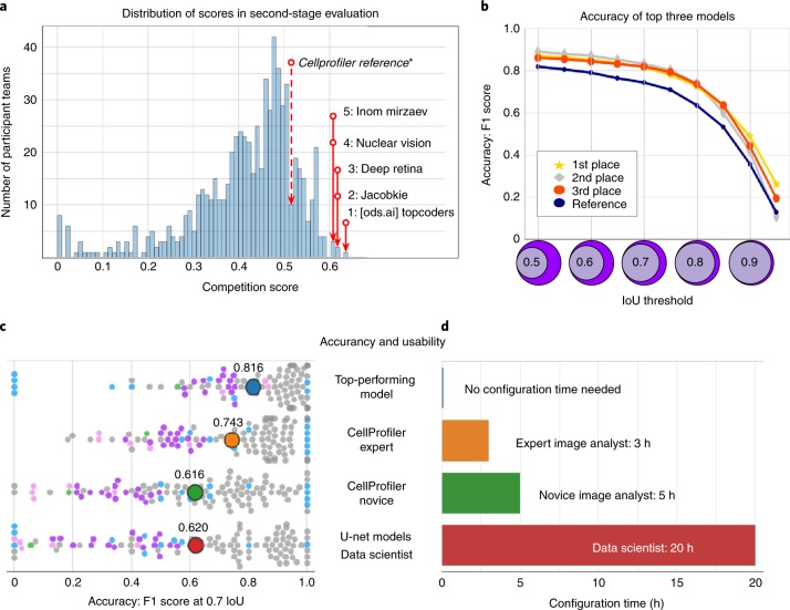
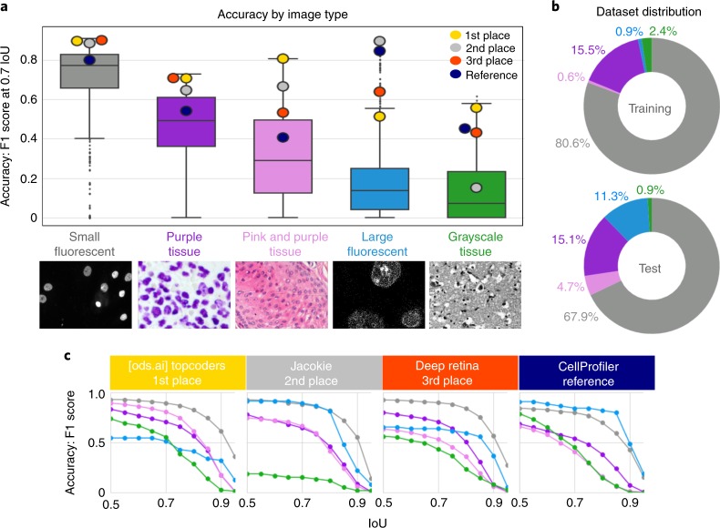
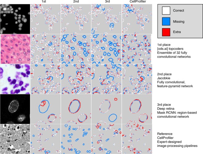
Segmenting the nuclei of cells in microscopy images is often the first step in the quantitative analysis of imaging data for biological and biomedical applications. Many bioimage analysis tools can segment nuclei in images but need to be selected and configured for every experiment. The 2018 Data Science Bowl attracted 3,891 teams worldwide to make the first attempt to build a segmentation method that could be applied to any two-dimensional light microscopy image of stained nuclei across experiments, with no human interaction. Top participants in the challenge succeeded in this task, developing deep-learning-based models that identified cell nuclei across many image types and experimental conditions without the need to manually adjust segmentation parameters. This represents an important step toward configuration-free bioimage analysis software tools.
Overview
The claims and statistics of this paper haven’t been checked yet.
Run the check — free, no account needed. It reads the open-access text and checks the sources it cites.
Integrity signals
Registry
Cites
Statistics
Figures
Verification
Claims the paper makes
The claims and statistics of this paper haven’t been checked yet.
Run the check — free, no account needed. It reads the open-access text and checks the sources it cites.
Claims other papers make about this one
Each is checked against this paper’s own text: does it say what the citing paper says it does?
The source backs part of the claim, not all of it — or backs it for a different population, condition or comparison.
In the citing paper
“However, we did not see an improvement for the ensemble of models compared to the generalist model (Extended Data Fig. 2), consistent with the results of ref. 27.”
From Cellpose 2.0: how to train your own model (2022) · cites this paper as [27]
Experimental — accuracy not yet measuredThe cited source says what the claim attributes to it.
In the citing paper
“Finally, we point out that the LiveCell study used a different average precision score from ours, which additionally requires a confidence score per ROI, while we used the average precision formulation from the Data Science Bowl challenge and other studies12,27,35.”
From Cellpose 2.0: how to train your own model (2022) · cites this paper as [27]
Self-check
Percentages, sums and reported differences re-computed from the paper’s own numbers.
The self-check needs the paper’s full text.
Citations
Cites
Deep Residual Learning for Image Recognition
He, K., Zhang, X., Ren, S., & Sun, J. (2016) · 2016 IEEE Conference on Computer Vision and Pattern Recognition (CVPR) · doi:10.1109/cvpr.2016.90 · Cited by 235,474
U-Net: Convolutional Networks for Biomedical Image Segmentation
Ronneberger, O., Fischer, P., & Brox, T. (2015) · Lecture notes in computer science · doi:10.1007/978-3-319-24574-4_28 · Cited by 99,404
LeCun, Y., Bengio, Y., & Hinton, G. (2015) · Nature · doi:10.1038/nature14539 · Cited by 84,126
Fiji: an open-source platform for biological-image analysis
Schindelin, J., Arganda-Carreras, I., Frise, E., Kaynig, V., Longair, M., Pietzsch, T., Preibisch, S., Rueden, C., Saalfeld, S., Schmid, B., et al. (2012) · Nature Methods · doi:10.1038/nmeth.2019 · Cited by 66,890
Microsoft COCO: Common Objects in Context
Lin, T.-Y., Maire, M., Belongie, S., Hays, J., Perona, P., Ramanan, D., Dollár, P., & Zitnick, C. L. (2014) · Lecture Notes in Computer Science · doi:10.1007/978-3-319-10602-1_48 · Cited by 45,460
A Threshold Selection Method from Gray-Level Histograms
Otsu, N. (1979) · IEEE Transactions on Systems, Man and Cybernetics · doi:10.1109/tsmc.1979.4310076 · Cited by 44,708
ImageNet Large Scale Visual Recognition Challenge
Russakovsky, O., Deng, J., Su, H., Krause, J., Satheesh, S., Ma, S., Huang, Z., Karpathy, A., Khosla, A., Bernstein, M., et al. (2015) · International Journal of Computer Vision · doi:10.1007/s11263-015-0816-y · Cited by 39,380
Feature Pyramid Networks for Object Detection
Lin, T.-Y., Dollar, P., Girshick, R., He, K., Hariharan, B., & Belongie, S. (2017) · 2017 IEEE Conference on Computer Vision and Pattern Recognition (CVPR) · doi:10.1109/cvpr.2017.106 · Cited by 30,939
He, K., Gkioxari, G., Dollar, P., & Girshick, R. (2017) · 2017 IEEE International Conference on Computer Vision (ICCV) · doi:10.1109/iccv.2017.322 · Cited by 29,137
Unpaired image-to-image translation using cycle-consistent adversarial networks
Zhu, J.-Y., Park, T., Isola, P., & Efros, A. A. (2017) · 2017 IEEE International Conference on Computer Vision (ICCV) · doi:10.1109/iccv.2017.244 · Cited by 23,602
Research works printed but not linked: 3
- Hollandi, R. et al. A deep learning framework for nucleus segmentation using image style transfer. Preprint at bioRxiv https://doi.org/10.1101/580605 (2019). · DOI
- Bolukbasi, T., Chang, K.-W., Zou, J. Y., Saligrama, V. & Kalai, A. T. in Advances in Neural Information Processing Systems (eds Lee, D. D. et al.) 4349–4357 (papers.nips.cc, 2016).
- Ren, S., He, K., Girshick, R. & Sun, J. in Advances in Neural Information Processing Systems 28 (eds. Cortes, C. et al.) 91–99 (Curran Associates, 2015).
Cited by
Sorted by citations
Items 201–300 of 811
High-throughput segmentation of unmyelinated axons by deep learning
Plebani, E., Biscola, N. P., Havton, L. A., Rajwa, B. P., Shemonti, A. S., Jaffey, D., Powley, T. L., Keast, J. R., Lu, K., & Dündar, M. (2022) · Scientific Reports · doi:10.1038/s41598-022-04854-3 · Cited by 24
Self-supervised machine learning for live cell imagery segmentation
Robitaille, M. C., Byers, J. M., Christodoulides, J. A., & Raphael, M. P. (2022) · Communications Biology · doi:10.1038/s42003-022-04117-x · Cited by 31
blob loss: Instance Imbalance Aware Loss Functions for Semantic Segmentation
Kofler, F., Shit, S., Ezhov, I., Fidon, L., Horvath, I., Al-Maskari, R., Li, H., Bhatia, H. S., Loehr, T., Piraud, M., et al. (2023) · Lecture notes in computer science · doi:10.1007/978-3-031-34048-2_58 · Cited by 24
SMTF: Sparse transformer with multiscale contextual fusion for medical image segmentation
Zhang, X., Zhang, X., Ouyang, L., Qin, C., Xiao, L., & Xiong, D. (2023) · Biomedical Signal Processing and Control · doi:10.1016/j.bspc.2023.105458 · Cited by 24
Kulyabin, M., Zhdanov, A. E., Pershin, A., Sokolov, G., Nikiforova, A., Ronkin, M., Борисов, В., & Maier, A. (2024) · Bioengineering · doi:10.3390/bioengineering11090940 · Cited by 23
Tay, F. R., Loveless, R., & Ravenel, T. D. (2026) · Dental Research · doi:10.1016/j.dtrs.2025.100005 · Cited by 25
Superhuman cell death detection with biomarker-optimized neural networks
Linsley, J. W., Linsley, D. A., Lamstein, J., Ryan, G., Shah, K. D., Castello, N. A., Oza, V. D., Kalra, J., Wang, S., Tokuno, Z., et al. (2021) · Science Advances · doi:10.1126/sciadv.abf8142 · Cited by 27
A Catalyst‐Like System Enables Efficient Perovskite Solar Cells
Yang, Y., Li, G., Zhao, L., Tan, P., Li, Y., Li, S., Tan, L., Deng, C., Wang, S., Zhao, Z., et al. (2024) · Advanced Materials · doi:10.1002/adma.202311145 · Cited by 23
Spatial genomics maps the structure, character and evolution of cancer clonesPreprint
Lomakin, A., Svedlund, J., Strell, C., Gatarić, M., Shmatko, A., Park, J. S., Ju, Y. S., Dentro, S. C., Kleshchevnikov, V., Vaskivskyi, V., et al. (2021) · bioRxiv (Cold Spring Harbor Laboratory) · doi:10.1101/2021.04.16.439912 · Cited by 23
RxRx3: Phenomics Map of BiologyPreprint
Fay, M. M., Kraus, O. Z., Victors, M. L., Arumugam, L., Vuggumudi, K., Urbanik, J., Hansen, K., Çelik, S., Cernek, N., Jagannathan, G., et al. (2023) · bioRxiv (Cold Spring Harbor Laboratory) · doi:10.1101/2023.02.07.527350 · Cited by 33
MaS-TransUNet: A Multiattention Swin Transformer U-Net for Medical Image Segmentation
Upadhyay, A. K., & Bhandari, A. K. (2024) · IEEE Transactions on Radiation and Plasma Medical Sciences · doi:10.1109/trpms.2024.3477528 · Cited by 22
Krikid, F., Rositi, H., & Vacavant, A. (2024) · Journal of Imaging · doi:10.3390/jimaging10120311 · Cited by 32
Wazir, S., & Kim, D. (2025) · IEEE Computer Society Conference on Computer Vision and Pattern Recognition/Proceedings - IEEE Computer Society Conference on Computer Vision and Pattern Recognition/Proceedings · doi:10.1109/cvpr52734.2025.02874 · Cited by 22
Multistar: Instance Segmentation Of Overlapping Objects With Star-Convex Polygons
Walter, F. C., Damrich, S., & Hamprecht, F. A. (2021) · IEEE International Symposium on Biomedical Imaging (ISBI) · doi:10.1109/isbi48211.2021.9433769 · Cited by 22
Bioimage analysis workflows: community resources to navigate through a complex ecosystem
Paul‐Gilloteaux, P., Tosi, S., Hèriché, J.-K., Alban Gaignard, A., Ménager, H., Marée, R., Baecker, V., Klemm, A. H., Kalaš, M., Zhang, C., et al. (2021) · F1000Research · doi:10.12688/f1000research.52569.1 · Cited by 22
Han, W., Cheung, A., Yaffe, M. J., & Martel, A. L. (2022) · Scientific Reports · doi:10.1038/s41598-022-08355-1 · Cited by 22
Biomedical Image Segmentation with Modified U-Net
Tatli, U., & Budak, C. (2023) · Traitement du signal · doi:10.18280/ts.400211 · Cited by 21
Maddalena, L., Antonelli, L., Albu, A., Hada, A., & Guarracino, M. R. (2022) · Algorithms · doi:10.3390/a15090313 · Cited by 24
Chen, B., Yin, Z., Ng, W., Wang, D. M., Tuan, R. S., Bise, R., & Ker, D. F. E. (2024) · npj Imaging · doi:10.1038/s44303-024-00046-y · Cited by 23
Olimov, B., Koh, S., & Kim, J. (2021) · IEEE Access · doi:10.1109/access.2021.3128607 · Cited by 20
Automated Annotator Variability Inspection for Biomedical Image Segmentation
Schilling, M. P., Scherr, T., Münke, F. R., Neumann, O., Schutera, M., Mikut, R., & Reischl, M. (2022) · IEEE Access · doi:10.1109/access.2022.3140378 · Cited by 21
Ahmed, M. R., Fahim, M. A. I., Islam, A. K. M. M., Islam, S., & Shatabda, S. (2023) · Neurocomputing · doi:10.1016/j.neucom.2023.126362 · Cited by 20
Yuan, H., & Peng, J. (2024) · Pattern Recognition · doi:10.1016/j.patcog.2024.110428 · Cited by 20
Vijayan, A., Mody, T. A., Yu, Q., Wolny, A., Cerrone, L., Strauss, S., Tsiantis, M., Smith, R. S., Hamprecht, F. A., Kreshuk, A., et al. (2024) · Development · doi:10.1242/dev.202800 · Cited by 24
Convolutional Blur Attention Network for Cell Nuclei Segmentation
Le, P. T., Pham, T. V., Hsu, Y., & Wang, J. (2022) · Sensors · doi:10.3390/s22041586 · Cited by 20
Zhou, Y., Li, L., Wang, C., Song, L., & Yang, G. (2024) · IEEE Transactions on Medical Imaging · doi:10.1109/tmi.2024.3474028 · Cited by 19
Deep learning-enabled analysis reveals distinct neuronal phenotypes induced by aging and cold-shock
Saberi‐Bosari, S., Flores, K., & San‐Miguel, A. (2020) · BMC Biology · doi:10.1186/s12915-020-00861-w · Cited by 18
Li, Z., Tang, Z., Hu, J., Wang, X., Jia, D., & Zhang, Y. (2023) · Biomedical Signal Processing and Control · doi:10.1016/j.bspc.2023.104785 · Cited by 22
Deep cell phenotyping and spatial analysis of multiplexed imaging with TRACERx-PHLEX
Magness, A. J., Colliver, E., Enfield, K. S. S., Lee, C., Shimato, M., Daly, E., Moore, D. A., Sivakumar, M., Valand, K., Levi, D., et al. (2024) · Nature Communications · doi:10.1038/s41467-024-48870-5 · Cited by 22
ASAM: Boosting Segment Anything Model with Adversarial Tuning
Li, B. H., Xiao, H., & Tang, L. (2024) · IEEE Computer Society Conference on Computer Vision and Pattern Recognition/Proceedings - IEEE Computer Society Conference on Computer Vision and Pattern Recognition/Proceedings · doi:10.1109/cvpr52733.2024.00355 · Cited by 24
A Deep Learning Pipeline for Nucleus Segmentation
Zaki, G., Gudla, P. R., Lee, K., Kim, J. J., Ozbun, L. L., Shachar, S., Gadkari, M., Sun, J., Fraser, I. D. C., Franco, L. M., et al. (2020) · Cytometry Part A · doi:10.1002/cyto.a.24257 · Cited by 19
Poźniak, J., Pedri, D., Landeloos, E., Herck, Y. V., Antoranz, A., Karras, P., Nowosad, A., Makhzami, S., Bervoets, G., Dewaele, M., et al. (2022) · bioRxiv (Cold Spring Harbor Laboratory) · doi:10.1101/2022.08.11.502598 · Cited by 21
The crucial role of bioimage analysts in scientific research and publication
Cimini, B. A., Bankhead, P., D’Antuono, R., Fazeli, E., Fernández-Rodrı́guez, J., Fuster-Barceló, C., Haase, R., Jambor, H. K., Jones, M. L., Jug, F., et al. (2024) · Journal of Cell Science · doi:10.1242/jcs.262322 · Cited by 17
Self-citationUN-SAM: Domain-adaptive self-prompt segmentation for universal nuclei images
Chen, Z., Xu, Q., Liu, X., & Yuan, Y. (2025) · Medical Image Analysis · doi:10.1016/j.media.2025.103607 · Cited by 57
DPCFN: Dual path cross fusion network for medical image segmentation
Jiang, S., Li, J., & Hua, Z. (2022) · Engineering Applications of Artificial Intelligence · doi:10.1016/j.engappai.2022.105420 · Cited by 18
OpSeF: Open Source Python Framework for Collaborative Instance Segmentation of Bioimages
Rasse, T. M., Hollandi, R., & Horváth, P. (2020) · Frontiers in Bioengineering and Biotechnology · doi:10.3389/fbioe.2020.558880 · Cited by 17
A survey of deep learning methods on cell instance segmentation
Wang, C., Lee, W.-T., & Su, T.-S. (2025) · Neural Computing and Applications · doi:10.1007/s00521-025-11119-3 · Cited by 21
Single cell imaging-based chromatin biomarkers for tumor progression
Venkatachalapathy, S., Jokhun, D. S., Andhari, M. D., & Shivashankar, G. V. (2021) · Scientific Reports · doi:10.1038/s41598-021-02441-6 · Cited by 17
MFA-Net: Multiple Feature Association Network for medical image segmentation
Li, Z., Zhang, N., Gong, H., Qiu, R., & Zhang, W. (2023) · Computers in Biology and Medicine · doi:10.1016/j.compbiomed.2023.106834 · Cited by 21
P-TransUNet: an improved parallel network for medical image segmentation
Chong, Y., Xie, N., Liu, X., & Pan, S. (2023) · BMC Bioinformatics · doi:10.1186/s12859-023-05409-7 · Cited by 13
AttResDU-Net: Medical Image Segmentation Using Attention-based Residual Double U-Net
Khan, A. M., Ashrafee, A., Khan, F. S., Hasan, M. B., & Kabir, M. H. (2023) · International Joint Conference on Neural Networks (IJCNN) · doi:10.1109/ijcnn54540.2023.10191528 · Cited by 18
Defining the boundaries: challenges and advances in identifying cells in microscopy images
Gogoberidze, N., & Cimini, B. A. (2023) · Current Opinion in Biotechnology · doi:10.1016/j.copbio.2023.103055 · Cited by 18
Self-citationHarnessing artificial intelligence to reduce phototoxicity in live imaging
Gómez‐de‐Mariscal, E., Rosario, M. D., Pylvänäinen, J. W., Jacquemet, G., & Henriques, R. (2024) · Journal of Cell Science · doi:10.1242/jcs.261545 · Cited by 17
Габдуллин, М. Т., Mukasheva, A., Koishiyeva, D., Umarov, T., Bissembayev, A., Kim, K.-S., & Kang, J. W. (2024) · Biotechnology and Bioprocess Engineering · doi:10.1007/s12257-024-00130-5 · Cited by 18
A survey on cell nuclei instance segmentation and classification: Leveraging context and attention
Nunes, J. D., Montezuma, D., Oliveira, D., Pereira, T., & Cardoso, J. S. (2024) · Medical Image Analysis · doi:10.1016/j.media.2024.103360 · Cited by 19
A Practical Guide to Supervised Deep Learning for Bioimage Analysis: Challenges and good practices
Uhlmann, V., Donati, L., & Sage, D. (2022) · IEEE Signal Processing Magazine · doi:10.1109/msp.2021.3123589 · Cited by 16
Xiaosu, Y., Tian, J., Wan, Y., Chen, M., Chen, L., & Chen, J. (2023) · Medical Physics · doi:10.1002/mp.16217 · Cited by 16
Liu, L., Chen, T., Zhang, Q., Zhang, W., Yang, H., Hu, X., Xiao, J., Liu, Q. S., & Jiang, G. (2023) · Environmental Science & Technology · doi:10.1021/acs.est.3c05252 · Cited by 17
SortedAP: Rethinking evaluation metrics for instance segmentation
Chen, L., Wu, Y., Stegmaier, J., & Merhof, D. (2023) · IEEE International Conference on Computer Vision Workshops · doi:10.1109/iccvw60793.2023.00424 · Cited by 16
A generic plug & play diffusion-based denosing module for medical image segmentation
Li, G., Jin, D., Zheng, Y., Cui, J., Gai, W., & Qi, M. (2024) · Neural Networks · doi:10.1016/j.neunet.2024.106096 · Cited by 18
Davidson, S. J., Saggese, T., & Krajňáková, J. (2024) · Frontiers in Plant Science · doi:10.3389/fpls.2024.1322920 · Cited by 16
Patino, C. A., Mukherjee, P., Lemaitre, V., Pathak, N., & Espinosa, H. D. (2021) · SLAS TECHNOLOGY · doi:10.1177/2472630320982320 · Cited by 16
Oral epithelial cell segmentation from fluorescent multichannel cytology images using deep learning
Sunny, S. P., Khan, A. I., Rangarajan, M., Hariharan, A., Birur, P. N., Pandya, H. J., Shah, N., Kuriakose, M. A., & Suresh, A. (2022) · Computer Methods and Programs in Biomedicine · doi:10.1016/j.cmpb.2022.107205 · Cited by 16
Wang, S., Zhang, X., Ren, L., & Li, J. (2026) · International Journal of Machine Learning and Cybernetics · doi:10.1007/s13042-025-02851-2 · Cited by 15
Histopathology image segmentation and classification for cancer revelation
Kurmi, Y., Chaurasia, V., & Kapoor, N. (2021) · Signal Image and Video Processing · doi:10.1007/s11760-021-01865-x · Cited by 15
An Enhanced Feature Extraction Network for Medical Image Segmentation
Gao, Y. J., Che, X., Xu, H., & Bie, M. (2023) · Applied Sciences · doi:10.3390/app13126977 · Cited by 15
Cellstitch: 3D cellular anisotropic image segmentation via optimal transport
Liu, Y., Jin, Y., Azizi, E., & Blumberg, A. J. (2023) · BMC Bioinformatics · doi:10.1186/s12859-023-05608-2 · Cited by 15
Software Tools for 2D Cell Segmentation
Liu, P., Li, J., Chang, J., Hu, P., Sun, Y., Jiang, Y., Zhang, F., & Shao, H. (2024) · Cells · doi:10.3390/cells13040352 · Cited by 17
In silico labeling in cell biology: Potential and limitations
Elmalam, N., Nedava, L. B., & Zaritsky, A. (2024) · Current Opinion in Cell Biology · doi:10.1016/j.ceb.2024.102378 · Cited by 14
Pan, F., Wu, Y., Cui, K., Chen, S., Li, Y., Liu, Y., Shakoor, A., Zhao, H., Lu, B., Zhi, S., et al. (2024) · Computers in Biology and Medicine · doi:10.1016/j.compbiomed.2024.109151 · Cited by 15
Nuclear instance segmentation and tracking for preimplantation mouse embryos
Nunley, H., Shao, B., Denberg, D. W., Grover, P., Singh, J., Avdeeva, M., Joyce, B. W., Kim, R., Kohrman, A. Q., Biswas, A. G., et al. (2024) · Development · doi:10.1242/dev.202817 · Cited by 15
Cao, Y., & Cheng, Y. (2025) · IEEE Access · doi:10.1109/access.2025.3526602 · Cited by 15
Screening as a strategy to drive regenerative medicine research
Vermeulen, S., & de Boer, J. (2020) · Methods · doi:10.1016/j.ymeth.2020.04.004 · Cited by 14
ACDC: Automated Cell Detection and Counting for Time-Lapse Fluorescence Microscopy
Rundo, L., Tangherloni, A., Tyson, D. R., Betta, R., Militello, C., Spolaor, S., Nobile, M. S., Besozzi, D., Lubbock, A. L. R., Quaranta, V., et al. (2020) · Applied Sciences · doi:10.3390/app10186187 · Cited by 15
Yu, M., Pei, K., Li, X., Wei, X., Wang, C., & Gao, J. (2022) · Computers in Biology and Medicine · doi:10.1016/j.compbiomed.2022.106161 · Cited by 15
PAANet: Progressive Alternating Attention for Automatic Medical Image Segmentation
Srivastava, A., Chanda, S., Jha, D., Riegler, M. A., Halvorsen, P., Johansen, D., & Pal, U. (2021) · International Conference on Bio-engineering for Smart Technologies (BioSMART) · doi:10.1109/biosmart54244.2021.9677844 · Cited by 15
Harland, L. T. G., Lohoff, T., Koulena, N., Pierson, N. G., Pape, C., Ameen, F., Griffiths, J. A., Theeuwes, B., Wilson, N. K., Kreshuk, A., et al. (2025) · Cell Reports · doi:10.1016/j.celrep.2025.116047 · Cited by 13
Cell segmentation and representation with shape priors
Hirling, D., & Horváth, P. (2022) · Computational and Structural Biotechnology Journal · doi:10.1016/j.csbj.2022.12.034 · Cited by 15
Lv, J., Hu, Y., Fu, Q., Zhang, Z., Hu, Y., Lv, L., Yang, G., Li, J., & Zhao, Y. (2022) · IEEE International Conference on Bioinformatics and Biomedicine (BIBM) · doi:10.1109/bibm55620.2022.9995348 · Cited by 14
Segment-then-Segment: Context-Preserving Crop-Based Segmentation for Large Biomedical Images
Benčević, M., Qiu, Y., Galić, I., & Pižurica, A. (2023) · Sensors · doi:10.3390/s23020633 · Cited by 14
A Depth-Wise Separable U-Net Architecture with Multiscale Filters to Detect Sinkholes
Alshawi, R., Hoque, M. T., & Flanagin, M. C. (2023) · Remote Sensing · doi:10.3390/rs15051384 · Cited by 14
Fourier Channel Attention Powered Lightweight Network for Image Segmentation
Zou, F., Liu, Y., Chen, Z., Zhanghao, K., & Jin, D. (2023) · IEEE Journal of Translational Engineering in Health and Medicine · doi:10.1109/jtehm.2023.3262841 · Cited by 14
Toward Source-Free Cross Tissues Histopathological Cell Segmentation via Target-Specific Finetuning
Li, Z., Li, C., Luo, X., Zhou, Y., Zhu, J., Xu, C., Yang, M., Wu, Y., & Chen, Y. (2023) · IEEE Transactions on Medical Imaging · doi:10.1109/tmi.2023.3263465 · Cited by 14
White Blood Cell Segmentation Using DeepLabv3+ for Improved Hematological Disease Detection
Joshi, V. C., & Mehta, M. A. (2023) · IEEE Region 10 Symposium (TENSYMP) · doi:10.1109/tensymp55890.2023.10223628 · Cited by 14
MPFC-Net: A multi-perspective feature compensation network for medical image segmentation
Wu, X.-H., Huang, S., Shu, X., Hu, C.-L., & Wu, X.-J. (2024) · Expert systems with applications · doi:10.1016/j.eswa.2024.123430 · Cited by 14
MFLUnet: multi-scale fusion lightweight Unet for medical image segmentation
Cao, D., Zhang, R., & Zhang, Y. (2024) · Biomedical Optics Express · doi:10.1364/boe.529505 · Cited by 14
Huang, J.-R., Zhou, Y., Luo, Y., Liu, G., Guo, H., & Yang, G. (2024) · Lecture notes in computer science · doi:10.1007/978-3-031-73404-5_9 · Cited by 14
Gu, Z., Wang, S., Rong, R., Zhao, Z., Wu, F., Zhou, Q., Wen, Z., Chi, Z., Fang, Y. V., Peng, Y., et al. (2024) · Laboratory Investigation · doi:10.1016/j.labinv.2024.102184 · Cited by 16
Metric-learning-assisted domain adaptation
Yin, Y., Yang, Z., Hu, H., & Wu, X. (2021) · Neurocomputing · doi:10.1016/j.neucom.2021.05.023 · Cited by 14
Pécot, T., Cuitiño, M. C., Johnson, R. H., Timmers, C. D., & Leone, G. W. (2022) · PLoS Computational Biology · doi:10.1371/journal.pcbi.1009949 · Cited by 17
User-Accessible Machine Learning Approaches for Cell Segmentation and Analysis in Tissue
Winfree, S. (2022) · Frontiers in Physiology · doi:10.3389/fphys.2022.833333 · Cited by 15
GIANI – open-source software for automated analysis of 3D microscopy images
Barry, D. J., Gerri, C., Bell, D. M., D’Antuono, R., & Niakan, K. K. (2022) · Journal of Cell Science · doi:10.1242/jcs.259511 · Cited by 14
Maimaitili, M., Chen, M., Febbraro, F., Ucuncu, E., Kelly, R. L., Niclis, J. C., Christiansen, J. R., Mermet-Joret, N., Niculescu, D., Lauritsen, J., et al. (2023) · Nature Communications · doi:10.1038/s41467-023-43471-0 · Cited by 14
Mela, C. A., & Liu, Y. (2021) · BMC Bioinformatics · doi:10.1186/s12859-021-04245-x · Cited by 14
Embedding-based Instance Segmentation in MicroscopyPreprint
Lalit, M., Tomančák, P., & Jug, F. (2021) · arXiv (Cornell University) · doi:10.48550/arxiv.2101.10033 · Cited by 19
LCRM: Layer-Wise Complexity Reduction Method for CNN Model Optimization on End Devices
Hussain, H., Tamizharasan, P. S., & Yadav, P. K. (2023) · IEEE Access · doi:10.1109/access.2023.3290620 · Cited by 16
Zhao, K., Zhang, Q., Wan, C., Pan, Q., & Qin, Y. (2025) · Scientific Reports · doi:10.1038/s41598-024-80977-z · Cited by 13
Liu, H., Zhao, Z., Ma, Q., Du, J., & Liu, X. (2025) · International Journal of Coal Science & Technology · doi:10.1007/s40789-025-00790-5 · Cited by 13
Modular deep learning enables automated identification of monoclonal cell lines
Fischbacher, B., Hedaya, S., Hartley, B. J., Wang, Z., Lallos, G., Hutson, D., Zimmer, M., Brammer, J., The NYSCF Global Stem Cell Array Team, & Paull, D. J. (2021) · Nature Machine Intelligence · doi:10.1038/s42256-021-00354-7 · Cited by 13
Deep Neural Networks Learn Meta-Structures from Noisy Labels in Semantic Segmentation
Luo, Y., Liu, G., Guo, Y., & Yang, G. (2022) · Proceedings of the AAAI Conference on Artificial Intelligence · doi:10.1609/aaai.v36i2.20085 · Cited by 13
Singha, A., & Bhowmik, M. K. (2022) · Multimedia Tools and Applications · doi:10.1007/s11042-022-14098-y · Cited by 13
DRI-UNet: dense residual-inception UNet for nuclei identification in microscopy cell images
Sharma, A., & Mishra, P. K. (2023) · Neural computing & applications (Print) · doi:10.1007/s00521-023-08729-0 · Cited by 14
Lin, A., & Lu, A. X. (2022) · bioRxiv (Cold Spring Harbor Laboratory) · doi:10.1101/2022.10.14.512286 · Cited by 13
Highly Multiplexed 3D Profiling of Cell States and Immune Niches in Human TumoursPreprint
Yapp, C., Nirmal, A. J., Zhou, F., Wong, A. Y. H., Tefft, J. B., Lu, Y. D., Shang, Z., Maliga, Z., Llopis, P. D. L. M. M., Murphy, G. F., et al. (2023) · bioRxiv (Cold Spring Harbor Laboratory) · doi:10.1101/2023.11.10.566670 · Cited by 13
CIHM: context-insight hybrid Mamba for efficient medical image segmentation
Ma, Y., Liu, Y., Cheng, J., Zhan, H., Qi, G., Zhang, X., & Zhu, Z. (2026) · Visual Intelligence · doi:10.1007/s44267-026-00113-5 · Cited by 12
Segmentation Technology of Nucleus Image Based on U-Net Network
Fang, J., Zhou, Q., & Wang, S. (2021) · Scientific Programming · doi:10.1155/2021/1892497 · Cited by 12
Scribble-Supervised Cell Segmentation Using Multiscale Contrastive Regularization
Oh, H.-J., Lee, K., & Jeong, W. (2022) · IEEE International Symposium on Biomedical Imaging (ISBI) · doi:10.1109/isbi52829.2022.9761608 · Cited by 12
Context Prior Guided Semantic Modeling for Biomedical Image Segmentation
Wu, H., Wang, Z., Li, Z., Wen, Z., & Qin, J. (2022) · ACM Transactions on Multimedia Computing Communications and Applications · doi:10.1145/3558520 · Cited by 12
Scherr, T., Seiffarth, J., Wollenhaupt, B., Neumann, O., Schilling, M. P., Kohlheyer, D., Scharr, H., Nöh, K., & Mikut, R. (2022) · PLoS ONE · doi:10.1371/journal.pone.0277601 · Cited by 13
Machine learning for cross-scale microscopy of viruses
Petkidis, A., Andriasyan, V., & Greber, U. F. (2023) · Cell Reports Methods · doi:10.1016/j.crmeth.2023.100557 · Cited by 13
Metadata
- Authors
- Caicedo, J. C., Goodman, A., Karhohs, K. W., Cimini, B. A., Ackerman, J., Haghighi, M., Heng, C., Becker, T., Doan, M., McQuin, C., Rohban, M., Singh, S., & Carpenter, A. E.
- Affiliations
- Broad Institute (US)
- Corresponding author
- Carpenter, Anne E.
- ORCID
- Venue
- Nature Methods
- Published
- 2019-10-21
- Volume
- 16
- Issue
- 12
- Pages
- 1247-1253
- ISSN
- 1548-7091 (ISSN-L)
- 1548-7105
- PMC
- PMC6919559
- OpenAlex
- W2980998394
- Cited by
- 809
- References
- 49
- Citations by year
- 2026: 86 · 2025: 142 · 2024: 150 · 2023: 144 · 2022: 124 · 2021: 114 · 2020: 44 · 2019: 4
- FWCI
- 34.63
- Type
- journal article
- Language
- en
- Update notices
- correction (2020-01-22)
- Open access
- hybrid
- doi.org
- Locations
- Nature Methods (publishedVersion, cc-by)
- Europe PMC (PubMed Central) (publishedVersion, cc-by)
- PubMed Central (publishedVersion, cc-by)
- PubMed (publishedVersion)
- Funders
- Eli Lilly and Company
- Nvidia
- Broad Institute
- National Institutes of Health
- National Institute of General Medical Sciences (R35GM122547)
- Topics
- Life Sciences › Biochemistry, Genetics and Molecular Biology › Biophysics › Cell Image Analysis Techniques
- Physical Sciences › Computer Science › Computer Vision and Pattern Recognition › Digital Imaging for Blood Diseases
- Physical Sciences › Computer Science › Computer Vision and Pattern Recognition › Advanced Neural Network Applications
- Keywords
- nuclei segmentation, bioimage analysis, deep learning, microscopy image analysis, automated image analysis
- MeSH terms
- Data Science, Cell Nucleus, Humans, Image Processing, Computer-Assisted, Microscopy, Fluorescence
About these checks
Registry, retraction, Cites and statistics results are computed, not judged, with one exception: a printed reference that no index lists is read once by a model before it counts as a citation error. Claims are extracted by a model and kept only when their quote appears verbatim in the text; each cited source is then checked, with a model’s help, for whether it says what the claim relies on. Those verdicts can be wrong — read the claim’s quote and the cited source before relying on one.
Statistics and arithmetic are recomputed, not judged.
Confidence bands are shown only for fields calibrated on our annotated test set. None is calibrated yet, so no band is shown.
What counts as a claim ›How we measure confidence ›How we check this ›
Are you an author, or do you see a problem with this page? Request a review
