

The DOI matches its Crossref record; 48 of the 49 references it cites were found in a registry or an index.
Nucleus segmentation across imaging experiments: the 2018 Data Science Bowl
Open accessAbstract
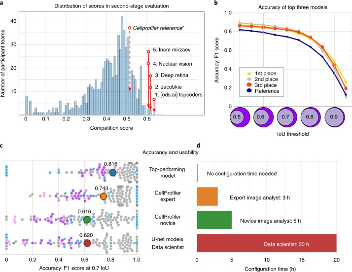
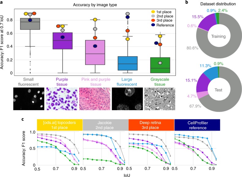
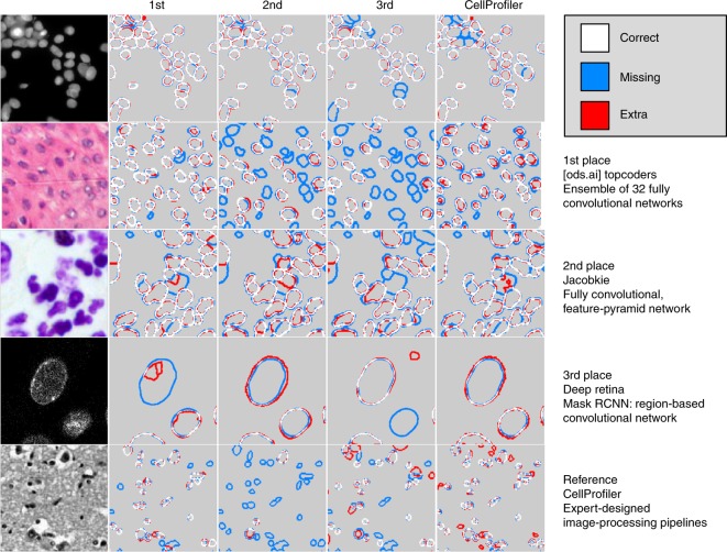
Segmenting the nuclei of cells in microscopy images is often the first step in the quantitative analysis of imaging data for biological and biomedical applications. Many bioimage analysis tools can segment nuclei in images but need to be selected and configured for every experiment. The 2018 Data Science Bowl attracted 3,891 teams worldwide to make the first attempt to build a segmentation method that could be applied to any two-dimensional light microscopy image of stained nuclei across experiments, with no human interaction. Top participants in the challenge succeeded in this task, developing deep-learning-based models that identified cell nuclei across many image types and experimental conditions without the need to manually adjust segmentation parameters. This represents an important step toward configuration-free bioimage analysis software tools.
Overview
The claims and statistics of this paper haven’t been checked yet.
Run the check — free, no account needed. It reads the open-access text and checks the sources it cites.
Integrity signals
Registry
Cites
Statistics
Figures
Verification
Claims the paper makes
The claims and statistics of this paper haven’t been checked yet.
Run the check — free, no account needed. It reads the open-access text and checks the sources it cites.
Claims other papers make about this one
Each is checked against this paper’s own text: does it say what the citing paper says it does?
The source backs part of the claim, not all of it — or backs it for a different population, condition or comparison.
In the citing paper
“However, we did not see an improvement for the ensemble of models compared to the generalist model (Extended Data Fig. 2), consistent with the results of ref. 27.”
From Cellpose 2.0: how to train your own model (2022) · cites this paper as [27]
Experimental — accuracy not yet measuredThe cited source says what the claim attributes to it.
In the citing paper
“Finally, we point out that the LiveCell study used a different average precision score from ours, which additionally requires a confidence score per ROI, while we used the average precision formulation from the Data Science Bowl challenge and other studies12,27,35.”
From Cellpose 2.0: how to train your own model (2022) · cites this paper as [27]
Self-check
Percentages, sums and reported differences re-computed from the paper’s own numbers.
The self-check needs the paper’s full text.
Citations
Cites
Deep Residual Learning for Image Recognition
He, K., Zhang, X., Ren, S., & Sun, J. (2016) · 2016 IEEE Conference on Computer Vision and Pattern Recognition (CVPR) · doi:10.1109/cvpr.2016.90 · Cited by 235,474
U-Net: Convolutional Networks for Biomedical Image Segmentation
Ronneberger, O., Fischer, P., & Brox, T. (2015) · Lecture notes in computer science · doi:10.1007/978-3-319-24574-4_28 · Cited by 99,404
LeCun, Y., Bengio, Y., & Hinton, G. (2015) · Nature · doi:10.1038/nature14539 · Cited by 84,126
Fiji: an open-source platform for biological-image analysis
Schindelin, J., Arganda-Carreras, I., Frise, E., Kaynig, V., Longair, M., Pietzsch, T., Preibisch, S., Rueden, C., Saalfeld, S., Schmid, B., et al. (2012) · Nature Methods · doi:10.1038/nmeth.2019 · Cited by 66,890
Microsoft COCO: Common Objects in Context
Lin, T.-Y., Maire, M., Belongie, S., Hays, J., Perona, P., Ramanan, D., Dollár, P., & Zitnick, C. L. (2014) · Lecture Notes in Computer Science · doi:10.1007/978-3-319-10602-1_48 · Cited by 45,460
A Threshold Selection Method from Gray-Level Histograms
Otsu, N. (1979) · IEEE Transactions on Systems, Man and Cybernetics · doi:10.1109/tsmc.1979.4310076 · Cited by 44,708
ImageNet Large Scale Visual Recognition Challenge
Russakovsky, O., Deng, J., Su, H., Krause, J., Satheesh, S., Ma, S., Huang, Z., Karpathy, A., Khosla, A., Bernstein, M., et al. (2015) · International Journal of Computer Vision · doi:10.1007/s11263-015-0816-y · Cited by 39,380
Feature Pyramid Networks for Object Detection
Lin, T.-Y., Dollar, P., Girshick, R., He, K., Hariharan, B., & Belongie, S. (2017) · 2017 IEEE Conference on Computer Vision and Pattern Recognition (CVPR) · doi:10.1109/cvpr.2017.106 · Cited by 30,939
He, K., Gkioxari, G., Dollar, P., & Girshick, R. (2017) · 2017 IEEE International Conference on Computer Vision (ICCV) · doi:10.1109/iccv.2017.322 · Cited by 29,137
Unpaired image-to-image translation using cycle-consistent adversarial networks
Zhu, J.-Y., Park, T., Isola, P., & Efros, A. A. (2017) · 2017 IEEE International Conference on Computer Vision (ICCV) · doi:10.1109/iccv.2017.244 · Cited by 23,602
Research works printed but not linked: 3
- Hollandi, R. et al. A deep learning framework for nucleus segmentation using image style transfer. Preprint at bioRxiv https://doi.org/10.1101/580605 (2019). · DOI
- Bolukbasi, T., Chang, K.-W., Zou, J. Y., Saligrama, V. & Kalai, A. T. in Advances in Neural Information Processing Systems (eds Lee, D. D. et al.) 4349–4357 (papers.nips.cc, 2016).
- Ren, S., He, K., Girshick, R. & Sun, J. in Advances in Neural Information Processing Systems 28 (eds. Cortes, C. et al.) 91–99 (Curran Associates, 2015).
Cited by
Sorted by citations
Items 601–700 of 811
A Multi-scale Cascaded Channel Attention Visual Mamba UNet model for medical image segmentation
Shi-li, W., Qiao, Y., Chao, Q., & Zhang, X. (2025) · Multimedia Systems · doi:10.1007/s00530-025-02090-z · Cited by 1
Yang, J., Pang, X., Lin, C., Wang, H., & Zhang, A. (2025) · Biomedical Signal Processing and Control · doi:10.1016/j.bspc.2025.109406 · Cited by 1
Egi, Y., Oter, T., Hajyzadeh, M., & Çatak, M. (2025) · Plants · doi:10.3390/plants15010047 · Cited by 1
AMAL-NET: A Lightweight Axial Attention-Based Model for Medical Image Segmentation
Yu, X., & Guo, L. (2025) · International Conference on New Trends in Computational Intelligence (NTCI) · doi:10.1109/ntci67886.2025.11308294 · Cited by 1
Abera, D. E., He, J., Liu, J., Zaki, N., & Qin, W. (2026) · Displays · doi:10.1016/j.displa.2025.103337 · Cited by 1
A Lightweight Modified Adaptive UNet for Nucleus Segmentation
Khan, M. R. K., Mohaidat, T., & Khalil, K. (2026) · Sensors · doi:10.3390/s26020665 · Cited by 1
Diffusion U-Net coupled with text-attention guided block for medical image segmentation
Li, J., Wang, C., Duan, P., & Li, J. (2026) · International Journal of Machine Learning and Cybernetics · doi:10.1007/s13042-025-02983-5 · Cited by 1
Shu, X., Shi, A., Guo, X., Fan, Y., & Zhang, X. M. (2026) · Applied Soft Computing · doi:10.1016/j.asoc.2026.114760 · Cited by 1
Protocol for automated analysis of biological images using Python code
Sacks, B., Mody, J., Huang, Y., Jung, S., Kimelman, R., Eubanks, E., Guadagno, E., Cevallos, J., Guntur, A. R., Suvvari, N. R., et al. (2026) · STAR Protocols · doi:10.1016/j.xpro.2026.104368 · Cited by 2
Image-based epigenetic profiling with deep learning and high-speed super-resolution microscopy
Wang, Y., Ghani, N. S. A., Dutta, M., Adachi, S., Katoh, K., Namihira, M., Mitsuyama, T., & Saito, Y. (2026) · Epigenetics & Chromatin · doi:10.1186/s13072-026-00662-5 · Cited by 1
Wang, H., Lu, Z., Ge, F., Yang, J., Li, H., & Yu, P. (2026) · Signal Image and Video Processing · doi:10.1007/s11760-026-05236-2 · Cited by 1
Genetic Programming Algorithm Evolving Robust Unary Costs for Efficient Graph Cut Segmentation
Mostafa, R. M., Mabrouk, E., Ayman, A., Zidan, H. Z., & Ibrahim, A. M. (2026) · Algorithms · doi:10.3390/a19040256 · Cited by 1
Cell Segmentation Beyond 2D—A Review of the State‐of‐the‐Art
Schmeisser, F., Muhammad, H., Younas, J., Lovell, G. F., Migliori, B., Dengel, A. R., & Ahmed, S. (2026) · Advanced Intelligent Discovery · doi:10.1002/aidi.202500206 · Cited by 1
CC-Net: A cross-hierarchical context-aware network for medical image segmentation
Zhang, X., Zhao, Z., Sun, W., Zhang, Y., Yu, C., & Meng, X. (2026) · Neural Networks · doi:10.1016/j.neunet.2026.108950 · Cited by 1
ColorSAM: teaching SAM to segment color medical images via quaternion decoding and prompt generation
Zhan, B., Wang, F., Song, E., & Zhang, R. (2026) · Expert Systems with Applications · doi:10.1016/j.eswa.2026.132857 · Cited by 1
Al-Hamadani, M. N. A., Szilágyi, P., Szemán-Nagy, G., Hajdú, A., & Harangi, B. (2026) · Array · doi:10.1016/j.array.2026.100946 · Cited by 1
Yang, L. T., & Yang, Z. (2026) · Journal of Translational Medicine · doi:10.1186/s12967-026-08577-1 · Cited by 1
Coughlan, M. F., Zhang, L., Perelman, R. T., Khan, U., Zhang, X., Upputuri, P. K., Zakharov, Y. N., Qiu, L., & Perelman, L. T. (2026) · Science Advances · doi:10.1126/sciadv.aei8554 · Cited by 1
ASW-Net: A Deep Learning-based Tool for Cell Nucleus Segmentation of Fluorescence Microscopy
Pan, W., Liu, Z., & Lin, G. N. (2021) · International Conference on Bioinformatics and Biomedical Science (ICBBS) · doi:10.1145/3498731.3498734 · Cited by 1
Hajiabadi, H., Hilbert, L., & Koziolek, A. (2022) · Euromicro Conference on Software Engineering and Advanced Applications (SEAA) · doi:10.1109/seaa56994.2022.00011 · Cited by 1
Bouilhol, E., Lefevre, E., Barry, T., Levet, F., Béghin, A., Viasnoff, V., Galindo, X., Galland, R., Sibarita, J., & Nikolski, M. (2023) · International Joint Conference on Biomedical Engineering Systems and Technologies (BIOSTEC) · doi:10.5220/0011623500003414 · Cited by 1
A Novel Approach for Nuclei Segmentation Using U-Net
Manju, P., Devassy, B. R., Rajan, V., & King, G. R. G. (2023) · International Conference on Networking and Communications (ICNWC) · doi:10.1109/icnwc57852.2023.10127290 · Cited by 1
Application and progress of highcontent imaging in molecular biology
Boyang, H., Yangyanqiu, W., Wenting, R., Chenxin, Y., Chu, J., Zhanbo, Q., Yanjun, Y., Qiang, Y., & Han, S. (2023) · Biotechnology Journal · doi:10.1002/biot.202300170 · Cited by 2
Chen, L., Wang, S. T., Wan, L., Su, J., & Wang, S. (2023) · IET Computer Vision · doi:10.1049/cvi2.12243 · Cited by 1
A comparative study for nuclei segmentation using latest deep learning optimizers
Eltayab, E. N., Al‐Atabany, W., Salem, N. M., & Mahmoud, L. N. (2023) · Novel Intelligent and Leading Emerging Sciences Conference (NILES) · doi:10.1109/niles59815.2023.10296591 · Cited by 1
Utz, J., Weise, T., Schlereth, M., Wagner, F., Thies, M., Gu, M., Uderhardt, S., & Breininger, K. (2023) · IEEE International Conference on Computer Vision Workshops · doi:10.1109/iccvw60793.2023.00417 · Cited by 1
Reinforcement learning for instance segmentation with high-level priors
Hilt, P., Zarvandi, M., Kaziakhmedov, E., Bhide, S., Laptin, M., Pape, C., & Kreshuk, A. (2023) · IEEE International Conference on Computer Vision Workshops · doi:10.1109/iccvw60793.2023.00423 · Cited by 1
TransCA: Transformer-Based Context Aggregator for Medical Image Segmentation
Sun, Y., Liu, Z., & Peng, X. (2023) · International Conference on Information Technology in Medicine and Education · doi:10.1109/itme60234.2023.00085 · Cited by 1
Centersam: Fully Automatic Prompt for Dense Nucleus Segmentation
Li, Y., Ren, H., Deng, J., Ma, X., & Xie, X. (2024) · IEEE International Symposium on Biomedical Imaging (ISBI) · doi:10.1109/isbi56570.2024.10635872 · Cited by 1
Borkar, K., Reen, A. S., Jawahar, C. V., & Arora, C. (2024) · Indian Conference on Computer Vision, Graphics and Image Processing · doi:10.1145/3702250.3702275 · Cited by 1
Seal, S., & Mukherjee, D. P. (2024) · International Conference on Computational Intelligence and Network Systems (CINS) · doi:10.1109/cins63881.2024.10864429 · Cited by 1
Zhang, X., Zhao, S., Liang, L., Yang, S., & Wang, Z. (2025) · Journal of Electronic Imaging · doi:10.1117/1.jei.34.3.033009 · Cited by 1
My Brother's Keeper: A Case Study in Evolutionary Biology and Animal Behavior.
Benson, K. E. (2004) · The journal of college science teaching · Cited by 1
Image processing of biofilms and its applications
Banerjee, S. (2022) · Elsevier eBooks · doi:10.1016/b978-0-323-88480-8.00012-1 · Cited by 1
[Medical nucleus image segmentation network based on convolution and attention mechanism].
Zhi, P., Deng, J., & Zhong, Z. (2022) · PubMed Central · doi:10.7507/1001-5515.202112013 · Cited by 1
A cellular segmentation algorithm with fast customization
(2022) · Nature Methods · doi:10.1038/s41592-022-01664-3 · Cited by 1
Automated Counting via Multicolumn Network and CytoSMART Exact FL Microscope
Flórez, S. L., González‐Briones, A., Hernández, G., & la Prieta, F. D. (2023) · Lecture notes in networks and systems · doi:10.1007/978-3-031-22356-3_20 · Cited by 1
Maylaa, T., Windal, F., Benhabiles, H., Maubon, G., Maubon, N., Vandenhaute, E., & Collard, D. (2023) · Lecture notes in networks and systems · doi:10.1007/978-981-19-7663-6_22 · Cited by 1
Incorporating Boundary Uncertainty into loss functions for biomedical image segmentationPreprint
Yeung, M., Yang, G., Sala, E., Schönlieb, C., & Rundo, L. (2021) · arXiv (Cornell University) · doi:10.48550/arxiv.2111.00533 · Cited by 3
Liqun, Z., Li, L., & Yang, G. (2022) · doi:10.36227/techrxiv.20188742.v1 · Cited by 1
GOAT: Deep learning-enhanced Generalized Organoid Annotation ToolPreprint
Bremer, J. P., Baumdick, M. E., Knorr, M. S., Wegner, L. H. M., Wesche, J., Jordan-Paiz, A., Jung, J. M., Highton, A. J., Jäger, J., Hinrichs, O., et al. (2022) · bioRxiv (Cold Spring Harbor Laboratory) · doi:10.1101/2022.09.06.506648 · Cited by 1
Bouilhol, E., Lefevre, E., Barry, T., Levet, F., Béghin, A., Viasnoff, V., Galindo, X., Galland, R., Sibarita, J., & Nikolski, M. (2022) · bioRxiv (Cold Spring Harbor Laboratory) · doi:10.1101/2022.10.27.514030 · Cited by 1
LIVECell---A large-scale dataset for label-free live cell segmentationPreprint
Khalid, N. (2023) · doi:10.36227/techrxiv.20375367.v1 · Cited by 1
Hong, B., Zeng, B., Feng, H., Liu, Z., Ni, Q., Wang, W., Li, M., Yang, M., Wang, M., Sun, L., et al. (2024) · bioRxiv (Cold Spring Harbor Laboratory) · doi:10.1101/2024.05.28.596350 · Cited by 1
MedSegBench: A Comprehensive Benchmark for Medical Image Segmentation in Diverse Data ModalitiesPreprint
Aydın, M., & Kuş, Z. (2024) · medRxiv · doi:10.1101/2024.08.26.24312619 · Cited by 1
Visualizing developmental dynamics of nuclear morphology and transport machinery in DrosophilaPreprint
Shindo, Y., Balachandra, S., & Amodeo, A. A. (2024) · bioRxiv (Cold Spring Harbor Laboratory) · doi:10.1101/2024.10.17.618964 · Cited by 1
Celldetective, an AI-enhanced image analysis tool for unraveling dynamic cell interactionsPreprint
Torro, R., Díaz-Bello, B., Arawi, D. E., Dervanova, K., Ammer, L., Dupuy, F., Chames, P., Sengupta, K., & Limozin, L. (2025) · eLife · doi:10.7554/elife.105302 · Cited by 3
Deep learning for microscopy without machine learning expertisePreprint
Zhou, X., & Wang, S. (2026) · bioRxiv (Cold Spring Harbor Laboratory) · doi:10.64898/2026.04.03.716415 · Cited by 1
A Decade of Deep Learning-based Biomedical Image SegmentationPreprint
Yu, S., Wang, H., Wang, N., Chen, S., Wu, J., Yuan, Z., Qi, T., Zhou, Z., Xia, F., Ma, J., et al. (2026) · bioRxiv (Cold Spring Harbor Laboratory) · doi:10.64898/2026.04.27.721127 · Cited by 1
Optimizing the Cell Painting assay for image-based profiling
Cimini, B. A., Chandrasekaran, S. N., Kost-Alimova, M., Miller, L., Goodale, A., Fritchman, B., Byrne, P., Garg, S., Jamali, N., Logan, D. J., et al. (2023) · Nature Protocols · doi:10.1038/s41596-023-00840-9 · Cited by 177
Self-citationMarx, V. (2019) · Nature Methods · doi:10.1038/s41592-019-0668-4
HistoFlow: Label-Efficient and Interactive Deep Learning Cell AnalysisPreprint
Henning, T., Bergner, B., & Lippert, C. (2020) · bioRxiv (Cold Spring Harbor Laboratory) · doi:10.1101/2020.07.15.204891
Deep Neural-Gas Clustering for Instance Segmentation across Imaging Experiments
Grüning, P., & Mamlouk, A. M. (2020) · International Joint Conference on Neural Networks (IJCNN) · doi:10.1109/ijcnn48605.2020.9207602
Spatially Dependent U-Nets: Highly Accurate Architectures for Medical Imaging SegmentationPreprint
Carvalho, J. B. S., Santinha, J., Miladinovic, D., & Buhmann, J. M. (2021) · arXiv (Cornell University) · doi:10.48550/arxiv.2103.11713
Inference with generalizable classifier predictionsPreprint
Evans, C., Weinberg, Z. Y., Puthenveedu, M. A., & G’Sell, M. (2021) · arXiv (Cornell University) · doi:10.48550/arxiv.2106.07623
A Size Adaptive Neural Network for Nucleus Segmentation
Shu, J., Gao, Q., Guan, Y., & Zhang, Q. (2021) · IEEE International Conference on Digital Signal Processing (DSP) · doi:10.1145/3458380.3458434
Multi-Modality Microscopy Image Style Transfer for Nuclei SegmentationPreprint
Liu, Y., Wagner, S. J., & Peng, T. (2021) · arXiv (Cornell University) · doi:10.48550/arxiv.2111.12138
Segmentation of Nucleus in Histopathological Images Using Deep Learning Architectures
Ayaz, O., Usta, H., & Bilgin, G. (2021) · Medical Technologies Congress (TIPTEKNO) · doi:10.1109/tiptekno53239.2021.9632996
Digital Image Analysis Tools Developed by the Indiana O’Brien Center
Dunn, K. W. (2021) · Frontiers in Physiology · doi:10.3389/fphys.2021.812170
Fully Automatic Cell Segmentation with Fourier DescriptorsPreprint
Hirling, D., & Horváth, P. (2021) · bioRxiv (Cold Spring Harbor Laboratory) · doi:10.1101/2021.12.17.472408
Automated Cell Segmentation for Phase-Contrast Images of Adhesion Cell Culture
Ye, G., & Kaya, M. (2021) · International Conference on Bio-engineering for Smart Technologies (BioSMART) · doi:10.1109/biosmart54244.2021.9677717
Applications of deep learning in single-cell analysis
Hollandi, R. (2021) · SZTE Repository of Dissertations (University of Szeged) · doi:10.14232/phd.10801
Local image fitting based active contour loss with deep learning for nuclei segmentation
Tran, V.-T. P. T. T.-T. (2022) · The University of Danang - Journal of Science and Technology · doi:10.31130/ud-jst.2022.100e
Efficient End-to-end Learning for Cell Segmentation with Machine Generated Incomplete AnnotationsPreprint
Shrestha, P., Kuang, N., & Yu, J. (2022) · bioRxiv (Cold Spring Harbor Laboratory) · doi:10.1101/2022.07.03.498609
Nguyen, V.-D., Nham, D.-H.-N., Pham, V.-T., & Tran, T.-T. (2022) · International Conference on Communications and Electronics (ICCE) · doi:10.1109/icce55644.2022.9852083
Tensor decomposition based networks for nuclei segmentation and classification
Chen, J., & Chen, Z. (2022) · Electronics Letters · doi:10.1049/ell2.12668
Fluorescence Microscopy Images Segmentation Based on Prototypical Networks with a Few Annotations
Guo, Y., Luo, Y., Li, W., & Yang, G. (2022) · Lecture notes in computer science · doi:10.1007/978-3-031-18910-4_14
Антонова, Е. И., Guskov, G., Yarushkina, N. G., Sapunkov, A., & Khambikova, A. (2023) · Studies in systems, decision and control · doi:10.1007/978-3-031-22938-1_43
Cell identification and phenotyping using classical machine learning and deep learning
Balassa, T. (2022) · Eötvös Loránd Tudományegyetem · doi:10.15476/elte.2021.207
Application of deep learning algorithms to single-cell segmentation and phenotypic profiling
Moshkov, N. (2022) · SZTE Repository of Dissertations (University of Szeged) · doi:10.14232/phd.11423
Evaluation of Cell Segmentation Using Pruning and Quantization
Polisi, X., Avdiu, D., Uka, A., Halili, A. N., Kollcaku, K., & Ciulla, C. (2023) · International Conference on Computing, Electronics & Communications Engineering (iCCECE) · doi:10.1109/iccece59400.2023.10238641
Collaborative region-boundary interaction network for medical image segmentation
Ta, N., Chen, H., Du, B., Wang, X., & Shi, Z. (2023) · Multimedia Tools and Applications · doi:10.1007/s11042-023-15505-8
Dasgupta, S., & Turban, S. (2021) · Journal of Emerging Investigators · doi:10.59720/21-032
LIVECell—A large-scale dataset for label-free live cell segmentationPreprint
Khalid, N. (2023) · doi:10.36227/techrxiv.20375367
NU-Net: a self-supervised smart filter for enhancing blobs in bioimages
Lim, S., Beaurepaire, E., & Chessel, A. (2023) · IEEE International Conference on Computer Vision Workshops · doi:10.1109/iccvw60793.2023.00420
Pseudotime analysis for time-series single-cell sequencing and imaging dataPreprint
Li, G., Kim, H., Pendyala, S., Zhang, R., Distèche, C. M., Vert, J., Deng, X., Fowler, D. M., & Noble, W. S. (2023) · bioRxiv (Cold Spring Harbor Laboratory) · doi:10.1101/2023.11.03.565575
The TYC Dataset for Understanding Instance-Level Semantics and Motions of Cells in Microstructures
Reich, C., Prangemeier, T., & Koeppl, H. (2023) · IEEE International Conference on Computer Vision Workshops · doi:10.1109/iccvw60793.2023.00426
Global and Local Aware Learning Network for Semi-Supervised Cell Image SegmentationPreprint
Zhao, L., Yang, J., Li, K., Ding, W., Hu, B., & Zhou, H. (2024) · SSRN Electronic Journal · doi:10.2139/ssrn.4681442
Li, Y., Wang, G., Li, Y., Ren, L., & Wang, H. (2024) · SSRN Electronic Journal · doi:10.2139/ssrn.4689418
Towards Automated Nanoenergetic Reaction Characterization with Computational Vision
Gaines, T., Hurt, J. A., Keller, J. M., Scott, G. J., Boyle, C., Maschmann, M. R., & Price, S. R. (2023) · IEEE Applied Imagery Pattern Recognition Workshop · doi:10.1109/aipr60534.2023.10440709
Noisy-label problems and datasets
Carneiro, G. (2024) · Elsevier eBooks · doi:10.1016/b978-0-44-315441-6.00011-3
(2024) · Elsevier eBooks · doi:10.1016/b978-0-44-315441-6.00016-2
Creating a universal cell segmentation algorithm
(2024) · Nature Methods · doi:10.1038/s41592-024-02254-1
Chen, A., Wu, L., Winfree, S., Dunn, K. W., Salama, P., & Delp, E. J. (2024) · SPIE Medical Imaging: Image Processing · doi:10.1117/12.3005440
Analysis of Energy-Efficient LCRM Optimization Algorithm in Computer Vision-based CNNs
Hussain, H., & Tamizharasan, P. S. (2024) · doi:10.1109/energycon58629.2024.10488814
Nartova, A. V., Matveev, A. V., Mashukov, M. Y., Belotserkovskii, V. A., Sankova, N. N., Kudinov, V. Y., & Okunev, A. G. (2023) · Kinetics and Catalysis · doi:10.31857/s045388112304010x
Active Learning Enabled Low-Cost Cell Image Segmentation Using Bounding Box AnnotationPreprint
Zhu, Y., Yang, Q., & Xu, L. (2024) · SSRN Electronic Journal · doi:10.2139/ssrn.4835620
Nuclei Segmentation Method based on CycleGAN-generated Images
Shu, J., Wang, Q., & Liu, S. (2023) · International Conference on Sensors, Signal and Image Processing (SSIP) · doi:10.1145/3653863.3653867
EDO-SANet: Shape-Aware Network with Edge Detection Operator for Polyp Segmentation
Zhao, Y., Wang, X., Li, Y., & Yin, J. (2024) · Asia Conference on Computer Vision, Image Processing and Pattern Recognition (CVIPPR) · doi:10.1145/3663976.3664239
Looverbosch, T. V. D., Beuckeleer, S. D., Smet, F. D., Sijbers, J., & Vos, W. H. D. (2024) · bioRxiv (Cold Spring Harbor Laboratory) · doi:10.1101/2024.07.18.602066
S3-TTA: Scale-Style Selection for Test-Time Augmentation in Biomedical Image Segmentation
Xie, K., Huang, S., Cajas, S. A., Pfister, H., & Wei, D. (2024) · IEEE International Symposium on Biomedical Imaging (ISBI) · doi:10.1109/isbi56570.2024.10635833
MGAU-ResNet-multi-grained attention U-shaped residual networks for medical image segmentation
Jing, W., & Sien, L. C. (2024) · AIP conference proceedings · doi:10.1063/5.0229757
ATU-NET: An Adaptive Transformation-Based U-NET for Medical Image Segmentation
Du, Q., Zhong, B., & Ma, K. (2024) · Proceedings - International Conference on Image Processing · doi:10.1109/icip51287.2024.10647750
Weng, W., & Zhu, X. (2024) · Lecture notes in computer science · doi:10.1007/978-3-031-72120-5_36
Elmeslimany, E. M., Kishk, S. S., & Altantawy, D. A. (2024) · SN Computer Science · doi:10.1007/s42979-024-03262-w
Image-based identification and isolation of micronucleated cells to dissect cellular consequencesPreprint
DiPeso, L., Pendyala, S., Huang, H. Z., Fowler, D. M., & Hatch, E. M. (2024) · eLife · doi:10.7554/elife.101579.1
LANet: Lightweight Attention Network for Medical Image Segmentation
Tang, Y., Pertsau, D., Zhao, D., Kupryianava, D., & Tatur, M. M. (2024) · Communications in computer and information science · doi:10.1007/978-3-031-73420-5_18
Learning Instance-Discriminative Pixel Embeddings Using Pixel Triplets
Chen, L., & Merhof, D. (2024) · Lecture notes in computer science · doi:10.1007/978-3-031-73284-3_29
A Lightweight-Based Architecture for Nuclei Segmentation in Histopathology Images
Pham, V.-T., Vu, N.-T., Pham, V.-T., & Tran, T.-T. (2024) · Lecture notes in networks and systems · doi:10.1007/978-3-031-74127-2_32
Metadata
- Authors
- Caicedo, J. C., Goodman, A., Karhohs, K. W., Cimini, B. A., Ackerman, J., Haghighi, M., Heng, C., Becker, T., Doan, M., McQuin, C., Rohban, M., Singh, S., & Carpenter, A. E.
- Affiliations
- Broad Institute (US)
- Corresponding author
- Carpenter, Anne E.
- ORCID
- Venue
- Nature Methods
- Published
- 2019-10-21
- Volume
- 16
- Issue
- 12
- Pages
- 1247-1253
- ISSN
- 1548-7091 (ISSN-L)
- 1548-7105
- PMC
- PMC6919559
- OpenAlex
- W2980998394
- Cited by
- 809
- References
- 49
- Citations by year
- 2026: 86 · 2025: 142 · 2024: 150 · 2023: 144 · 2022: 124 · 2021: 114 · 2020: 44 · 2019: 4
- FWCI
- 34.63
- Type
- journal article
- Language
- en
- Update notices
- correction (2020-01-22)
- Open access
- hybrid
- doi.org
- Locations
- Nature Methods (publishedVersion, cc-by)
- Europe PMC (PubMed Central) (publishedVersion, cc-by)
- PubMed Central (publishedVersion, cc-by)
- PubMed (publishedVersion)
- Funders
- Eli Lilly and Company
- Nvidia
- Broad Institute
- National Institutes of Health
- National Institute of General Medical Sciences (R35GM122547)
- Topics
- Life Sciences › Biochemistry, Genetics and Molecular Biology › Biophysics › Cell Image Analysis Techniques
- Physical Sciences › Computer Science › Computer Vision and Pattern Recognition › Digital Imaging for Blood Diseases
- Physical Sciences › Computer Science › Computer Vision and Pattern Recognition › Advanced Neural Network Applications
- Keywords
- nuclei segmentation, bioimage analysis, deep learning, microscopy image analysis, automated image analysis
- MeSH terms
- Data Science, Cell Nucleus, Humans, Image Processing, Computer-Assisted, Microscopy, Fluorescence
About these checks
Registry, retraction, Cites and statistics results are computed, not judged, with one exception: a printed reference that no index lists is read once by a model before it counts as a citation error. Claims are extracted by a model and kept only when their quote appears verbatim in the text; each cited source is then checked, with a model’s help, for whether it says what the claim relies on. Those verdicts can be wrong — read the claim’s quote and the cited source before relying on one.
Statistics and arithmetic are recomputed, not judged.
Confidence bands are shown only for fields calibrated on our annotated test set. None is calibrated yet, so no band is shown.
What counts as a claim ›How we measure confidence ›How we check this ›
Are you an author, or do you see a problem with this page? Request a review
