

The DOI matches its Crossref record; 29 of the 29 references it cites were found in a registry or an index.
LIVECell—A large-scale dataset for label-free live cell segmentation
Open accessAbstract
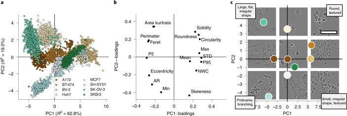
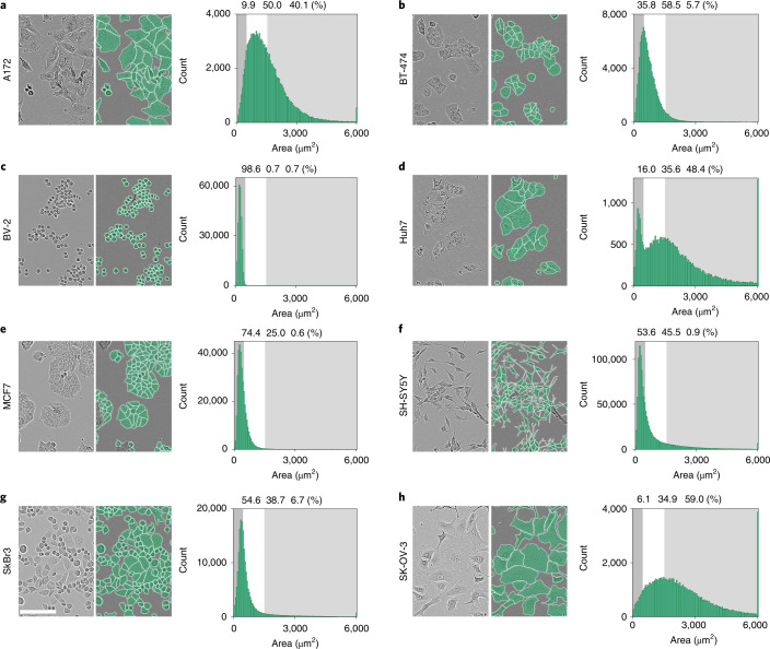
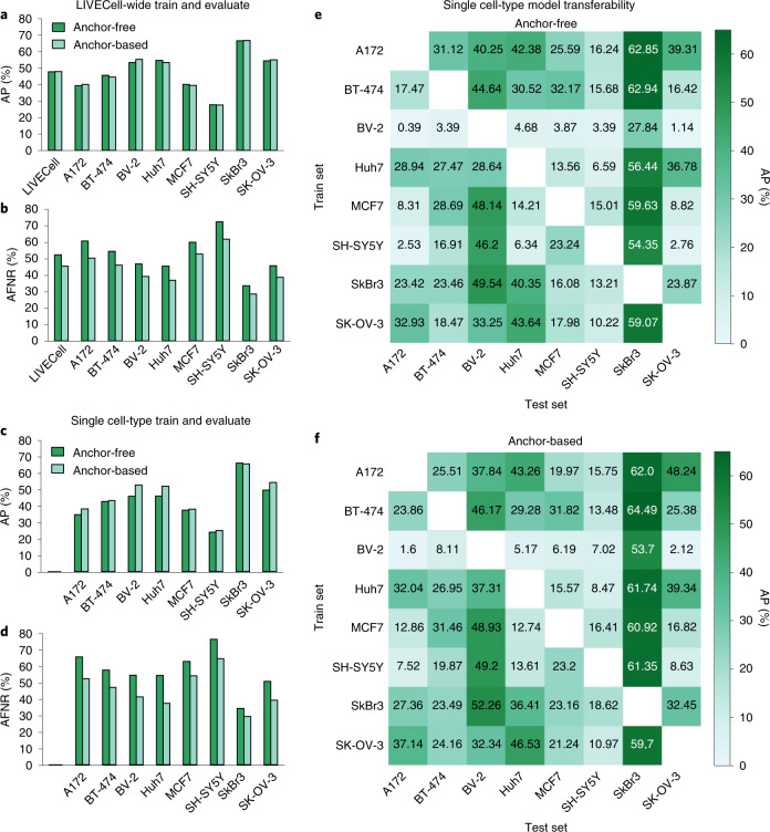
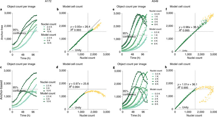
Light microscopy combined with well-established protocols of two-dimensional cell culture facilitates high-throughput quantitative imaging to study biological phenomena. Accurate segmentation of individual cells in images enables exploration of complex biological questions, but can require sophisticated imaging processing pipelines in cases of low contrast and high object density. Deep learning-based methods are considered state-of-the-art for image segmentation but typically require vast amounts of annotated data, for which there is no suitable resource available in the field of label-free cellular imaging. Here, we present LIVECell, a large, high-quality, manually annotated and expert-validated dataset of phase-contrast images, consisting of over 1.6 million cells from a diverse set of cell morphologies and culture densities. To further demonstrate its use, we train convolutional neural network-based models using LIVECell and evaluate model segmentation accuracy with a proposed a suite of benchmarks.
Overview
The claims and statistics of this paper haven’t been checked yet.
Run the check — free, no account needed. It reads the open-access text and checks the sources it cites.
Integrity signals
Registry
Cites
Statistics
Figures
Verification
Claims the paper makes
The claims and statistics of this paper haven’t been checked yet.
Run the check — free, no account needed. It reads the open-access text and checks the sources it cites.
Claims other papers make about this one
Each is checked against this paper’s own text: does it say what the citing paper says it does?
The source backs part of the claim, not all of it — or backs it for a different population, condition or comparison.
In the citing paper
“The LiveCell dataset consists of 3,188 training and 1,516 test images of eight different cell lines collected using phase-contrast microscopy, and each image has manual segmentations of the cells (https://sartorius-research.github.io/LIVECell/)7.”
From Cellpose 2.0: how to train your own model (2022) · cites this paper as [7]
Experimental — accuracy not yet measured
Self-check
Percentages, sums and reported differences re-computed from the paper’s own numbers.
The self-check needs the paper’s full text.
Citations
Cites
U-Net: Convolutional Networks for Biomedical Image Segmentation
Ronneberger, O., Fischer, P., & Brox, T. (2015) · Lecture notes in computer science · doi:10.1007/978-3-319-24574-4_28 · Cited by 99,404
An Image is Worth 16x16 Words: Transformers for Image Recognition at Scale
Dosovitskiy, A., Beyer, L., Kolesnikov, A., Weissenborn, D., Zhai, X., Unterthiner, T., Dehghani, M., Minderer, M., Heigold, G., Gelly, S., et al. (2020) · arXiv · doi:10.48550/arxiv.2010.11929 · Cited by 47,472
Microsoft COCO: Common Objects in Context
Lin, T.-Y., Maire, M., Belongie, S., Hays, J., Perona, P., Ramanan, D., Dollár, P., & Zitnick, C. L. (2014) · Lecture Notes in Computer Science · doi:10.1007/978-3-319-10602-1_48 · Cited by 45,460
PyTorch: An Imperative Style, High-Performance Deep Learning Library
Paszke, A., Gross, S., Massa, F., Lerer, A., Bradbury, J., Chanan, G., Killeen, T., Lin, Z., Gimelshein, N., Antiga, L., et al. (2019) · arXiv · doi:10.48550/arxiv.1912.01703 · Cited by 31,473
FCOS: Fully Convolutional One-Stage Object Detection
Tian, Z., Shen, C., Chen, H., & He, T. (2019) · 2019 IEEE/CVF International Conference on Computer Vision (ICCV) · doi:10.1109/iccv.2019.00972 · Cited by 7,013
CellProfiler: image analysis software for identifying and quantifying cell phenotypes
Carpenter, A. E., Jones, T. R., Lamprecht, M. R., Clarke, C., Kang, I. H., Friman, O., Guertin, D. A., Chang, J. H., Lindquist, R. A., Moffat, J., et al. (2006) · Genome biology · doi:10.1186/gb-2006-7-10-r100 · Cited by 5,212
Cellpose: a generalist algorithm for cellular segmentation
Stringer, C., Wang, T., Michaelos, M., & Pachitariu, M. (2021) · Nature Methods · doi:10.1038/s41592-020-01018-x · Cited by 4,369
Kuznetsova, A., Rom, H., Alldrin, N., Uijlings, J., Krasin, I., Pont-Tuset, J., Kamali, S., Popov, S., Malloci, M., Kolesnikov, A., et al. (2018) · arXiv (Cornell University) · doi:10.48550/arxiv.1811.00982 · Cited by 2,194
ResNeSt: Split-Attention Networks
Zhang, H., Wu, C., Zhang, Z., Zhu, Y., Lin, H., Zhang, Z., Sun, Y., He, T., Mueller, J. W., Manmatha, R., et al. (2022) · IEEE/CVF Conference on Computer Vision and Pattern Recognition Workshops (CVPRW) · doi:10.1109/cvprw56347.2022.00309 · Cited by 2,115
Cascade R-CNN: High Quality Object Detection and Instance Segmentation
Cai, Z., & Vasconcelos, N. M. (2019) · IEEE Transactions on Pattern Analysis and Machine Intelligence · doi:10.1109/tpami.2019.2956516 · Cited by 2,062
Research works printed but not linked: 1
- Levene, H. in Contributions to Probability and Statistics: Essays in Honor of Harold Hotelling (eds Olkin, I. et al.) 279–292 (Stanford Univ. Press, 1961).
Cited by
Sorted by citations
Items 101–200 of 304
Self-supervised learning using diverse cell images for cervical cancer classification
Hemalatha, K., & Vetriselvi, V. (2024) · Measurement · doi:10.1016/j.measurement.2024.116413 · Cited by 8
Nishimura, K., & Bise, R. (2023) · IEEE Winter Conference on Applications of Computer Vision · doi:10.1109/wacv56688.2023.00320 · Cited by 7
Initial refinement of data from video‐based single‐cell tracking
Korsnes, M. S., & Korsnes, R. (2023) · Cancer Innovation · doi:10.1002/cai2.88 · Cited by 6
Transfer Learning-Based Neuronal Cell Instance Segmentation With Pointwise Attentive Path Fusion
Li, G., & Xiao, Z. (2022) · IEEE Access · doi:10.1109/access.2022.3176956 · Cited by 7
Celldetective: an AI-enhanced image analysis tool for unraveling dynamic cell interactionsPreprint
Torro, R., Díaz-Bello, B., Arawi, D. E., Dervanova, K., Ammer, L., Dupuy, F., Chames, P., Sengupta, K., & Limozin, L. (2025) · eLife · doi:10.7554/elife.105302.1 · Cited by 9
Fiore, P., Terlizzi, A., Bardozzo, F., Lió, P., & Tagliaferri, R. (2025) · Computers in Biology and Medicine · doi:10.1016/j.compbiomed.2025.110274 · Cited by 6
Prompt-Tuning SAM: From Generalist to Specialist with Only 2,048 Parameters and 16 Training Images
Piater, T., Barz, B., & Freytag, A. (2025) · IEEE/CVF Conference on Computer Vision and Pattern Recognition Workshops (CVPRW) · doi:10.1109/cvprw67362.2025.00455 · Cited by 6
Improving flat fluorescence microscopy in scattering tissue through deep learning strategies
Feshki, M., Martel, S., Koninck, Y. D., & Gosselin, B. J. (2023) · Optics Express · doi:10.1364/oe.489677 · Cited by 6
Advances in QSAR through artificial intelligence and machine learning methods
Selvaraj, C., Elango, E., Prabhu, P., Velmurugan, D., & Singh, S. K. (2023) · Elsevier eBooks · doi:10.1016/b978-0-443-15339-6.00033-3 · Cited by 6
CLANet: A comprehensive framework for cross-batch cell line identification using brightfield images
Tong, L., Corrigan, A. M., Kumar, N. R., Hallbrook, K., Orme, J. P., Wang, Y., & Zhou, H. (2024) · Medical Image Analysis · doi:10.1016/j.media.2024.103123 · Cited by 6
Schilling, M. P., Faraj, R. E. K. E., Gómez, J. E. U., Sonnentag, S. J., Wang, F., Nestler, B., Orian‐Rousseau, V., Popova, A. A., Levkin, P. A., & Reischl, M. (2023) · Scientific Reports · doi:10.1038/s41598-023-32144-z · Cited by 6
Mallongi, A., Stang, Ernyasih, E., Palutturi, S., Rauf, A. U., Astuti, R. D. P., & Birawida, A. B. (2023) · Journal of Law and Sustainable Development · doi:10.55908/sdgs.v11i9.1048 · Cited by 6
Adversarial Attacks on Medical Segmentation Model via Transformation of Feature Statistics
Lee, W., Ju, M., Sim, Y., Jung, Y. K., Kim, T. H., & Kim, Y. (2024) · Applied Sciences · doi:10.3390/app14062576 · Cited by 6
Optimizing deep learning-based segmentation of densely packed cells using cell surface markers
Han, S., Phasouk, K., Zhu, J., & Fong, Y. (2024) · BMC Medical Informatics and Decision Making · doi:10.1186/s12911-024-02502-6 · Cited by 12
Liu, B., & Du, Y. (2024) · Computers in Biology and Medicine · doi:10.1016/j.compbiomed.2024.109456 · Cited by 6
A U-Net Based Progressive GAN for Microscopic Image Augmentation
Zhou, Q., & Yin, H. (2022) · Lecture notes in computer science · doi:10.1007/978-3-031-12053-4_34 · Cited by 6
Robust virtual staining of landmark organellesPreprint
Liu, Z., Hirata-Miyasaki, E., Pradeep, S., Rahm, J. V., Foley, C., Chandler, T., Ivanov, I. E., Woosley, H. O., Lao, T., Balasubramanian, A., et al. (2024) · bioRxiv (Cold Spring Harbor Laboratory) · doi:10.1101/2024.05.31.596901 · Cited by 6
Schurman, C. A., Bons, J., Kumaar, P. V. A., Fang, J., Roberts, A., Hormazabal, G. V., Riley, R. R., Tao, N., Verdin, E. M., & Schilling, B. (2025) · bioRxiv (Cold Spring Harbor Laboratory) · doi:10.1101/2025.05.25.655991 · Cited by 6
Automated image segmentation for accelerated nanoparticle characterization
Day, A. L., Wahl, C. B., dos Reis, R., Liao, W., Li, Y., Kilic, M. N. T., Mirkin, C. A., Dravid, V. P., Choudhary, A., & Agrawal, A. (2025) · Scientific Reports · doi:10.1038/s41598-025-01337-z · Cited by 5
Munck, J. D., Grootaert, C., Magdalenic, K., Deveci, D., Gansemans, Y., Nieuwerburgh, F. V., Boon, N., Skirtach, A., Rajkovic, A., D’hooghe, M., et al. (2025) · Biomedicine & Pharmacotherapy · doi:10.1016/j.biopha.2025.118926 · Cited by 5
Goyal, V., Schaub, N. J., Voss, T. C., & Hotaling, N. A. (2023) · BMC Bioinformatics · doi:10.1186/s12859-023-05486-8 · Cited by 6
Self-supervised learning for generalizable particle picking in cryo-EM micrographs
Zamanos, A., Koromilas, P., Bouritsas, G., Kastritis, P. L., & Panagakis, Y. (2025) · Cell Reports Methods · doi:10.1016/j.crmeth.2025.101089 · Cited by 4
FineBio: A Fine-Grained Video Dataset of Biological Experiments with Hierarchical Annotation
Yagi, T., Ohashi, M., Huang, Y., Furuta, R., Adachi, S., Mitsuyama, T., & Sato, Y. (2025) · International Journal of Computer Vision · doi:10.1007/s11263-025-02523-2 · Cited by 5
Nuclei-Net: a multi-stage fusion model for nuclei segmentation in microscopy images
Roy, K., Saha, S., Banik, D., & Bhattacharjee, D. (2023) · Innovations in Systems and Software Engineering · doi:10.1007/s11334-023-00537-y · Cited by 5
SEG: Segmentation Evaluation in absence of Ground truth labelsPreprint
Sims, Z., Strgar, L., Thirumalaisamy, D., Heussner, R. T., Thibault, G., & Chang, Y. H. (2023) · bioRxiv (Cold Spring Harbor Laboratory) · doi:10.1101/2023.02.23.529809 · Cited by 8
Benchmarking cellular segmentation methods against CellposePreprint
Stringer, C., & Pachitariu, M. (2024) · bioRxiv (Cold Spring Harbor Laboratory) · doi:10.1101/2024.04.06.587952 · Cited by 9
Hasan, E., Fuad, M. T. H., Sharif, O., & Wagler, A. E. (2026) · Journal of Imaging · doi:10.3390/jimaging12030098 · Cited by 4
Molina-Moreno, M., Schilling, M. P., Reischl, M., & Mikut, R. (2025) · IEEE Access · doi:10.1109/access.2025.3555020 · Cited by 4
A Deep Learning Multimodal Fusion-Based Method for Cell and Nucleus Segmentation
Shen, B., Gu, Z., Zhou, J., Yan, B., Fang, Y., & Wang, H. (2025) · IEEE Transactions on Medical Imaging · doi:10.1109/tmi.2025.3592625 · Cited by 5
Deckers, T., Aerts, J.-M., & Bloemen, V. (2022) · Scientific Reports · doi:10.1038/s41598-022-22561-x · Cited by 4
Optical microscopic imaging, manipulation, and analysis methods for morphogenesis research
Katoh, T. A., Fukai, Y. T., & Ishibashi, T. (2023) · Microscopy · doi:10.1093/jmicro/dfad059 · Cited by 5
Celldetective, an AI-enhanced image analysis tool for unraveling dynamic cell interactions
Torro, R., Díaz-Bello, B., Arawi, D. E., Dervanova, K., Ammer, L., Dupuy, F., Chames, P., Sengupta, K., & Limozin, L. (2026) · eLife · doi:10.7554/elife.105302.3 · Cited by 4
Owen, R., Nasir, A., Amer, M. H., Nie, C., Xue, X., Burroughs, L., Denning, C. N., Wildman, R. D., Khan, F. A., Alexander, M. R., et al. (2024) · Advanced Intelligent Systems · doi:10.1002/aisy.202400573 · Cited by 4
A scalable, data analytics workflow for image-based morphological profiles
Forsgren, E., Cloarec, O., Jonsson, P., Lovell, G. F., & Trygg, J. (2024) · Chemometrics and Intelligent Laboratory Systems · doi:10.1016/j.chemolab.2024.105232 · Cited by 4
Self-citationEfficient segmentation algorithm for complex cellular image analysis system
Liu, Y., Wang, C., Wen, Y., Huo, Y., & Liu, J. (2023) · IET Control Theory and Applications · doi:10.1049/cth2.12466 · Cited by 4
Increasing the sensitivity, recovery, and integrality of spatially resolved proteomics by LCM-MTAPreprint
Gu, L., Li, X., Li, Z., Wang, Q.-Q., Zheng, K., Yu, G., Dai, C., Li, J., Zhao, B., Zhang, H., et al. (2022) · bioRxiv (Cold Spring Harbor Laboratory) · doi:10.1101/2022.08.21.504675 · Cited by 4
Spatial omics of acute myocardial infarction reveals a novel mode of immune cell infiltrationPreprint
Wünnemann, F., Sicklinger, F., Bestak, K., Nimo, J., Thiemann, T., Amrute, J., Nordbeck, M., Hartmann, N., Ibarra-Arellano, M. A., Tanevski, J., et al. (2024) · bioRxiv (Cold Spring Harbor Laboratory) · doi:10.1101/2024.05.20.594955 · Cited by 4
Dong, Y., Saglietti, C., Bayard, Q., Espín-Pérez, A., Carpentier, S., Buszta, D., Tissot-Renaud, S., Dubois, R., Kamburov, A., Kang, S., et al. (2024) · bioRxiv (Cold Spring Harbor Laboratory) · doi:10.1101/2024.11.01.621259 · Cited by 4
CellSpot: Deep Learning-Based Efficient Cell Center Detection in Microscopic Images
Khalid, N., Caroprese, M., Lovell, G. F., Trygg, J., Dengel, A. R., & Ahmed, S. (2024) · Lecture notes in computer science · doi:10.1007/978-3-031-72353-7_16 · Cited by 3
Self-citationPrytula, Y., Tsiporenko, I., Zeynalli, A., & Fishman, D. (2025) · IEEE/CVF Conference on Computer Vision and Pattern Recognition Workshops (CVPRW) · doi:10.1109/cvprw67362.2025.00460 · Cited by 4
Systematic evaluation of computational methods for cell segmentation
Yang, R., Xue, G., Wang, Z., Cai, Y., Yang, W., Que, J., Tan, R., Sun, H., Wang, P., Xu, Z., et al. (2026) · Briefings in Bioinformatics · doi:10.1093/bib/bbag066 · Cited by 4
Huynh, H. N., Ha, Q. T., Tran, A. T., Tran, H. D. T., Bui, P. A., & Tran, T. N. (2022) · IEEE Eurasia Conference on Biomedical Engineering, Healthcare and Sustainability (ECBIOS) · doi:10.1109/ecbios54627.2022.9945050 · Cited by 3
IDCIA: Immunocytochemistry Dataset for Cellular Image Analysis
Mohammed, A. A., Fonder, C., Sakaguchi, D. S., Tavanapong, W., Mallapragada, S. K., & Idris, A. (2023) · ACM Multimedia Systems Conference (MMSys) · doi:10.1145/3587819.3592558 · Cited by 3
Quantitative Evaluation of Neurite Morphology Using Graph Structure
Chikudo, F., Baar, S., Ota, A., Kuragano, M., Tokuraku, K., & Watanabe, S. (2023) · Electronics · doi:10.3390/electronics12234750 · Cited by 3
DLBCL cells with ferroptosis morphology can be detected with a deep convolutional neural network
Kotkaranta, P. H., Chan, M. C., Vuolio, T., Miinalainen, I., Kuitunen, H. K., Turpeenniemi-Hujanen, T., Teppo, H., Kuittinen, O. M., & Kuusisto, M. E. L. (2024) · Biomedicine & Pharmacotherapy · doi:10.1016/j.biopha.2024.117785 · Cited by 4
A quality assessment algorithm for no-reference images based on transfer learning
Yang, Y., Liu, C., Wu, H., & Yu, D. (2025) · PeerJ Computer Science · doi:10.7717/peerj-cs.2654 · Cited by 3
Zhang, T., McCourty, H., Sánchez-Tafolla, B. M., Nikolaev, A., & Mihaylova, L. S. (2025) · Neurocomputing · doi:10.1016/j.neucom.2025.130511 · Cited by 4
CUPre: Cross-domain Unsupervised Pre-training for few-shot cell segmentation
Liao, W., Li, X., Wang, Q., Xu, Y., Yin, Z., & Xiong, H. (2025) · Information Fusion · doi:10.1016/j.inffus.2025.103641 · Cited by 5
Cell-APP: A generalizable method for cell annotation and cell-segmentation model training
Virdi, A. J., & Joglekar, A. P. (2025) · Molecular Biology of the Cell · doi:10.1091/mbc.e25-02-0076 · Cited by 3
Probabilistic Domain Adaptation for Biomedical Image Segmentation
Archit, A., & Pape, C. (2025) · IEEE International Conference on Computer Vision Workshops · doi:10.1109/iccvw69036.2025.00603 · Cited by 3
A pH Monitoring Algorithm for Orifice Plate Culture Medium
Li, Y., Huang, A., Zhang, T., Wen, L., Shi, Z., & Shi, L. (2022) · Applied Sciences · doi:10.3390/app12157560 · Cited by 3
Jiang, J., Zeng, Z., Xu, J., Wang, W., Shi, B., Lan, Z., Chen, Y., Yao, W., Wang, Y., & Zhang, H. (2023) · Heliyon · doi:10.1016/j.heliyon.2023.e20181 · Cited by 3
Lboukili, I., Stamatas, G. N., & Descombes, X. (2024) · Journal of Biomedical Optics · doi:10.1117/1.jbo.29.8.086003 · Cited by 3
aiSEGcell: User-friendly deep learning-based segmentation of nuclei in transmitted light images
Schirmacher, D., Armagan, Ü., Zhang, Y., Kull, T., Auler, M. M. K., & Schroeder, T. (2024) · PLoS Computational Biology · doi:10.1371/journal.pcbi.1012361 · Cited by 3
Occlusion Segmentation: Restore and Segment Invisible Areas for Particle Objects
Liu, J., Jiang, Z., Gui, W., Chen, Z., & Zhang, C. (2024) · IEEE Transactions on Automation Science and Engineering · doi:10.1109/tase.2024.3450900 · Cited by 4
Feature Enhanced Semi-Supervised Attention U-Net for Cell Segmentation
Raj, A. S., Asha, S. B., & Gopakumar, G. (2024) · International Conference on Computing, Communication and Networking Technologies (ICCCNT) · doi:10.1109/icccnt61001.2024.10726083 · Cited by 3
BCSnet: A U-Net-Based Model for Segmentation of Brain Cells in Trypan Blue Images
Kudryavtsev, A., Simkin, I. V., Dragun, M. A., Alexandrova, O. P., Malashin, I. P., Sukhanov, D. A., Nelyub, V., Бородулин, А. С., Yurchenko, S. O., & Тынченко, В. С. (2024) · IEEE Access · doi:10.1109/access.2024.3519893 · Cited by 3
Seiffarth, J., Blöbaum, L., Paul, R. D., Friederich, N., Sitcheu, A. J. Y., Mikut, R., Scharr, H., Grünberger, A., & Nöh, K. (2025) · Lecture notes in computer science · doi:10.1007/978-3-031-91721-9_20 · Cited by 3
Characterization of regeneration initiating cells during Xenopus laevis tail regenerationPreprint
Radek Sindelka, E., Abaffy, P., Žucha, D., Naraine, R., Daniel, K., Jiri, N., Smetana, K., Lacina, L., Endaya, B., Neužil, J., et al. (2023) · bioRxiv (Cold Spring Harbor Laboratory) · doi:10.1101/2023.03.30.534908 · Cited by 3
Optimizing Deep Learning-Based Segmentation of Densely Packed Cells using Cell Surface MarkersPreprint
Han, S., Phasouk, K., Zhu, J., & Fong, Y. (2023) · Research Square · doi:10.21203/rs.3.rs-3307496/v1 · Cited by 3
Pizzagalli, D. U., Carrillo-Barberà, P., Palladino, E., Ceni, K., Thelen, B., Pulfer, A., Moscatello, E., Cabini, R. F., Textor, J. C., Wortel, I. M. N., et al. (2024) · bioRxiv (Cold Spring Harbor Laboratory) · doi:10.1101/2024.12.02.626343 · Cited by 3
Сергеевич, В. Н., Putlyaev, E., Сергеевна, Ч. Е., Usachev, V., Karagyaur, M. N., Дмитриевич, Б. К., Grigorieva, O., Tyurin‐Kuzmin, P. A., & Kulebyakin, K. Y. (2025) · bioRxiv (Cold Spring Harbor Laboratory) · doi:10.1101/2025.05.13.653705 · Cited by 3
Bounding Box Is All You Need: Learning to Segment Cells in 2D Microscopic Images via Box Annotations
Khalid, N., Caroprese, M., Lovell, G. F., Porto, D. A., Trygg, J., Dengel, A. R., & Ahmed, S. (2024) · Lecture notes in computer science · doi:10.1007/978-3-031-66955-2_22 · Cited by 2
Self-citationValentino, M., D’Agostino, M., Licitra, F., Pirone, D., Vitale, F., Micco, A. D., Pavone, L. M., Miccio, L., Bianco, V., & Ferraro, P. (2025) · Biomedical Optics Express · doi:10.1364/boe.566028 · Cited by 2
Deep Multi-Scale and Attention-Based Architectures for Semantic Segmentation in Biomedical Imaging
Harouni, M., Goyal, V., Feldman, G., Michael, S., & Voss, T. C. (2025) · Computers, materials & continua/Computers, materials & continua (Print) · doi:10.32604/cmc.2025.067915 · Cited by 5
HusMorph: a simple machine learning app for automated morphometric landmarking
Kristiansen, H. H., Metz, M., Silva‐Garay, L., Jutfelt, F., & Leeuwis, R. H. J. (2025) · Conservation Physiology · doi:10.1093/conphys/coaf073 · Cited by 3
Altendorfer, S., Walker, S. J., & Daub, C. O. (2026) · International Journal of Cancer · doi:10.1002/ijc.70733 · Cited by 2
Cell segmentation in microscopy images using a SAM-based U-Net architecture and a novel dataset
Alam, M. S., Jackson, M., Lord, M., & Meijering, E. H. W. (2026) · Computer Methods and Programs in Biomedicine · doi:10.1016/j.cmpb.2026.109470 · Cited by 2
Wu, Y., Spies, S., Bo, Q., Zhang, Y., Qi, J., Wesseling, P., Groot, M. L., & Zhang, A. (2026) · Matter & Light · doi:10.1016/j.matlit.2026.100044 · Cited by 2
Brain Cell Segmentation from the LIVECell Dataset Using Deep Learning
Huynh, H. N., Tran, A. T., Tran, H. D. T., & Tran, T. N. (2023) · World Congress on Medical Physics and Biomedical Engineering, September 7 - 12, 2009, Munich, Germany · doi:10.1007/978-3-031-44630-6_40 · Cited by 2
Schilling, M. P., Klinger, L., Schumacher, U., Schmelzer, S., López, M. B., Nestler, B., & Reischl, M. (2023) · Annual International Conference of the IEEE Engineering in Medicine and Biology Society (EMBC) · doi:10.1109/embc40787.2023.10341074 · Cited by 2
DCSN: A Flexible and Efficient Lightweight Network for Dense Cell Segmentation
Pan, F., & Wang, F. (2023) · IEEE International Conference on Medical Artificial Intelligence (MedAI) · doi:10.1109/medai59581.2023.00052 · Cited by 2
Gil, E. M., Steelman, Z. A., Владимировна, С. А., & Bixler, J. N. (2022) · Quantitative Phase Imaging · doi:10.1117/12.2608770 · Cited by 2
Automated Single Particle Growth Measurement using Segmentation
Rafique, M. A., Hussain, M. I., Hassan, M. A., Jung, W., Kim, B., & Jeon, M. (2022) · IEEE International Conference on Advanced Video and Signal Based Surveillance (AVSS) · doi:10.1109/avss56176.2022.9959296 · Cited by 2
Deep Learning Empowered Fresnel-based Lensless Fluorescence Microscopy*
Feshki, M., Koninck, Y. D., & Gosselin, B. J. (2023) · Annual International Conference of the IEEE Engineering in Medicine and Biology Society (EMBC) · doi:10.1109/embc40787.2023.10339990 · Cited by 2
Du, Y., Liu, B., Chen, H., & Fu, Y. (2024) · Biomedical Signal Processing and Control · doi:10.1016/j.bspc.2024.105973 · Cited by 2
Chu, H., Wang, K., Cheng, H., Ma, W., Dong, L., Gou, Y., Yang, J., & Cai, H. (2023) · bioRxiv (Cold Spring Harbor Laboratory) · doi:10.1101/2023.04.20.537419 · Cited by 2
Point2Mask: A Weakly Supervised Approach for Cell Segmentation Using Point AnnotationPreprint
Khalid, N. (2023) · doi:10.36227/techrxiv.20368959.v1 · Cited by 2
Self-citation(2025) · Nature Methods · doi:10.1038/s41592-025-02759-3 · Cited by 2
Deep Learning-Based Multi-task Approach for Neuronal Cells Classification and Segmentation
Belghiti, K. A., Mikram, M., Rhanoui, M., & Yousfi, S. (2023) · Lecture notes in networks and systems · doi:10.1007/978-981-99-3243-6_25 · Cited by 1
A Perspective on Developing Modeling and Image Analysis Tools to Investigate Mechanosensing Proteins
Ouderkirk, S. N., Sedley, A., Ong, M., Shifflet, M. R., Harkrider, Q. C., Wright, N. T., & Miller, C. J. (2023) · Integrative and Comparative Biology · doi:10.1093/icb/icad107 · Cited by 1
Khalid, N., Koochali, M., Lesmes-Leon, D. N., Caroprese, M., Lovell, G. F., Porto, D. A., Trygg, J., Dengel, A. R., & Ahmed, S. (2024) · Lecture notes in computer science · doi:10.1007/978-3-031-66955-2_27 · Cited by 1
Self-citationNguyen, T.-N.-T., Ong, X.-H., Hoang-Thien-Nguyen, Bui, V.-H., Vu, H.-N., Nguyen, M. T., Nguyen, Q., & Nguyen, H.-H. (2025) · IEEE International Conference on Image Processing, Applications and Systems (IPAS) · doi:10.1109/ipas63548.2025.10924518 · Cited by 1
Representation of high-dimensional cell morphology and morphodynamics in 2D latent space
Cunningham, C. T., & Sun, B. (2025) · Physical Biology · doi:10.1088/1478-3975/adcd37 · Cited by 1
Leveraging foundation models and goal-dependent annotations for automated cell confluence assessment
Joas, M. J., Freund, D., Haase, R., Rahm, E., & Ewald, J. (2025) · Frontiers in Computer Science · doi:10.3389/fcomp.2025.1562358 · Cited by 1
CellStyle: Improved Zero-Shot Cell Segmentation via Style Transfer
Yilmaz, R., Chen, Z., Wu, Y., & Stegmaier, J. (2025) · Lecture notes in computer science · doi:10.1007/978-3-032-04971-1_7 · Cited by 1
Challenges in explaining deep learning models for data with biological variation
Tětková, L., Dreier, E. S., Malm, R., & Hansen, L. K. (2025) · PLoS ONE · doi:10.1371/journal.pone.0333965 · Cited by 1
Advances in deep learning for bacterial image segmentation in optical microscopy
Tan, Z., Ding, Y., Ma, H., Li, J., Zheng, D., Bai, H., Xin, W., Li, L., & Peng, B. (2025) · Journal of Innovative Optical Health Sciences · doi:10.1142/s1793545825300113 · Cited by 1
de Oliveira, H. Q., da Rosa, S. A., Pereira, L. C., de Mendonça, L. B., Scholl, J. N., Figueiró, F., Bitencourt, T. C., Echevarria, D. P., Buzzo, J. L. A., D'Agostin, J. B., et al. (2025) · FEBS Journal · doi:10.1111/febs.70339 · Cited by 1
A weakly supervised framework for automated biological assay assessment
Jiang, H., Guo, Q., Zhi, X., Li, H., & Chen, Y. (2025) · Virus Research · doi:10.1016/j.virusres.2025.199677 · Cited by 1
Huynh, S. T., Nguyen, H. X., Duong, D. Q., Nguyen, T. T., Nguyen, N. K. P., Nguyen, H. T. L., Vu, P. N., Nguyen, N. M., Vu, K. D., Ho, H. Q. N., et al. (2025) · IEEE Journal of Selected Topics in Signal Processing · doi:10.1109/jstsp.2025.3648286 · Cited by 1
The time dimension matters: Improving mode of action classification with live-cell imaging
Forsgren, E., Rietdijk, J., Holmberg, D., Juneblad, J., Migliori, B., Johansson, M. M., Puigvert, J. C., Trygg, J., Lovell, G. F., Spjuth, O., et al. (2025) · Artificial Intelligence in the Life Sciences · doi:10.1016/j.ailsci.2025.100152 · Cited by 1
Self-citationLeveraging AI for cell biology discovery
Simizo, A., de Morais, M., Vesco, M. F. [., & Nakaya, H. I. (2026) · Biochemical Society Transactions · doi:10.1042/bst20253023 · Cited by 1
Hos, Y. E., & Cicek, G. (2026) · Springer eBooks · doi:10.1007/978-3-032-07267-2_14 · Cited by 1
Deng, P., Jang, D., Berryman, S. G., Duffy, S. P., & Ma, H. (2026) · Small Methods · doi:10.1002/smtd.202501369 · Cited by 1
Cell Segmentation Beyond 2D—A Review of the State‐of‐the‐Art
Schmeisser, F., Muhammad, H., Younas, J., Lovell, G. F., Migliori, B., Dengel, A. R., & Ahmed, S. (2026) · Advanced Intelligent Discovery · doi:10.1002/aidi.202500206 · Cited by 1
Self-citationAl-Hamadani, M. N. A., Szilágyi, P., Szemán-Nagy, G., Hajdú, A., & Harangi, B. (2026) · Array · doi:10.1016/j.array.2026.100946 · Cited by 1
Yang, L. T., & Yang, Z. (2026) · Journal of Translational Medicine · doi:10.1186/s12967-026-08577-1 · Cited by 1
Gao, Z., Ge, J., Lu, J., Wu, L., Clayton, H., Zhang, D., Liu, J., Machado, I. P., Chen, Y., Crispin‐Ortuzar, M., et al. (2026) · Intelligent Oncology · doi:10.1016/j.intonc.2026.100074 · Cited by 1
Metadata
- Authors
- Edlund, C., Jackson, T. R., Khalid, N., Bevan, N., Dale, T., Dengel, A., Ahmed, S., Trygg, J., & Sjögren, R.
- Affiliations
- Sartorius (United Kingdom) (GB)
- German Research Centre for Artificial Intelligence (DE)
- Umeå University (SE)
- Corresponding author
- Sjögren, Rickard
- ORCID
- Venue
- Nature Methods
- Published
- 2021-08-30
- Volume
- 18
- Issue
- 9
- Pages
- 1038-1045
- ISSN
- 1548-7091 (ISSN-L)
- 1548-7105
- PMC
- PMC8440198
- OpenAlex
- W3196890347
- Cited by
- 304
- References
- 29
- Citations by year
- 2026: 56 · 2025: 88 · 2024: 63 · 2023: 65 · 2022: 31 · 2021: 1
- FWCI
- 18.09
- Type
- journal article
- Language
- en
- Open access
- hybrid
- doi.org
- Locations
- Nature Methods (publishedVersion, cc-by)
- Europe PMC (PubMed Central) (publishedVersion, other-oa)
- PubMed Central (publishedVersion, cc-by)
- PubMed (publishedVersion)
- DiVA at Umeå University (Umeå University) (submittedVersion, other-oa)
- Topics
- Life Sciences › Biochemistry, Genetics and Molecular Biology › Biophysics › Cell Image Analysis Techniques
- Physical Sciences › Computer Science › Computer Vision and Pattern Recognition › Advanced Neural Network Applications
- Physical Sciences › Computer Science › Computer Vision and Pattern Recognition › Medical Image Segmentation Techniques
- Keywords
- cell segmentation, label-free imaging, phase contrast microscopy, convolutional neural networks, segmentation benchmark, cell culture, high-throughput imaging, cell morphology
- MeSH terms
- Humans, Image Processing, Computer-Assisted, Microscopy, Models, Biological, Databases, Factual, Neural Networks, Computer, Cell Culture Techniques
About these checks
Registry, retraction, Cites and statistics results are computed, not judged, with one exception: a printed reference that no index lists is read once by a model before it counts as a citation error. Claims are extracted by a model and kept only when their quote appears verbatim in the text; each cited source is then checked, with a model’s help, for whether it says what the claim relies on. Those verdicts can be wrong — read the claim’s quote and the cited source before relying on one.
Statistics and arithmetic are recomputed, not judged.
Confidence bands are shown only for fields calibrated on our annotated test set. None is calibrated yet, so no band is shown.
What counts as a claim ›How we measure confidence ›How we check this ›
Are you an author, or do you see a problem with this page? Request a review
