

The DOI matches its Crossref record; 29 of the 29 references it cites were found in a registry or an index.
LIVECell—A large-scale dataset for label-free live cell segmentation
Open accessAbstract
Light microscopy combined with well-established protocols of two-dimensional cell culture facilitates high-throughput quantitative imaging to study biological phenomena. Accurate segmentation of individual cells in images enables exploration of complex biological questions, but can require sophisticated imaging processing pipelines in cases of low contrast and high object density. Deep learning-based methods are considered state-of-the-art for image segmentation but typically require vast amounts of annotated data, for which there is no suitable resource available in the field of label-free cellular imaging. Here, we present LIVECell, a large, high-quality, manually annotated and expert-validated dataset of phase-contrast images, consisting of over 1.6 million cells from a diverse set of cell morphologies and culture densities. To further demonstrate its use, we train convolutional neural network-based models using LIVECell and evaluate model segmentation accuracy with a proposed a suite of benchmarks.
Overview
The claims and statistics of this paper haven’t been checked yet.
Run the check — free, no account needed. It reads the open-access text and checks the sources it cites.
Integrity signals
Registry
Cites
Statistics
Figures
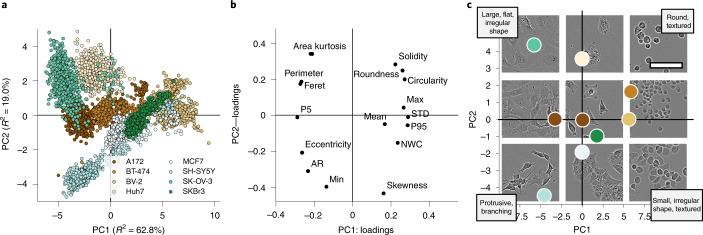
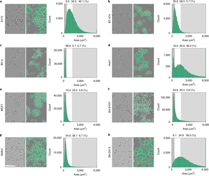
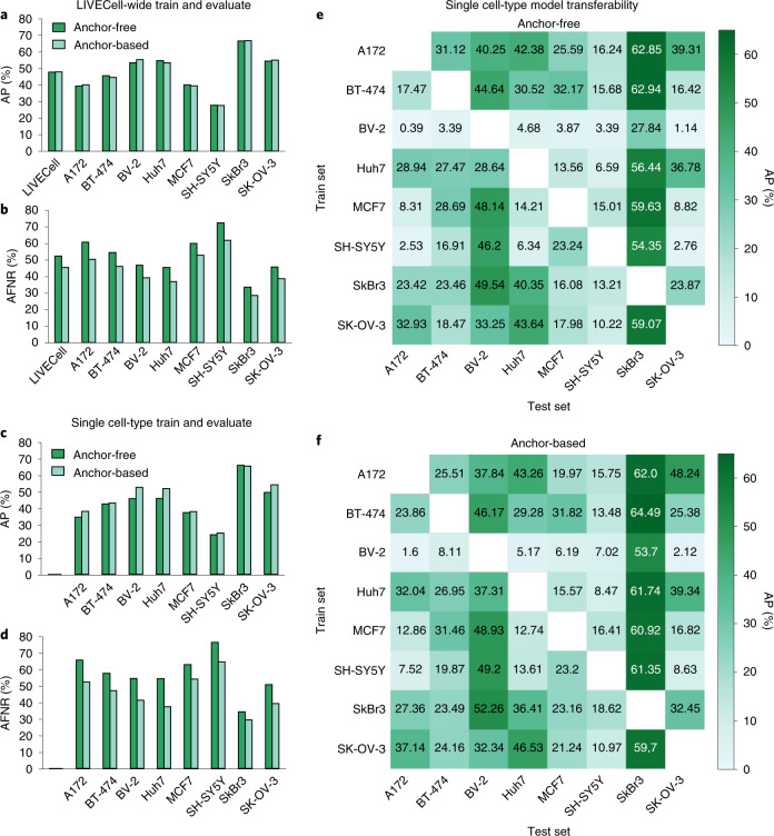
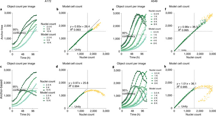
Verification
Claims the paper makes
The claims and statistics of this paper haven’t been checked yet.
Run the check — free, no account needed. It reads the open-access text and checks the sources it cites.
Claims other papers make about this one
Each is checked against this paper’s own text: does it say what the citing paper says it does?
The source backs part of the claim, not all of it — or backs it for a different population, condition or comparison.
In the citing paper
“The LiveCell dataset consists of 3,188 training and 1,516 test images of eight different cell lines collected using phase-contrast microscopy, and each image has manual segmentations of the cells (https://sartorius-research.github.io/LIVECell/)7.”
From Cellpose 2.0: how to train your own model (2022) · cites this paper as [7]
Experimental — accuracy not yet measured
Self-check
Percentages, sums and reported differences re-computed from the paper’s own numbers.
The self-check needs the paper’s full text.
Citations
Cites
U-Net: Convolutional Networks for Biomedical Image Segmentation
Ronneberger, O., Fischer, P., & Brox, T. (2015) · Lecture notes in computer science · doi:10.1007/978-3-319-24574-4_28 · Cited by 99,404
An Image is Worth 16x16 Words: Transformers for Image Recognition at Scale
Dosovitskiy, A., Beyer, L., Kolesnikov, A., Weissenborn, D., Zhai, X., Unterthiner, T., Dehghani, M., Minderer, M., Heigold, G., Gelly, S., et al. (2020) · arXiv · doi:10.48550/arxiv.2010.11929 · Cited by 47,472
Microsoft COCO: Common Objects in Context
Lin, T.-Y., Maire, M., Belongie, S., Hays, J., Perona, P., Ramanan, D., Dollár, P., & Zitnick, C. L. (2014) · Lecture Notes in Computer Science · doi:10.1007/978-3-319-10602-1_48 · Cited by 45,460
PyTorch: An Imperative Style, High-Performance Deep Learning Library
Paszke, A., Gross, S., Massa, F., Lerer, A., Bradbury, J., Chanan, G., Killeen, T., Lin, Z., Gimelshein, N., Antiga, L., et al. (2019) · arXiv · doi:10.48550/arxiv.1912.01703 · Cited by 31,473
FCOS: Fully Convolutional One-Stage Object Detection
Tian, Z., Shen, C., Chen, H., & He, T. (2019) · 2019 IEEE/CVF International Conference on Computer Vision (ICCV) · doi:10.1109/iccv.2019.00972 · Cited by 7,013
CellProfiler: image analysis software for identifying and quantifying cell phenotypes
Carpenter, A. E., Jones, T. R., Lamprecht, M. R., Clarke, C., Kang, I. H., Friman, O., Guertin, D. A., Chang, J. H., Lindquist, R. A., Moffat, J., et al. (2006) · Genome biology · doi:10.1186/gb-2006-7-10-r100 · Cited by 5,212
Cellpose: a generalist algorithm for cellular segmentation
Stringer, C., Wang, T., Michaelos, M., & Pachitariu, M. (2021) · Nature Methods · doi:10.1038/s41592-020-01018-x · Cited by 4,369
Kuznetsova, A., Rom, H., Alldrin, N., Uijlings, J., Krasin, I., Pont-Tuset, J., Kamali, S., Popov, S., Malloci, M., Kolesnikov, A., et al. (2018) · arXiv (Cornell University) · doi:10.48550/arxiv.1811.00982 · Cited by 2,194
ResNeSt: Split-Attention Networks
Zhang, H., Wu, C., Zhang, Z., Zhu, Y., Lin, H., Zhang, Z., Sun, Y., He, T., Mueller, J. W., Manmatha, R., et al. (2022) · IEEE/CVF Conference on Computer Vision and Pattern Recognition Workshops (CVPRW) · doi:10.1109/cvprw56347.2022.00309 · Cited by 2,115
Cascade R-CNN: High Quality Object Detection and Instance Segmentation
Cai, Z., & Vasconcelos, N. M. (2019) · IEEE Transactions on Pattern Analysis and Machine Intelligence · doi:10.1109/tpami.2019.2956516 · Cited by 2,062
Research works printed but not linked: 1
- Levene, H. in Contributions to Probability and Statistics: Essays in Honor of Harold Hotelling (eds Olkin, I. et al.) 279–292 (Stanford Univ. Press, 1961).
Cited by
Sorted by citations
Items 201–300 of 304
EA-UNet: A Macrophages Image Segmentation Model Based on U-Net with External Attention
Liu, Y., Nie, Y., Liu, N., Yao, F., Zhu, J., & Wang, S. (2022) · International Conference on Image, Vision and Computing (ICIVC) · doi:10.1109/icivc55077.2022.9886758 · Cited by 1
Deep Learning and Mosaic algorithm: An efficient combination for single cell segmentation
Luu, K., Nguyen, M., Toai, T. Q., Pham, T., & Do, T. (2022) · RIVF International Conference on Computing and Communication Technologies (RIVF) · doi:10.1109/rivf55975.2022.10013808 · Cited by 1
CWL-PLAS: Task Workflows Assisted by Data Science Cloud Platforms
Detti, A., Funari, L., Petrucci, L., D’Orazio, M., Mencattini, A., & Martinelli, E. (2023) · IEEE Access · doi:10.1109/access.2023.3272619 · Cited by 1
Attention U-net for Cell Instance Segmentation
Liu, Q., Mo, T., Yao, Y., & Zhang, Y. (2023) · China Automation Congress (CAC) · doi:10.1109/cac59555.2023.10450733 · Cited by 1
Label-Free Nervous System Single Cell Classification Using Pretrained VGG and ResNet Networks
Fiore, P., Bardozzo, F., & Tagliaferri, R. (2024) · IEEE International Forum on Research and Technology for Society and Industry (RTSI) · doi:10.1109/rtsi61910.2024.10761642 · Cited by 1
Stamped counting for biomedical images
Jeremias, J., & Pape, C. (2025) · Methods in microscopy · doi:10.1515/mim-2024-0021 · Cited by 1
Accuracy Improvement of Cell Image Segmentation Using Feedback Former
Mitsuoka, H., & Hotta, K. (2025) · Lecture notes in computer science · doi:10.1007/978-3-031-91578-9_21 · Cited by 1
Miyaki, S., Morikura, T., Nishimoto, S., Tokuoka, Y., Yamada, T., & Funahashi, A. (2026) · IEEE Access · doi:10.1109/access.2026.3657610 · Cited by 1
A cellular segmentation algorithm with fast customization
(2022) · Nature Methods · doi:10.1038/s41592-022-01664-3 · Cited by 1
Khalid, N., Koochali, M., Rajashekhar, V., Munir, M., Edlund, C., Jackson, T., Trygg, J., Sjögren, R., Dengel, A. R., & Ahmed, S. (2022) · doi:10.36227/techrxiv.19181552.v1 · Cited by 1
Self-citationMechanics and Motility of Epithelial Cells: From Single Cell Behavior to Collective Migration
Skamrahl, M. (2023) · doi:10.53846/goediss-9668 · Cited by 1
Chromosome-scale Genome Assembly of the Allopolyploid Arundo donaxPreprint
Ren, M., Liu, F., Han, X., Wu, D., & Peng, H. (2023) · bioRxiv (Cold Spring Harbor Laboratory) · doi:10.1101/2023.06.18.544523 · Cited by 1
PACE: Point Annotation-Based Cell Segmentation for Efficient Microscopic Image AnalysisPreprint
Khalid, N. (2023) · doi:10.36227/techrxiv.23815473.v1 · Cited by 1
Self-citationPACE: Point Annotation-Based Cell Segmentation for Efficient Microscopic Image AnalysisPreprint
Khalid, N. (2023) · doi:10.36227/techrxiv.23815473 · Cited by 1
Self-citationGPT-4V exhibits human-like performance in biomedical image classificationPreprint
Ji, Z., & Hou, W. (2024) · Research Square · doi:10.21203/rs.3.rs-3826740/v1 · Cited by 1
Yu, J. (2024) · bioRxiv (Cold Spring Harbor Laboratory) · doi:10.1101/2024.06.03.597195 · Cited by 1
Celldetective, an AI-enhanced image analysis tool for unraveling dynamic cell interactionsPreprint
Torro, R., Díaz-Bello, B., Arawi, D. E., Dervanova, K., Ammer, L., Dupuy, F., Chames, P., Sengupta, K., & Limozin, L. (2025) · eLife · doi:10.7554/elife.105302 · Cited by 3
Brown, A. D., Scaramozza, A., Zhang, H., Mahin, S., Suresh, N., Tsusaka, T., Eliazer, S., Liu, X., Feeley, B. T., & Brack, A. S. (2025) · bioRxiv (Cold Spring Harbor Laboratory) · doi:10.1101/2025.09.26.678822 · Cited by 1
Glass, D. R., Dornisch, E. M., Yin, H., Ludmann, S. A., Samudre, A., Kuhl, S., Malone, J., Chander, A., Kaul, N., Phalen, C. G., et al. (2026) · bioRxiv (Cold Spring Harbor Laboratory) · doi:10.64898/2026.04.13.717827 · Cited by 1
Instance segmentation of low contrast and high density cell images using mask R-CNN
Huang, H., & Chen, Z. (2022) · International Conference on Informatics Engineering & Information Science (ICIEIS) · doi:10.1117/12.2627206
Khalid, N., Koochali, M., Rajashekhar, V., Munir, M., Edlund, C., Jackson, T. R., Trygg, J., Sjögren, R., Dengel, A., & Ahmed, S. (2022) · doi:10.36227/techrxiv.19181552
Self-citationEfficient End-to-end Learning for Cell Segmentation with Machine Generated Incomplete AnnotationsPreprint
Shrestha, P., Kuang, N., & Yu, J. (2022) · bioRxiv (Cold Spring Harbor Laboratory) · doi:10.1101/2022.07.03.498609
Application of deep learning algorithms to single-cell segmentation and phenotypic profiling
Moshkov, N. (2022) · SZTE Repository of Dissertations (University of Szeged) · doi:10.14232/phd.11423
Yidong, 王. W., Yongzhao, 杜. D., Ling, 黎. L., Yuqing, 傅. F., & Yong, 刁. D. (2023) · Laser & Optoelectronics Progress · doi:10.3788/lop222437
A Lightweight Framework for Semantic Segmentation of Biomedical Images
Münke, F. R., Rettenberger, L., Popova, A. A., & Reischl, M. (2023) · Current Directions in Biomedical Engineering · doi:10.1515/cdbme-2023-1048
Point2Mask: A Weakly Supervised Approach for Cell Segmentation Using Point AnnotationPreprint
Khalid, N. (2023) · doi:10.36227/techrxiv.20368959
Self-citationRaina, M., Cheng, H., Suryadevara, H. N. S. K., Stransfield, T., Xu, D. H., Ma, Q., Eadon, M. T., & Wang, J. (2023) · bioRxiv (Cold Spring Harbor Laboratory) · doi:10.1101/2023.11.09.566479
The TYC Dataset for Understanding Instance-Level Semantics and Motions of Cells in Microstructures
Reich, C., Prangemeier, T., & Koeppl, H. (2023) · IEEE International Conference on Computer Vision Workshops · doi:10.1109/iccvw60793.2023.00426
Study on Fine-Grained Visual Classification of Low-Resolution Urinary Erythrocyte
Ji, Q., Yin, T., Zhang, P., Liu, Q., & Hou, C. (2024) · Journal of Imaging Informatics in Medicine · doi:10.1007/s10278-024-01082-1
A Line Is All You Need: Weak Supervision for 2.5D Cell Segmentation
Schmeisser, F., Thomann, C., Petiot, E., Lovell, G. F., Caroprese, M., Dengel, A. R., & Ahmed, S. (2024) · Lecture notes in computer science · doi:10.1007/978-3-031-66955-2_28
Self-citationU-Net as a deep learning-based method for platelets segmentation in microscopic imagesPreprint
de Sousa, E. M. V., Kumar, A., Coupland, C. A., Vaz, T. F., Jones, W., Valcarce-Diñeiro, R., & Calaminus, S. D. J. (2024) · medRxiv · doi:10.1101/2024.08.23.24312502
Human Brain-Inspired Network Using Transformer and Feedback Processing for Cell Image Segmentation
Mitsuoka, H., & Hotta, K. (2025) · IEEE Access · doi:10.1109/access.2025.3552847
Point-Based Weakly Supervised 2.5D Cell Segmentation
Schmeisser, F., Dengel, A. R., & Ahmed, S. (2024) · Lecture notes in computer science · doi:10.1007/978-3-031-72353-7_25
Self-citationSimple quantitation and spatial characterization of label free cellular images
de Boer, V. C. J., & Zhang, X. (2024) · Heliyon · doi:10.1016/j.heliyon.2024.e40684 · Cited by 1
Haojie, G., Li, Z., & Shen, Q. (2025) · Holography, Diffractive Optics, and Applications · doi:10.1117/12.3036800
Contrastive Learning for Limited Medical Data – A Bipartite Strategy for Detection Tasks
Zhou, Q., & Yin, H. (2024) · Advances in intelligent systems and computing · doi:10.1007/978-3-031-78857-4_4
Zero-Shot, Big-Shot, Active-Shot - How to estimate cell confluence, lazilyPreprint
Joas, M. J., Freund, D., Haase, R., Rahm, E., & Ewald, J. (2025) · bioRxiv (Cold Spring Harbor Laboratory) · doi:10.1101/2025.01.17.633501
Medical image segmentation with hybrid neural networks
LI, C. X., Li, L., Wang, X., & Luo, M.-X. (2025) · Physica Scripta · doi:10.1088/1402-4896/adadc8
You, Z., Jiang, M., Shi, Z., Shi, C., Du, S., Zhao, M., Hérard, A., Souedet, N., & Delzescaux, T. (2025) · Neurocomputing · doi:10.1016/j.neucom.2025.129559
BESTDR: Bayesian quantification of mechanism-specific drug response in cell culturePreprint
McDonald, T. O., Bruno, S., Roney, J. P., Zervantonakis, I. K., & Franziska Michor, A. (2025) · bioRxiv (Cold Spring Harbor Laboratory) · doi:10.1101/2025.02.05.636681
Improve accuracy of cell segmentation based on a cascaded network for edge enhancement
Gong, S., Karl, Z., Yu, Z., & Shen, M. (2025) · Advanced Fiber Laser Conference (AFL) · doi:10.1117/12.3058681
Look in different views: Multi-scheme regression guided cell instance segmentation
Huang, Y., Feng, W., Lyu, S., Cheng, G., Zhao, Q., Li, L., & Chen, L. (2025) · Knowledge-Based Systems · doi:10.1016/j.knosys.2025.113779
Live-cell analyses with unsegmented images to study cancer cell response to modified T cell therapyPreprint
Epstein, L., Weiner, A. C., Verma, A., Saedi, M., Carnevale, J. C., Marson, A., & Engelhardt, B. E. (2025) · bioRxiv (Cold Spring Harbor Laboratory) · doi:10.1101/2025.06.04.657687
Zheng, J., Liu, Q., Ding, Y., Cao, Y., Hu, Y., & Wang, Z. (2025) · Medical Image Analysis · doi:10.1016/j.media.2025.103675
Dong, Z., McIntosh, C., & Schwartz, G. W. (2025) · bioRxiv (Cold Spring Harbor Laboratory) · doi:10.1101/2025.08.27.672636
Razzaq, T., Qazi, A., & Iqbal, A. (2025) · IEEE/CVF Conference on Computer Vision and Pattern Recognition Workshops (CVPRW) · doi:10.1109/cvprw67362.2025.00413
Machine Learning and Artificial Intelligence for Cell Biology Imaging
Maddalena, L., Antonelli, L., Polverino, F., Asteriti, I. A., Degrassi, F., Guarguaglini, G., Albu, A., Hada, A., & Guarracino, M. R. (2025) · Springer eBooks · doi:10.1007/978-3-031-97461-8_11
Sokol, Y., Божок, Г. А., Galuza, A. A., Akhiiezer, О., & Tatariants, M. (2025) · World Congress on Medical Physics and Biomedical Engineering, September 7 - 12, 2009, Munich, Germany · doi:10.1007/978-3-032-06494-3_54
Dong, C., Xie, X., Yue, S., Ba, W., Li, W., Zhang, G., Rong, X., Ai, H., Fang, J., & Jiang, X. (2025) · Computer Methods and Programs in Biomedicine · doi:10.1016/j.cmpb.2025.109156
BESTDR Enables Bayesian Quantification of Mechanism-Specific Drug Responses
McDonald, T. O., Bruno, S., Roney, J. P., Zervantonakis, I. K., & Franziska Michor, A. (2025) · Cancer Research · doi:10.1158/0008-5472.can-25-1181
Du, Y., Sun, H., Zhang, F., Lei, H., & Fu, Y. (2025) · Applied Optics · doi:10.1364/ao.574542
Castaño-Martín, R., Métais, A., Carbone, F., Bakouh, N., Balducci, E., Conrozier, D., Ameur, S., Varlet, P., Nabbout, R., Kabashi, E., et al. (2025) · bioRxiv (Cold Spring Harbor Laboratory) · doi:10.1101/2025.11.24.690101
Label-Free Adherent Cell Counting Based on Deep Learning
Yu, Z., Hao, Q., Sun, Y., & Tian, H. (2025) · International Conference on Advanced Algorithms and Neural Networks (AANN) · doi:10.1109/aann66429.2025.11257582
Zhou, F., Norton-Steele, A., Marsh, L., Byrne, H. M., Harrington, H. A., & Lü, X. (2025) · bioRxiv (Cold Spring Harbor Laboratory) · doi:10.1101/2025.11.28.691105
Enhanced stem cell image segmentation by leveraging visual processing mechanisms
Zhang, Z.-M., Wang, H., Liang, X., Liu, Z.-Y., Hu, J., An, G., & Liu, M. (2025) · Frontiers in Bioengineering and Biotechnology · doi:10.3389/fbioe.2025.1699691
Transfer learning methods for glioma C6 cell segmentation
Ilyukhin, D., Yachnaya, V. O., Malashin, R. O., Ermachenkova, M. K., Volkov, A. V., Георгиевна, П. С., & Denisov, A. A. (2025) · Computer Optics · doi:10.18287/2412-6179-co-1609
Goyal, V., Schaub, N. J., Voss, T. C., & Hotaling, N. A. (2024) · Figshare · doi:10.6084/m9.figshare.26626147
Goyal, V., Schaub, N. J., Voss, T. C., & Hotaling, N. A. (2024) · Figshare · doi:10.6084/m9.figshare.26626144
Goyal, V., Schaub, N. J., Voss, T. C., & Hotaling, N. A. (2024) · Figshare · doi:10.6084/m9.figshare.26626150
Han, S., Phasouk, K., Zhu, J., & Fong, Y. (2024) · Figshare · doi:10.6084/m9.figshare.25834170
An Active Learning Plugin In Napari To Fine-Tune Models For Large-scale Bioimage Analysis
Cervantes‐Sanchez, F. (2025) · Proceedings of the Python in Science Conferences · doi:10.25080/ectn7568
A deep learning-based method for counting high-density adherent cellsPreprint
Tian, H., Yu, Z., Duan, X., Ma, Y., Hao, Q., Sun, Y., & Cao, Y. (2025) · SSRN Electronic Journal · doi:10.2139/ssrn.5643033
Glioma C6: a novel dataset for training and benchmarking cell segmentation
Malashin, R., Георгиевна, П. С., Ilyukhin, D., Volkov, A., Yachnaya, V., Denisov, A., & Mikhalkova, M. (2026) · Machine Vision and Applications · doi:10.1007/s00138-026-01898-7 · Cited by 1
Box it and Track it: A Weakly Supervised Framework for Cell Tracking
Khalid, N., Koochali, M., Naseem, K., Lovell, G. F., Migliori, B., Porto, D. A., Trygg, J., Dengel, A. R., & Ahmed, S. (2026) · Lecture notes in computer science · doi:10.1007/978-3-032-12840-9_1
Self-citationIsaev, D., Liu, W. P., Cuevas, M., Tang, Y., Ang, K. K. H., Wilson, C., Mai, Q., Adra, J., Cruz, A., Nguyen, K., et al. (2026) · Scientific Reports · doi:10.1038/s41598-025-32685-5
How to Train Custom Cell Segmentation Models Using Cell-APP
Virdi, A. J., & Joglekar, A. P. (2026) · BIO-PROTOCOL · doi:10.21769/bioprotoc.5618
Todkar, A. S., Kotha, S. R., Bai, L., Mandula, S., Wilson, H. B., Meyer, D., Berdeaux, R., Roysam, B., & Varadarajan, N. (2026) · Frontiers in Bioinformatics · doi:10.3389/fbinf.2026.1711797
SelfAdapt: Unsupervised Domain Adaptation of Cell Segmentation Models
Reith, F. H., Franzen, J., Rumberger, J. L., & Kainmueller, D. (2025) · IEEE International Conference on Computer Vision Workshops · doi:10.1109/iccvw69036.2025.00612 · Cited by 1
High-resolution fluorescence microscopy dataset for neuron segmentation and morphological analysis
Alnemri, B., Basbrain, A., & Basabrain, M. S. (2026) · Biomedical Signal Processing and Control · doi:10.1016/j.bspc.2026.110005
Chambost, A. J. (2022) · theses.fr (ABES) · doi:10.70675/85bd0efcz09c5z484eza756z7d52a9d4329a
Meng, J., Zhang, X., Hou, J., Qi, S., Ma, F., Wang, Z., & Li, D. (2026) · Engineering Applications of Artificial Intelligence · doi:10.1016/j.engappai.2026.114747
Hybrid adversarial distilled defense for deep learning-based breast cancer detection
Das, S., Noor, F., Raim, T. Y., Das, B., Bhowmik, A., Nandi, D., Rahman, M. S., & Karmaker, D. (2026) · Engineering Research Express · doi:10.1088/2631-8695/ae60c9
Yang, R., Yang, Y., Dan, D., Wu, T., Yu, X. H., Min, J., Peng, T., Bai, C., Zhang, Y., Dai, T. Q., et al. (2026) · Optics and Lasers in Engineering · doi:10.1016/j.optlaseng.2026.109818
Bridging annotated microscopy imaging data and analysis method development for scientific discovery
Yamauchi, K. A., & Uhlmann, V. (2026) · Patterns · doi:10.1016/j.patter.2026.101542
Dheeraj, M., E, D. B., Benedict Antony, S., Yutisha, E., J, V. M., & Kodipalli, A. (2026) · Contemporary Computing Innovations Conference (CCIC) · doi:10.1109/ccic68129.2026.11485956
Synthetic data enables human-grade microtubule analysis with foundation models for segmentation
Koddenbrock, M., Westerhoff, J., Fachet, D., Reber, S., Gers, F. A., & Rodner, E. (2026) · PLoS Computational Biology · doi:10.1371/journal.pcbi.1013901
Single Tensor Cell Segmentation Using Scalar Field Representations
Vargas, K. I. R., Galdino, G. G., Ren, T. I., & Cunha, A. (2026) · IEEE International Symposium on Biomedical Imaging (ISBI) · doi:10.1109/isbi61048.2026.11515645
The Emerging Role of Artificial Intelligence in Advancing Molecular Precision Medicine for Cancer
Cerchia, C., & Lavecchia, A. (2026) · Cancer drug discovery and develogment/Cancer drug discovery and development · doi:10.1007/978-3-032-24071-2_13
Dynamic U-shaped convolutional network for mouse cardiac image segmentation and quantification
Wang, Y., Zhang, W., Zhang, W., Huang, C., Zhang, M., Xiao, N., & Xu, S. (2026) · Frontiers in Medicine · doi:10.3389/fmed.2026.1836537
LFCT: A Benchmark Dataset for Low-Frame-Rate Cell Tracking in Long-Term Live-Cell MicroscopyPreprint
Gachloo, M., Biswas, T., Lu, X., Greene, C. M., Hargett, C. K., Simancik, K. R., Birtwistle, M. R., & Iuricich, F. (2026) · bioRxiv (Cold Spring Harbor Laboratory) · doi:10.64898/2026.05.30.728955
Xu, F., Yu, J., Zhang, Z., Chen, J., & Wang, L. (2026) · Nanoscale · doi:10.1039/d6nr00509h
DyMamba: dynamic Mamba for microscopy image semantic segmentation
Cai, B., Wang, X., Jia, Z., Zhang, F., Hu, B., & Wan, X. (2026) · Bioinformatics · doi:10.1093/bioinformatics/btag391
Epstein, L., Weiner, A. C., Verma, A., Saeidi, M., Carnevale, J. C., Marson, A., & Engelhardt, B. E. (2026) · Scientific Reports · doi:10.1038/s41598-026-50029-9
Adaptive MoE Routing and KAN-Transformer with Distillation for Robust Cell Image Classification
Ding, Y., Wang, Z., Cao, Y., & Chen, Y. (2026) · Lecture notes in computer science · doi:10.1007/978-981-92-3485-1_28
Naseem, K., Khalid, N., Bertgen, L., Herrmann, J. M., Dengel, A. R., & Ahmed, S. (2026) · Lecture notes in computer science · doi:10.1007/978-3-032-25032-2_20
Self-citationPointTrack: A Weakly-Supervised Segment-and-Track Anything Framework for Microscopy
Khalid, N., Koochali, M., Naseem, K., Caroprese, M., Lovell, G. F., Porto, D. A., Trygg, J., Dengel, A. R., & Ahmed, S. (2026) · Lecture notes in computer science · doi:10.1007/978-3-032-25032-2_8
Self-citationTinelli, C., Scotti, C., Casaccio, F., Zlatic, M., Baroni, G., Morelli, L., & Paganelli, C. (2026) · Comput. Methods Programs Biomed. · doi:10.1016/j.cmpb.2026.109551
Ishioka, H., Iwamura, Y., Konta, Y., Sato, K., Kato, K., Tanaka, T., Nakai, T., & Suzuki, N. (2026) · Life Sciences · doi:10.1016/j.lfs.2026.124599
Mearri, M., & Töreyın, B. U. (2026) · Signal Processing and Communications Applications Conference (SIU) · doi:10.1109/siu71813.2026.11636900
Benoit, R., Bolopion, A., & Daguerre, H. (2026) · Lab on a Chip · doi:10.1039/d6lc00506c
DMQPMNet: Dual Modality Fusion for Cell Segmentation in Single Shot Quantitative Phase Microscopy
Chakraborty, R., Espinosa-Momox, A., Haskin, R., Xu, D., & Porras-Aguilar, R. (2026) · Proceedings - International Conference on Image Processing · doi:10.1109/icip61757.2026.11630052
dbverse scales spatial omics analysis with embedded analytical databasesPreprint
Ruiz, E. C., Jarzabek, V., Chen, J. G., Rizvanov, T., Amin, I., & Dries, R. (2026) · bioRxiv (Cold Spring Harbor Laboratory) · doi:10.64898/2026.08.09.743742
Zhou, F., Norton-Steele, A., Marsh, L., Byrne, H. M., Harrington, H. A., & Lü, X. (2026) · Nature Communications · doi:10.1038/s41467-026-75505-8
Self-driving labs: A new paradigm in cell culture
Grzelak, N. L., Gadsden, S. A., & Selvaganapathy, P. R. (2026) · Expert Systems with Applications · doi:10.1016/j.eswa.2026.134110
Zhu, C., Wang, Y., He, Y., Mo, S., 邵景仁, Song, H., Chen, Y., Chen, M., Liang, T., Peng, T., et al. (2026) · Research Square · doi:10.21203/rs.3.rs-10555359/v1
Decker, L., Olisov, D., Schleußner, N., Wiethoff, H., Schmidt, T., Nienhueser, H., Pausch, T. M., Korbel, J. O., & Diz-Muñoz, A. (2026) · bioRxiv (Cold Spring Harbor Laboratory) · doi:10.64898/2026.08.29.747727
Xun, D., Zhang, Z., Wang, H., Wang, Y., Fan, X., & Wáng, Y. (2026) · Molecules · doi:10.3390/molecules31173075
Shchechkin, I. D., Rodimova, S., Bobrov, N., Elagin, V., Ermakova, P., Mozherov, A., Kashina, A., Shcheslavskiy, V., Zagainov, V., Zagaynova, E., et al. (2026) · International Journal of Molecular Sciences · doi:10.3390/ijms27188162
A Secure Open Platform for Automated Nasal Cytology Analysis
Barletta, V. S., Caivano, D., Dimauro, A., & Lomonte, N. (2026) · International Conference on Smart and Sustainable Technologies (SpliTech) · doi:10.23919/splitech70133.2026.11685614
Towards Computational Sub-Diffraction Single Vesicle Detection in Fluorescence Microscopy
Schuiki, J., Zhang, H., Xin, D., Dlugosch, A., Schürz, M., Meisner-Kober, N., & Uhl, A. (2026) · Proceedings - IEEE Symposium on Computer-Based Medical Systems · doi:10.1109/cbms69103.2026.00246
Metadata
- Authors
- Edlund, C., Jackson, T. R., Khalid, N., Bevan, N., Dale, T., Dengel, A., Ahmed, S., Trygg, J., & Sjögren, R.
- Affiliations
- Sartorius (United Kingdom) (GB)
- German Research Centre for Artificial Intelligence (DE)
- Umeå University (SE)
- Corresponding author
- Sjögren, Rickard
- ORCID
- Venue
- Nature Methods
- Published
- 2021-08-30
- Volume
- 18
- Issue
- 9
- Pages
- 1038-1045
- ISSN
- 1548-7091 (ISSN-L)
- 1548-7105
- PMC
- PMC8440198
- OpenAlex
- W3196890347
- Cited by
- 304
- References
- 29
- Citations by year
- 2026: 56 · 2025: 88 · 2024: 63 · 2023: 65 · 2022: 31 · 2021: 1
- FWCI
- 18.09
- Type
- journal article
- Language
- en
- Open access
- hybrid
- doi.org
- Locations
- Nature Methods (publishedVersion, cc-by)
- Europe PMC (PubMed Central) (publishedVersion, other-oa)
- PubMed Central (publishedVersion, cc-by)
- PubMed (publishedVersion)
- DiVA at Umeå University (Umeå University) (submittedVersion, other-oa)
- Topics
- Life Sciences › Biochemistry, Genetics and Molecular Biology › Biophysics › Cell Image Analysis Techniques
- Physical Sciences › Computer Science › Computer Vision and Pattern Recognition › Advanced Neural Network Applications
- Physical Sciences › Computer Science › Computer Vision and Pattern Recognition › Medical Image Segmentation Techniques
- Keywords
- cell segmentation, label-free imaging, phase contrast microscopy, convolutional neural networks, segmentation benchmark, cell culture, high-throughput imaging, cell morphology
- MeSH terms
- Humans, Image Processing, Computer-Assisted, Microscopy, Models, Biological, Databases, Factual, Neural Networks, Computer, Cell Culture Techniques
About these checks
Registry, retraction, Cites and statistics results are computed, not judged, with one exception: a printed reference that no index lists is read once by a model before it counts as a citation error. Claims are extracted by a model and kept only when their quote appears verbatim in the text; each cited source is then checked, with a model’s help, for whether it says what the claim relies on. Those verdicts can be wrong — read the claim’s quote and the cited source before relying on one.
Statistics and arithmetic are recomputed, not judged.
Confidence bands are shown only for fields calibrated on our annotated test set. None is calibrated yet, so no band is shown.
What counts as a claim ›How we measure confidence ›How we check this ›
Are you an author, or do you see a problem with this page? Request a review
