

The DOI matches its Crossref record; 64 of the 64 references it cites were found in a registry or an index.
Precision Radiology: Predicting longevity using feature engineering and deep learning methods in a radiomics framework
Open accessAbstract
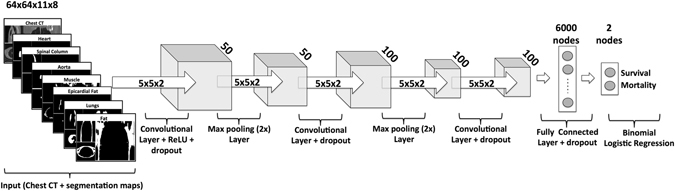
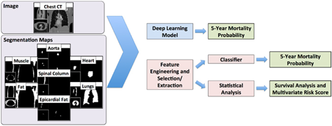
Precision medicine approaches rely on obtaining precise knowledge of the true state of health of an individual patient, which results from a combination of their genetic risks and environmental exposures. This approach is currently limited by the lack of effective and efficient non-invasive medical tests to define the full range of phenotypic variation associated with individual health. Such knowledge is critical for improved early intervention, for better treatment decisions, and for ameliorating the steadily worsening epidemic of chronic disease. We present proof-of-concept experiments to demonstrate how routinely acquired cross-sectional CT imaging may be used to predict patient longevity as a proxy for overall individual health and disease status using computer image analysis techniques. Despite the limitations of a modest dataset and the use of off-the-shelf machine learning methods, our results are comparable to previous 'manual' clinical methods for longevity prediction. This work demonstrates that radiomics techniques can be used to extract biomarkers relevant to one of the most widely used outcomes in epidemiological and clinical research - mortality, and that deep learning with convolutional neural networks can be usefully applied to radiomics research. Computer image analysis applied to routinely collected medical images offers substantial potential to enhance precision medicine initiatives.
Overview
The claims and statistics of this paper haven’t been checked yet.
Run the check — free, no account needed. It reads the open-access text and checks the sources it cites.
Integrity signals
Registry
Cites
Statistics
Figures
Verification
Claims the paper makes
The claims and statistics of this paper haven’t been checked yet.
Run the check — free, no account needed. It reads the open-access text and checks the sources it cites.
Claims other papers make about this one
Each is checked against this paper’s own text: does it say what the citing paper says it does?
In the citing paper
“Deep learning is applied in radiotherapy [89], in PET-MRI attenuation correction [90, 91], in radiomics [92, 93]”
From An overview of deep learning in medical imaging focusing on MRI (2019) · cites this paper as [93]
Self-check
Percentages, sums and reported differences re-computed from the paper’s own numbers.
The self-check needs the paper’s full text.
Citations
Cites
Breiman, L. (2001) · Machine Learning · doi:10.1023/a:1010933404324 · Cited by 133,538
LeCun, Y., Bengio, Y., & Hinton, G. (2015) · Nature · doi:10.1038/nature14539 · Cited by 84,126
Boyd, S., & Vandenberghe, L. (2004) · doi:10.1017/cbo9780511804441 · Cited by 56,601
Regression shrinkage and selection via the lasso
Tibshirani, R. (1996) · Journal of the Royal Statistical Society Series B: Statistical Methodology · doi:10.1111/j.2517-6161.1996.tb02080.x · Cited by 51,482
Charlson, M. E., Pompei, P., Ales, K. L., & MacKenzie, C. R. (1987) · Journal of Chronic Diseases · doi:10.1016/0021-9681(87)90171-8 · Cited by 49,742
(2014) · Cited by 33,419
The meaning and use of the area under a receiver operating characteristic (ROC) curve.
Hanley, J. A., & McNeil, B. J. (1982) · Radiology · doi:10.1148/radiology.143.1.7063747 · Cited by 21,856
Dermatologist-level classification of skin cancer with deep neural networks
Esteva, A., Kuprel, B., Novoa, R. A., Ko, J., Swetter, S. M., Blau, H. M., & Thrun, S. (2017) · Nature · doi:10.1038/nature21056 · Cited by 14,362
Peng, H., Long, F., & Ding, C. (2005) · IEEE Transactions on Pattern Analysis and Machine Intelligence · doi:10.1109/tpami.2005.159 · Cited by 10,818
Analysis of a complex of statistical variables into principal components.
Hotelling, H. (1933) · Journal of Educational Psychology · doi:10.1037/h0071325 · Cited by 9,691
Research works printed but not linked: 3
- Wilkens, L. R. & Lee, J. Nutritional epidemiology. (Wiley Online Library, 1998).
- Zeiler, M. D. & Fergus, R. In European Conference on Computer Vision. 818–833 (Springer).
- Nair, V. & Hinton, G. E. In Proceedings of the 27th International Conference on Machine Learning (ICML-10). 807–814.
Cited by
Sorted by citations
An overview of clinical decision support systems: Benefits, risks, and strategies for success
Sutton, R. T., Pincock, D., Baumgart, D. C., Sadowski, D. C., Fedorak, R. N., & Kroeker, K. I. (2020) · npj Digital Medicine · doi:10.1038/s41746-020-0221-y · Cited by 3,019
An overview of deep learning in medical imaging focusing on MRI
Lundervold, A. S., & Lundervold, A. (2019) · Zeitschrift für Medizinische Physik · doi:10.1016/j.zemedi.2018.11.002 · Cited by 2,058
Current Applications and Future Impact of Machine Learning in Radiology
Choy, G., Khalilzadeh, O., Michalski, M., Do, S., Samir, A. E., Pianykh, O. S., Geis, J. R., Pandharipande, P. V., Brink, J. A., & Dreyer, K. J. (2018) · Radiology · doi:10.1148/radiol.2018171820 · Cited by 800
Bien, N., Rajpurkar, P., Ball, R. L., Irvin, J., Park, A., Jones, E., Bereket, M., Patel, B. N., Yeom, K. W., Shpanskaya, K., et al. (2018) · PLOS Medicine · doi:10.1371/journal.pmed.1002699 · Cited by 734
AI in Medical Imaging Informatics: Current Challenges and Future Directions
Panayides, A. S., Amini, A., Filipovic, N. D., Sharma, A., Tsaftaris, S. A., Young, A., Foran, D., Do, N., Golemati, S., Kurc, T., et al. (2020) · IEEE Journal of Biomedical and Health Informatics · doi:10.1109/jbhi.2020.2991043 · Cited by 713
Surgical data science for next-generation interventions
Maier‐Hein, L., Vedula, S. S., Speidel, S., Navab, N., Kikinis, R., Park, A., Eisenmann, M., Feußner, H., Forestier, G., Giannarou, S., et al. (2017) · Nature Biomedical Engineering · doi:10.1038/s41551-017-0132-7 · Cited by 655
Hofmanninger, J., Prayer, F., Pan, J., Röhrich, S., Prosch, H., & Langs, G. (2020) · European Radiology Experimental · doi:10.1186/s41747-020-00173-2 · Cited by 647
Convolutional Neural Networks for Radiologic Images: A Radiologist’s Guide
Soffer, S., Ben-Cohen, A., Shimon, O., Amitai, M. M., Greenspan, H. K., & Klang, E. (2019) · Radiology · doi:10.1148/radiol.2018180547 · Cited by 528
Applications of Machine Learning Predictive Models in the Chronic Disease Diagnosis
Battineni, G., Sagaro, G. G., Chintalapudi, N., & Amenta, F. (2020) · Journal of Personalized Medicine · doi:10.3390/jpm10020021 · Cited by 418
From Handcrafted to Deep-Learning-Based Cancer Radiomics: Challenges and Opportunities
Afshar, P., Mohammadi, A., Plataniotis, K. N., Oikonomou, A., & Benali, H. (2019) · IEEE Signal Processing Magazine · doi:10.1109/msp.2019.2900993 · Cited by 334
Machine Learning in Radiology: Applications Beyond Image Interpretation
Lakhani, P. C., Prater, A. B., Hutson, R. K., Andriole, K. P., Dreyer, K. J., Morey, J., Prevedello, L. M. S., Clark, T. J., Geis, J. R., Itri, J. N., et al. (2017) · Journal of the American College of Radiology · doi:10.1016/j.jacr.2017.09.044 · Cited by 270
Bibault, J., Giraud, P., Housset, M., Durdux, C., Taı̈eb, J., Berger, A., Coriat, R., Chaussade, S., Dousset, B. E., Nordlinger, B., et al. (2018) · Scientific Reports · doi:10.1038/s41598-018-30657-6 · Cited by 229
Wang, X., Yang, W. H., Weinreb, J. C., Han, J., Li, Q., Kong, X., Yan, Y., Ke, Z., Luo, B., Liu, T., et al. (2017) · Scientific Reports · doi:10.1038/s41598-017-15720-y · Cited by 200
Deep Learning With Radiomics for Disease Diagnosis and Treatment: Challenges and Potential
Zhang, X., Zhang, Y., Zhang, G., Qiu, X., Tan, W., Yin, X., & Liao, L. (2022) · Frontiers in Oncology · doi:10.3389/fonc.2022.773840 · Cited by 201
Ghaderzadeh, M., Asadi, F., Hosseini, A., Bashash, D., Abolghasemi, H., & Roshanpoor, A. (2021) · Scientific Programming · doi:10.1155/2021/9933481 · Cited by 184
Bewes, J., Low, A., Morphett, A., Pate, F. D., & Henneberg, M. (2019) · Journal of Forensic and Legal Medicine · doi:10.1016/j.jflm.2019.01.004 · Cited by 174
Value of MRI in medicine: More than just another test?
van Beek, E. J. R., Kuhl, C. K., Anzai, Y., Desmond, P. M., Ehman, R. L., Gong, Q., Gold, G. E., Gulani, V., Hall-Craggs, M. A., Leiner, T., et al. (2018) · Journal of Magnetic Resonance Imaging · doi:10.1002/jmri.26211 · Cited by 170
Patient similarity for precision medicine: A systematic review
Parimbelli, E., Marini, S., Sacchi, L., & Bellazzi, R. (2018) · Journal of Biomedical Informatics · doi:10.1016/j.jbi.2018.06.001 · Cited by 151
Artificial intelligence in diagnostic and interventional radiology: Where are we now?
Boeken, T., Feydy, J., Lecler, A., Soyer, P. A., Feydy, A., Barat, M., & Duron, L. (2022) · Diagnostic and Interventional Imaging · doi:10.1016/j.diii.2022.11.004 · Cited by 152
Deep learning how to fit an intravoxel incoherent motion model to diffusion‐weighted MRI
Barbieri, S., Gurney‐Champion, O. J., Klaassen, R., & Thoeny, H. C. (2019) · Magnetic Resonance in Medicine · doi:10.1002/mrm.27910 · Cited by 150
Machine Learning Approaches in Cardiovascular Imaging
Henglin, M., Stein, G., Hushcha, P. V., Snoek, J., Wiltschko, A. B., & Cheng, S. C. (2017) · Circulation Cardiovascular Imaging · doi:10.1161/circimaging.117.005614 · Cited by 146
Hussain, L., Nguyen, T., Li, H., Abbasi, A. A., Lone, K. J., Zhao, Z., Zaib, M., Chen, A. L., & Duong, T. Q. (2020) · BioMedical Engineering OnLine · doi:10.1186/s12938-020-00831-x · Cited by 135
Alizadehsani, R., Khosravi, A., Roshanzamir, M., Abdar, M., Sarrafzadegan, N., Shafie, D., Khozeimeh, F., Shoeibi, A., Nahavandi, S., Panahiazar, M., et al. (2020) · Computers in Biology and Medicine · doi:10.1016/j.compbiomed.2020.104095 · Cited by 121
The pace of artificial intelligence innovations: Speed, talent, and trial-and-error
Tang, X., Li, X., Ding, Y., Song, M., & Bu, Y. (2020) · Journal of Informetrics · doi:10.1016/j.joi.2020.101094 · Cited by 131
Mohd Faizal, A. S., Thevarajah, T. M., Khor, S. M., & Chang, S.-W. (2021) · Computer Methods and Programs in Biomedicine · doi:10.1016/j.cmpb.2021.106190 · Cited by 113
Machine Learning in Radiation Oncology: Opportunities, Requirements, and Needs
Feng, M. U.-S., Valdés, G., Dixit, N., & Solberg, T. D. (2018) · Frontiers in Oncology · doi:10.3389/fonc.2018.00110 · Cited by 116
Extracting biological age from biomedical data via deep learning: too much of a good thing?
Pyrkov, T. V., Slipensky, K., Barg, M., Kondrashin, A., Zhurov, B., Zenin, A., Pyatnitskiy, M., Menshikov, L., Markov, S., & Fedichev, P. O. (2018) · Scientific Reports · doi:10.1038/s41598-018-23534-9 · Cited by 111
Fournier, L. S., Costaridou, L. I., Bidaut, L. M., Michoux, N., Lecouvet, F. E., de Geus‐Oei, L., Boellaard, R., Oprea‐Lager, D. E., Obuchowski, N. A., Caroli, A., et al. (2021) · European Radiology · doi:10.1007/s00330-020-07598-8 · Cited by 110
Artificial intelligence at the intersection of pathology and radiology in prostate cancer
Harmon, S. A., Azamat, S., Sanford, T., Choyke, P. L., & Türkbey, B. (2019) · Diagnostic and Interventional Radiology · doi:10.5152/dir.2019.19125 · Cited by 100
Radiomics in radiooncology – challenging the medical physicist
Peeken, J. C., Bernhofer, M., Wiestler, B., Goldberg, T., Cremers, D., Rost, B., Wilkens, J. J., Combs, S. E., & Nüsslin, F. (2018) · Physica Medica · doi:10.1016/j.ejmp.2018.03.012 · Cited by 98
Ensemble Learners of Multiple Deep CNNs for Pulmonary Nodules Classification Using CT Images
Zhang, B., Qi, S., Monkam, P., Li, C., Yang, F., Yao, Y., & Qian, W. (2019) · IEEE Access · doi:10.1109/access.2019.2933670 · Cited by 99
Non-destructive, high-content analysis of wheat grain traits using X-ray micro computed tomography
Hughes, A., Askew, K., Scotson, C. P., Williams, K. S., Sauzé, C., Corke, F. M. K., Doonan, J. H., & Nibau, C. (2017) · Plant Methods · doi:10.1186/s13007-017-0229-8 · Cited by 95
Abbaoui, W., Retal, S., Bhiri, B. E., Kharmoum, N., & Ziti, S. (2024) · Informatics in Medicine Unlocked · doi:10.1016/j.imu.2024.101475 · Cited by 95
Schreuder, A., Scholten, E. T., van Ginneken, B., & Jacobs, C. (2021) · Translational Lung Cancer Research · doi:10.21037/tlcr-2020-lcs-06 · Cited by 94
Artificial Intelligence Applications for Workflow, Process Optimization and Predictive Analytics
Guillon, L. L., Camirand, D., Guilbert, F., & Forghani, R. (2020) · Neuroimaging Clinics of North America · doi:10.1016/j.nic.2020.08.008 · Cited by 88
Machine Learning Principles for Radiology Investigators
Borstelmann, S. M. (2019) · Academic Radiology · doi:10.1016/j.acra.2019.07.030 · Cited by 82
Sharma, S. (2023) · SICOT-J · doi:10.1051/sicotj/2023018 · Cited by 82
Prospects of deep learning for medical imaging
Kim, J., Hong, J., & Park, H. (2018) · Precision and Future Medicine · doi:10.23838/pfm.2018.00030 · Cited by 82
Deep Learning in Nuclear Medicine and Molecular Imaging: Current Perspectives and Future Directions
Choi, H. (2017) · Nuclear Medicine and Molecular Imaging · doi:10.1007/s13139-017-0504-7 · Cited by 75
Heutink, F., Koch, V., Verbist, B. M., van der Woude, W.-J., Mylanus, E. A. M., Huinck, W. J., Sechopoulos, I., & Caballo, M. (2020) · Computer Methods and Programs in Biomedicine · doi:10.1016/j.cmpb.2020.105387 · Cited by 74
Du, D., Feng, H., Lv, W., Ashrafinia, S., Yuan, Q., Wang, Q., Yang, W., Feng, Q., Chen, W., Rahmim, A., et al. (2019) · Molecular Imaging and Biology · doi:10.1007/s11307-019-01411-9 · Cited by 70
3D-MCN: A 3D Multi-scale Capsule Network for Lung Nodule Malignancy Prediction
Afshar, P., Oikonomou, A., Naderkhani, F., Tyrrell, P. N., Plataniotis, K. N., Farahani, K., & Mohammadi, A. (2020) · Scientific Reports · doi:10.1038/s41598-020-64824-5 · Cited by 68
SigRep: Toward Robust Wearable Emotion Recognition With Contrastive Representation Learning
Dissanayake, V., Seneviratne, S., Rana, R. K., Wen, E., Kaluarachchi, T. I., & Nanayakkara, S. C. (2022) · IEEE Access · doi:10.1109/access.2022.3149509 · Cited by 66
Analyzing chronic disease biomarkers using electrochemical sensors and artificial neural networks
Sinha, K., Uddin, Z., Kawsar, H. I., Islam, S., Deen, M. J., & Howlader, M. M. R. (2022) · TrAC Trends in Analytical Chemistry · doi:10.1016/j.trac.2022.116861 · Cited by 69
Radiogenomics for Precision Medicine With a Big Data Analytics Perspective
Panayides, A. S., Pattichis, M. S., Leandrou, S., Pitris, C., Constantinidou, A., & Pattichis, C. S. (2019) · IEEE Journal of Biomedical and Health Informatics · doi:10.1109/jbhi.2018.2879381 · Cited by 66
MIXCAPS: A capsule network-based mixture of experts for lung nodule malignancy prediction
Afshar, P., Naderkhani, F., Oikonomou, A., Rafiee, M. J., Mohammadi, A., & Plataniotis, K. N. (2021) · Pattern Recognition · doi:10.1016/j.patcog.2021.107942 · Cited by 62
Technology Can Augment, but Not Replace, Critical Human Skills Needed for Patient Care
Alrassi, J., Katsufrakis, P. J., & Chandran, L. (2021) · Academic Medicine · doi:10.1097/acm.0000000000003733 · Cited by 60
Fournier, L. S., Costaridou, L. I., Bidaut, L. M., Michoux, N., Lecouvet, F. E., de Geus‐Oei, L., Boellaard, R., Oprea‐Lager, D. E., Obuchowski, N. A., Caroli, A., et al. (2021) · European Radiology · doi:10.1007/s00330-021-07721-3 · Cited by 60
Clinical judgement in the era of big data and predictive analytics
Chin‐Yee, B., & Upshur, R. (2018) · Journal of Evaluation in Clinical Practice · doi:10.1111/jep.12852 · Cited by 59
MR-Guided Radiotherapy for Liver Malignancies
Boldrini, L., Corradini, S., Gani, C., Henke, L. E., Hosni, A., Romano, A., & Dawson, L. A. (2021) · Frontiers in Oncology · doi:10.3389/fonc.2021.616027 · Cited by 59
Medical imaging dose optimisation from ground up: expert opinion of an international summit
Samei, E., Järvinen, H., Kortesniemi, M., Simantirakis, G., Goh, C. X. Y., Wallace, A. B., Vano, E., Bejan, A., Rehani, M. M., & Vassileva, J. (2018) · Journal of Radiological Protection · doi:10.1088/1361-6498/aac575 · Cited by 59
He, K., Liu, X., Li, M., Li, X., Yang, H., & Zhang, H. (2020) · BMC Medical Imaging · doi:10.1186/s12880-020-00457-4 · Cited by 60
Association Between Coffee Intake and Incident Heart Failure Risk
Stevens, L. M., Linstead, E. J., Hall, J. L., & Kao, D. P. (2021) · Circulation Heart Failure · doi:10.1161/circheartfailure.119.006799 · Cited by 53
Artificial intelligence and the future of radiographic scoring in rheumatoid arthritis: a viewpoint
Bird, A., Oakden‐Rayner, L., McMaster, C., Smith, L., Zeng, M., Wechalekar, M. D., Ray, S., Proudman, S., & Palmer, L. J. (2022) · Arthritis Research & Therapy · doi:10.1186/s13075-022-02972-x · Cited by 51
Self-citationKim, D. H., Wit, H., & Thurston, M. (2018) · Nuclear Medicine Communications · doi:10.1097/mnm.0000000000000890 · Cited by 49
Kortesniemi, M., Tsapaki, V., Trianni, A., Russo, P., Maas, A. J. J., Källman, H., Brambilla, M., & Damilakis, J. E. (2018) · Physica Medica · doi:10.1016/j.ejmp.2018.11.005 · Cited by 47
Mubarak, A. S., Serte, S., Al‐Turjman, F., Ameen, Z. S., & Özsöz, M. (2021) · Expert Systems · doi:10.1111/exsy.12842 · Cited by 47
Fernandes, C. D., Dinh, C. V., Walraven, I., Heijmink, S. W. T. J. P., Smolic, M., van Griethuysen, J. J. M., Simões, R., Losnegård, A., van der Poel, H. G., Pos, F. J., et al. (2018) · Physics and Imaging in Radiation Oncology · doi:10.1016/j.phro.2018.06.005 · Cited by 46
Dong, T., Yang, C., Cui, B., Zhang, T., Sun, X., Song, K., Wang, L., Kong, B., & Yang, X. (2020) · Frontiers in Oncology · doi:10.3389/fonc.2020.00464 · Cited by 46
Prime Time for Artificial Intelligence in Interventional Radiology
Seah, J. C. Y., Boeken, T., Sapoval, M. R., & Goh, G. S. (2022) · CardioVascular and Interventional Radiology · doi:10.1007/s00270-021-03044-4 · Cited by 46
Advances in artificial intelligence for the diagnosis and treatment of ovarian cancer (Review)
Wang, Y., Lin, W., Zhuang, X., Wang, X., He, Y., Li, L., & Lyu, G. (2024) · Oncology Reports · doi:10.3892/or.2024.8705 · Cited by 46
Piffer, S., Ubaldi, L., Tangaro, S., Retico, A., & Talamonti, C. (2024) · Progress in Biomedical Engineering · doi:10.1088/2516-1091/ad525b · Cited by 45
Stuckey, T., Gammon, R. S., Goswami, R., Depta, J. P., Steuter, J., Meine, F. J., Roberts, M. C., Singh, N., Ramchandani, S., Burton, T., et al. (2018) · PLoS ONE · doi:10.1371/journal.pone.0198603 · Cited by 37
Kostas Marias, A. (2021) · Journal of Imaging · doi:10.3390/jimaging7080124 · Cited by 35
The Aging Imageomics Study: rationale, design and baseline characteristics of the study population
Puig, J., Biarnés, C., Pedraza, S., Vilanova, J. C., Pamplona, R., Fernández‐Real, J. M., Brugada, R., Ramos, R., de Tuero, G. C., Calvó‐Perxas, L., et al. (2020) · Mechanisms of Ageing and Development · doi:10.1016/j.mad.2020.111257 · Cited by 32
Review of Applications of Artificial Intelligence Algorithms in Pavement Management
Yang, X., & Zhang, Z. (2022) · Journal of Transportation Engineering Part B Pavements · doi:10.1061/jpeodx.0000373 · Cited by 34
Artificial Intelligence at the Service of Precision Medicine
Abbaoui, W., Retal, S., Kharmoum, N., & Ziti, S. (2023) · Lecture notes in networks and systems · doi:10.1007/978-3-031-35248-5_9 · Cited by 29
Lee, J. H., Shin, J., Min, J. H., Jeong, W. K., Kim, H., Choi, S.-Y., Lee, J., Hong, S., & Kim, K. (2024) · Cancer Imaging · doi:10.1186/s40644-024-00653-3 · Cited by 28
Chen, M., Kong, C., Qiao, E., Chen, Y., Chen, W., Jiang, X., Fang, S., Zhang, D., Chen, W., Chen, W., et al. (2023) · Insights into Imaging · doi:10.1186/s13244-023-01380-2 · Cited by 27
Vallières, M., Laberge, S., Diamant, A., & Naqa, I. E. (2017) · Physics in Medicine and Biology · doi:10.1088/1361-6560/aa8a49 · Cited by 26
de Vos, B. D., Leßmann, N., de Jong, P. A., & Išgum, I. (2021) · Radiology Cardiothoracic Imaging · doi:10.1148/ryct.2021190219 · Cited by 26
Jung, K.-H., Park, H., & Hwang, W. (2017) · Hanyang Medical Reviews · doi:10.7599/hmr.2017.37.2.61 · Cited by 26
Arimura, H., Soufi, M., Ninomiya, K., Kamezawa, H., & Yamada, M. (2018) · Radiological Physics and Technology · doi:10.1007/s12194-018-0486-x · Cited by 26
Radiomics in Head and Neck Cancer Outcome Predictions
Gonçalves, M., Gsaxner, C., Ferreira, A. F., Li, J., Puladi, B., Kleesiek, J., Egger, J., & Alves, V. (2022) · Diagnostics · doi:10.3390/diagnostics12112733 · Cited by 24
Base station traffic prediction using XGBoost‐LSTM with feature enhancement
Du, Q., Yin, F., & Li, Z. (2019) · IET Networks · doi:10.1049/iet-net.2019.0103 · Cited by 23
Chen, M., Yang, J., Lu, J., Zhou, Z., Huang, K., Zhang, S.-H., Yuan, G., Zhang, Q., & Li, Z. (2022) · European Radiology · doi:10.1007/s00330-022-08882-5 · Cited by 23
Understanding the Research Landscape of Deep Learning in Biomedical Science: Scientometric Analysis
Nam, S., Kim, D., Jung, W., & Zhu, Y. (2022) · Journal of Medical Internet Research · doi:10.2196/28114 · Cited by 21
Kondou, H., Morohashi, R., Kimura, S., Idota, N., Matsunari, R., Ichioka, H., Bandou, R., Kawamoto, M., Deng, T., & Ikegaya, H. (2023) · Scientific Reports · doi:10.1038/s41598-023-48363-3 · Cited by 19
Hussain, L., Qureshi, S. A., Aldweesh, A. Y., Pirzada, J.-U.-R., Butt, F. M., Eldin, E. T., Ali, M., Algarni, A., & Nadim, M. A. (2022) · Connection Science · doi:10.1080/09540091.2022.2151566 · Cited by 18
Machine‐learning algorithm that can improve the diagnostic accuracy of septic arthritis of the knee
Choi, E.-S., Sim, J. A., Na, Y. G., Seon, J. K., & Shin, H. D. (2021) · Knee Surgery Sports Traumatology Arthroscopy · doi:10.1007/s00167-020-06418-2 · Cited by 16
Artificial Intelligence and Health Care
Peyrou, B., Vignaux, J.-J., & André, A. (2018) · Health informatics · doi:10.1007/978-3-319-98216-8_3 · Cited by 16
Li, S., Zhang, L., Guo, P., Pan, H.-Y., Chen, P.-Z., Xie, H.-F., Xie, B.-K., Chen, J.-Y., Lai, Q.-Q., Li, Y., et al. (2023) · Journal of Radiation Research and Applied Sciences · doi:10.1016/j.jrras.2023.100636 · Cited by 15
Artificial intelligence in radiation oncology
Yakar, M., & Etiz, D. (2021) · Artificial Intelligence in Medical Imaging · doi:10.35711/aimi.v2.i2.13 · Cited by 15
Nascimento, J. C., & Carneiro, G. (2019) · IEEE Transactions on Pattern Analysis and Machine Intelligence · doi:10.1109/tpami.2019.2922959 · Cited by 14
Self-citationDehbaghi, H. A., & Khoshgard, K. (2025) · International Journal of Emergency Medicine · doi:10.1186/s12245-025-01049-1 · Cited by 16
Wang, G., Zhang, X., Gao, Y., Yee, A. L., & Wang, X. (2023) · Journal of Organizational and End User Computing · doi:10.4018/joeuc.322553 · Cited by 11
A machine learning-based sonomics for prediction of thyroid nodule malignancies
Arabi, M., Nazari, M., Salahshour, A., Jenabi, E., Hajianfar, G., Khateri, M., & Shayesteh, S. P. (2023) · Endocrine · doi:10.1007/s12020-023-03407-6 · Cited by 11
Malone, C., Fennell, L., Folliard, T., & Kelly, C. (2019) · Physica Medica · doi:10.1016/j.ejmp.2019.08.014 · Cited by 11
Deep Neural Networks-Based Age Estimation of Cadavers Using CT Imaging of Vertebrae
Kondou, H., Morohashi, R., Ichioka, H., Bandou, R., Matsunari, R., Kawamoto, M., Idota, N., Ting, D., Kimura, S., & Ikegaya, H. (2023) · International Journal of Environmental Research and Public Health · doi:10.3390/ijerph20064806 · Cited by 10
Hayashi, Y. (2018) · Neural Computation · doi:10.1162/neco_a_01139 · Cited by 10
Waite, S., Davenport, M. S., Graber, M. L., Banja, J. D., Sheppard, B., & Bruno, M. A. (2024) · American Journal of Roentgenology · doi:10.2214/ajr.24.31686 · Cited by 9
The evolution of computer-based analysis of high-resolution CT of the chest in patients with IPF
Calandriello, L., & Walsh, S. L. (2021) · British Journal of Radiology · doi:10.1259/bjr.20200944 · Cited by 9
Deep Learning Models for Predicting Phenotypic Traits and Diseases from Omics Data
Islam, M. M., Wang, Y., & Hu, P. (2018) · InTech eBooks · doi:10.5772/intechopen.75311 · Cited by 9
Kim, Y. S., Lee, S. E., Chang, J. M., Kim, S.-Y., & Bae, Y. K. (2022) · Medicine · doi:10.1097/md.0000000000028621 · Cited by 8
Artificial intelligence in forensic anthropology: State of the art and Skeleton-ID project
Martos, R., Ibáñez, O. C., & Mesejo, P. (2024) · Elsevier eBooks · doi:10.1016/b978-0-12-819394-5.00006-7 · Cited by 7
Probing an AI regression model for hand bone age determination using gradient-based saliency mapping
Wang, Z. (2021) · Scientific Reports · doi:10.1038/s41598-021-90157-y · Cited by 7
Zhou, C., Chan, H., Chughtai, A. R., Hadjiiski, L. M., Kazerooni, E. A., & Wei, J. (2020) · European Journal of Radiology · doi:10.1016/j.ejrad.2020.109106 · Cited by 7
Iraji, M. S. (2019) · Journal of Applied Biomedicine · doi:10.32725/jab.2018.007 · Cited by 7
Tang, A., Hunsaker, A., & Hammer, M. M. (2021) · Journal of Thoracic Imaging · doi:10.1097/rti.0000000000000630 · Cited by 6
Saha, P., De, A., Roy, S. D., & Bhowmik, M. K. (2024) · Multimedia Tools and Applications · doi:10.1007/s11042-024-18953-y · Cited by 6
Metadata
- Authors
- Oakden-Rayner, L., Carneiro, G., Bessen, T., Nascimento, J. C., Bradley, A. P., & Palmer, L. J.
- Affiliations
- The University of Adelaide (AU)
- Royal Adelaide Hospital (AU)
- Instituto Superior Técnico (PT)
- The University of Queensland (AU)
- Venue
- Scientific Reports
- Published
- 2017-05-10
- Volume
- 7
- Issue
- 1
- Pages
- 1648-1648
- ISSN
- 2045-2322 (ISSN-L)
- PMC
- PMC5431941
- OpenAlex
- W2611902073
- Cited by
- 154
- References
- 64
- Citations by year
- 2026: 6 · 2025: 7 · 2024: 11 · 2023: 19 · 2022: 19 · 2021: 24 · 2020: 20 · 2019: 13 · 2018: 25 · 2017: 10
- FWCI
- 14.27
- Type
- journal article
- Language
- en
- Open access
- gold
- doi.org
- Locations
- Scientific Reports (publishedVersion, cc-by)
- PubMed Central (publishedVersion, cc-by)
- PubMed (publishedVersion)
- QUT ePrints (Queensland University of Technology) (submittedVersion, cc-by)
- DOAJ (DOAJ: Directory of Open Access Journals) (submittedVersion, cc-by-sa)
- Topics
- Health Sciences › Medicine › Radiology, Nuclear Medicine and Imaging › Radiomics and Machine Learning in Medical Imaging
- Physical Sciences › Computer Science › Artificial Intelligence › Machine Learning in Healthcare
- Health Sciences › Medicine › Health Informatics › Artificial Intelligence in Healthcare and Education
- Keywords
- radiomics, longevity prediction, mortality prediction, feature engineering, convolutional neural networks, precision medicine, biomarkers, computer image analysis, chronic disease
- MeSH terms
- Deep Learning, Diagnostic Imaging, Humans, Image Processing, Computer-Assisted, Longevity, Mortality, Phenotype, Radiology, Risk Factors, ROC Curve, Task Performance and Analysis, Tomography, X-Ray Computed, Reproducibility of Results, Area Under Curve, Kaplan-Meier Estimate, Precision Medicine
About these checks
Registry, retraction, Cites and statistics results are computed, not judged, with one exception: a printed reference that no index lists is read once by a model before it counts as a citation error. Claims are extracted by a model and kept only when their quote appears verbatim in the text; each cited source is then checked, with a model’s help, for whether it says what the claim relies on. Those verdicts can be wrong — read the claim’s quote and the cited source before relying on one.
Statistics and arithmetic are recomputed, not judged.
Confidence bands are shown only for fields calibrated on our annotated test set. None is calibrated yet, so no band is shown.
What counts as a claim ›How we measure confidence ›How we check this ›
Are you an author, or do you see a problem with this page? Request a review
