

The DOI matches its Crossref record; 154 of the 154 references it cites were found in a registry or an index.
Elongation factor Tu is a multifunctional and processed moonlighting protein
Open accessAbstract
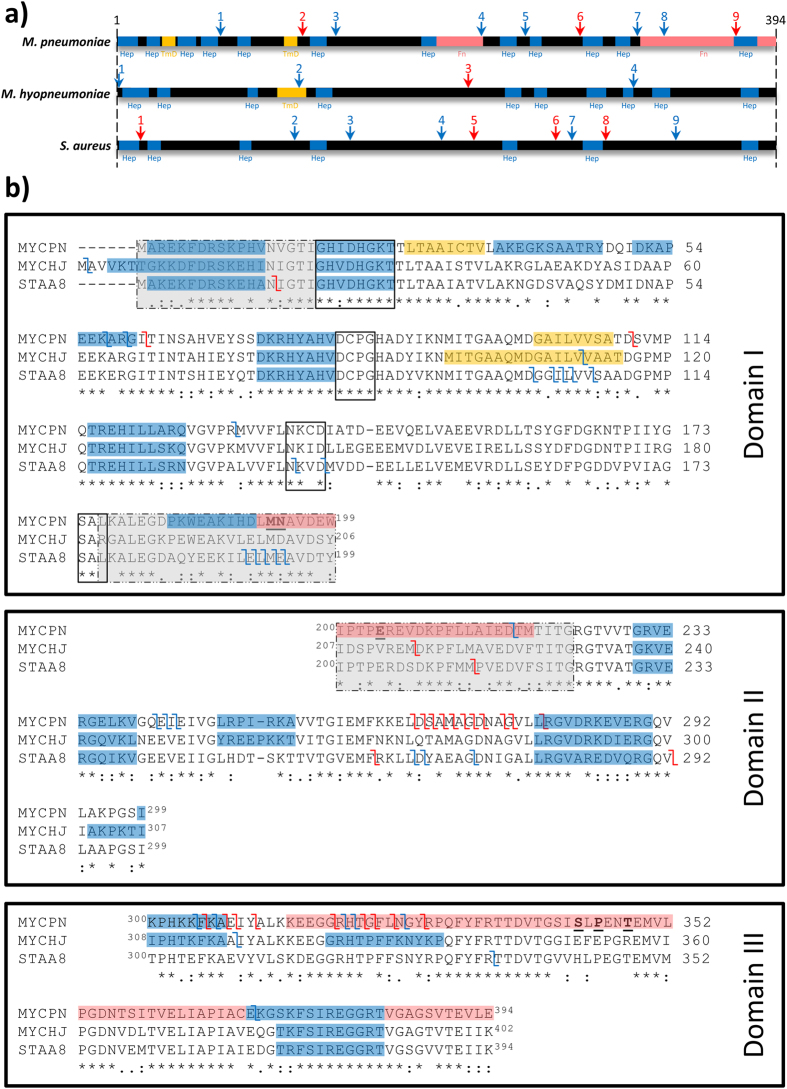
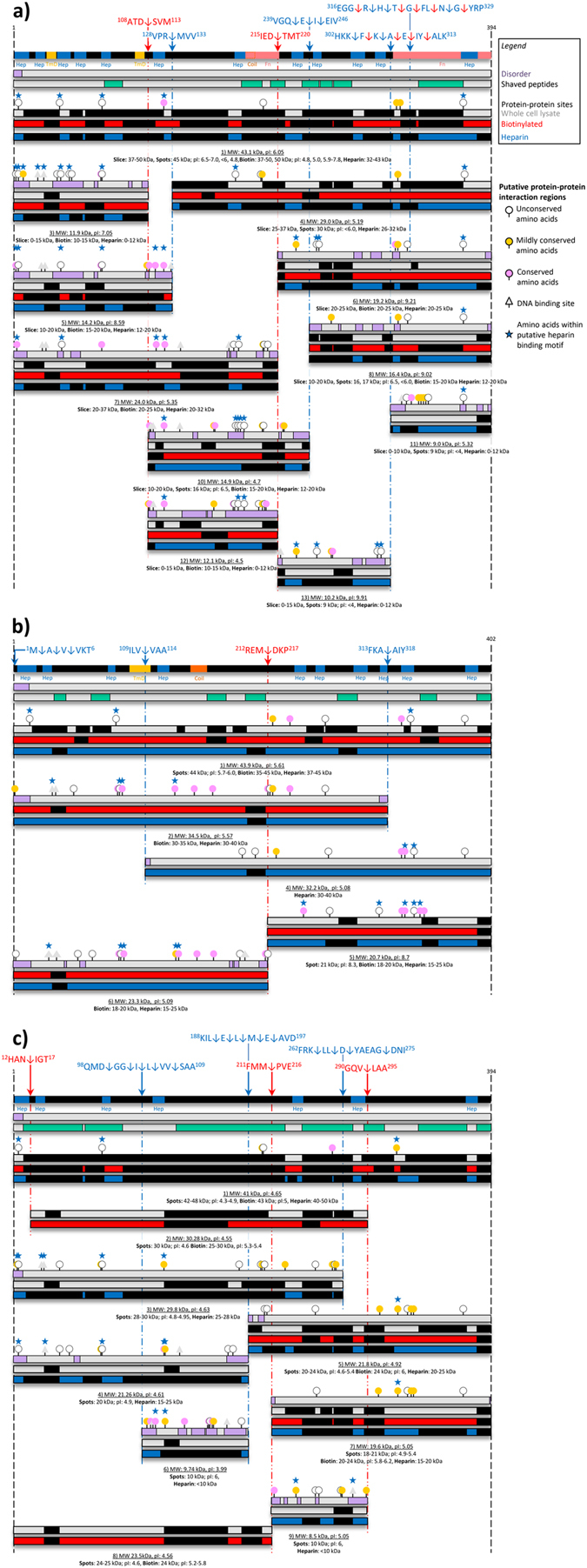
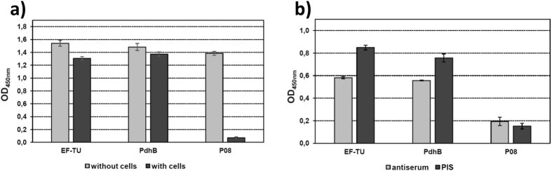
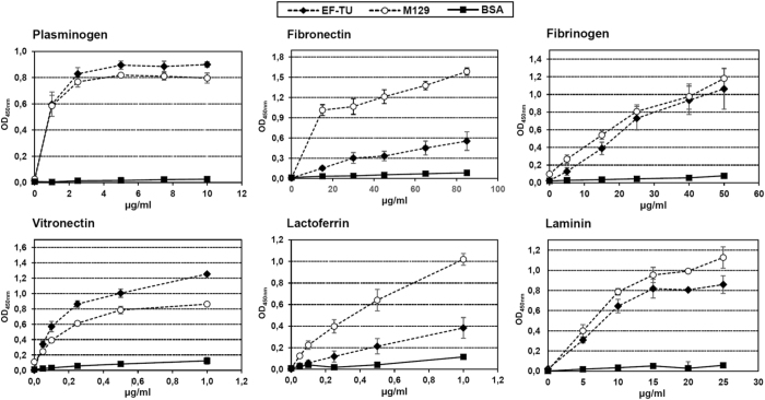
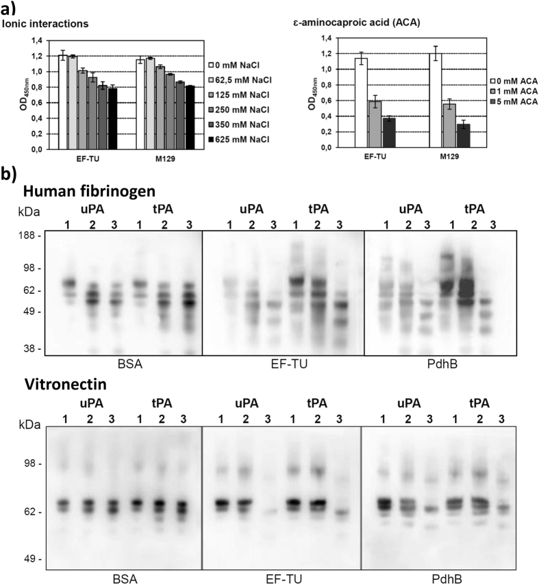
Many bacterial moonlighting proteins were originally described in medically, agriculturally, and commercially important members of the low G + C Firmicutes. We show Elongation factor Tu (Ef-Tu) moonlights on the surface of the human pathogens Staphylococcus aureus (Sa Ef-Tu) and Mycoplasma pneumoniae (Mpn Ef-Tu), and the porcine pathogen Mycoplasma hyopneumoniae (Mhp Ef-Tu). Ef-Tu is also a target of multiple processing events on the cell surface and these were characterised using an N-terminomics pipeline. Recombinant Mpn Ef-Tu bound strongly to a diverse range of host molecules, and when bound to plasminogen, was able to convert plasminogen to plasmin in the presence of plasminogen activators. Fragments of Ef-Tu retain binding capabilities to host proteins. Bioinformatics and structural modelling studies indicate that the accumulation of positively charged amino acids in short linear motifs (SLiMs), and protein processing promote multifunctional behaviour. Codon bias engendered by an A + T rich genome may influence how positively-charged residues accumulate in SLiMs.
Overview
The claims and statistics of this paper haven’t been checked yet.
Run the check — free, no account needed. It reads the open-access text and checks the sources it cites.
Integrity signals
Registry
Cites
Statistics
Figures
Verification
Claims the paper makes
The claims and statistics of this paper haven’t been checked yet.
Run the check — free, no account needed. It reads the open-access text and checks the sources it cites.
Claims other papers make about this one
Each is checked against this paper’s own text: does it say what the citing paper says it does?
The source backs part of the claim, not all of it — or backs it for a different population, condition or comparison.
In the citing paper
“Because of evolutionary pressure to delete nonessential genes, mycoplasmas are known for having such multifunctional proteins.114−117”
From Toward the Complete Functional Characterization of a Minimal Bacterial Proteome (2022) · cites this paper as [117]
Experimental — accuracy not yet measured
Self-check
Percentages, sums and reported differences re-computed from the paper’s own numbers.
The self-check needs the paper’s full text.
Citations
Cites
Fast, scalable generation of high‐quality protein multiple sequence alignments using Clustal Omega
Sievers, F., Wilm, A., Dineen, D. G., Gibson, T. J., Karplus, K. J., Li, W., López, R., McWilliam, H., Remmert, M., Söding, J., et al. (2011) · Molecular Systems Biology · doi:10.1038/msb.2011.75 · Cited by 15,426
SignalP 4.0: discriminating signal peptides from transmembrane regions
Petersen, T. N., Brunak, S., von Heijne, G., & Nielsen, H. (2011) · Nature Methods · doi:10.1038/nmeth.1701 · Cited by 8,847
Protein Identification and Analysis Tools in the ExPASy Server
Wilkins, M. R., Gasteiger, E., Bairoch, A., Sanchez, J., Williams, K. L., Appel, R. D., & Hochstrasser, D. F. (2003) · Humana Press eBooks · doi:10.1385/1-59259-584-7:531 · Cited by 6,244
Comparative Protein Structure Modeling Using Modeller
Eswar, N., Webb, B., Martı́-Renom, M. A., Madhusudhan, M. S., Eramian, D., Shen, M., Pieper, U., & Šali, A. (2006) · Current Protocols in Bioinformatics · doi:10.1002/0471250953.bi0506s15 · Cited by 4,618
Comparative Protein Structure Modeling Using MODELLER
Webb, B., & Šali, A. (2016) · Current Protocols in Bioinformatics · doi:10.1002/cpbi.3 · Cited by 4,404
Predicting coiled coils from protein sequences.
Lupas, A., Van Dyke, M., & Stock, J. (1991) · Science · doi:10.1126/science.252.5009.1162 · Cited by 3,953
Boller, T., & Felix, G. (2009) · Annual Review of Plant Biology · doi:10.1146/annurev.arplant.57.032905.105346 · Cited by 3,514
Phase separation of integral membrane proteins in Triton X-114 solution.
Bordier, C. (1981) · Journal of Biological Chemistry · doi:10.1016/s0021-9258(19)69848-0 · Cited by 3,324
Comparative Protein Structure Modeling Using MODELLER
Eswar, N., Webb, B., Martı́-Renom, M. A., Madhusudhan, M. S., Eramian, D., Shen, M., Pieper, U., & Šali, A. (2007) · Current Protocols in Protein Science · doi:10.1002/0471140864.ps0209s50 · Cited by 2,874
Gómez‐Gómez, L., & Boller, T. (2000) · Molecular Cell · doi:10.1016/s1097-2765(00)80265-8 · Cited by 2,319
Research works printed but not linked: 2
- Tacchi, J. L. et al. Post-translational processing targets functionally diverse proteins in Mycoplasma hyopneumoniae. Open biology 6, 150210, doi:10.1098/rsob.150210 (2016). · DOI · Paper page
- P, H. Diploma thesis, Technische Universität München, (2012).
Cited by
Sorted by citations
Nitrogen-fixing organelle in a marine alga
Coale, T. H., Loconte, V., Turk‐Kubo, K. A., Vanslembrouck, B., Mak, E. W. K., Cheung, S., Ekman, A. A., Chen, J.-H., Hagino, K., Takano, Y., et al. (2024) · Science · doi:10.1126/science.adk1075 · Cited by 249
The Diverse Functional Roles of Elongation Factor Tu (EF-Tu) in Microbial Pathogenesis
Harvey, K. L., Jarocki, V. M., Charles, I. G., & Djordjevic, S. P. (2019) · Frontiers in Microbiology · doi:10.3389/fmicb.2019.02351 · Cited by 218
Self-citationMycoplasma pneumoniae Infections: Pathogenesis and Vaccine Development
Jiang, Z., Li, S., Zhu, C., Zhou, R., & Leung, P. H. M. (2021) · Pathogens · doi:10.3390/pathogens10020119 · Cited by 184
Intracellular proteins moonlighting as bacterial adhesion factors
Jeffery, C. J. (2018) · AIMS Microbiology · doi:10.3934/microbiol.2018.2.362 · Cited by 94
Infection strategies of mycoplasmas: Unraveling the panoply of virulence factors
Yi-wen, C., Wu, Y., Qin, L., Cuiming, Z., & You, X. (2021) · Virulence · doi:10.1080/21505594.2021.1889813 · Cited by 84
Bacterial Excretion of Cytoplasmic Proteins (ECP): Occurrence, Mechanism, and Function
Ebner, P., & Götz, F. (2018) · Trends in Microbiology · doi:10.1016/j.tim.2018.10.006 · Cited by 79
Tartaglia, N. R., Breyne, K., Meyer, E., Cauty, C., Jardin, J., Chrétien, D., Dupont, A., Demeyere, K., Berkova, N., Azevedo, V., et al. (2018) · Frontiers in Cellular and Infection Microbiology · doi:10.3389/fcimb.2018.00277 · Cited by 83
Pathogenicity & virulence ofMycoplasma hyopneumoniae
Zimmer, F. M. A. L., Paes, J. A., Zaha, A., & Ferreira, H. B. (2020) · Virulence · doi:10.1080/21505594.2020.1842659 · Cited by 77
Hiltunen, A. K., Savijoki, K., Nyman, T. A., Miettinen, I., Ihalainen, P., Peltonen, J. P. K., & Fallarero, A. (2019) · Microorganisms · doi:10.3390/microorganisms7120584 · Cited by 72
A novel decoy strategy for polymyxin resistance in Acinetobacter baumannii
Park, J.-E., Kim, M.-S., Shin, B., Kang, M., Yang, J.-H., Lee, T. K., & Park, W. (2021) · eLife · doi:10.7554/elife.66988 · Cited by 68
Tartaglia, N. R., Nicolas, A., Rodovalho, V. D. R., da Luz, B. S. R., Briard‐Bion, V., Krupova, Z., Thierry, A., Coste, F., Burel, A., Martin, P., et al. (2020) · Scientific Reports · doi:10.1038/s41598-020-64952-y · Cited by 72
Mycoplasma hyopneumoniae resides intracellularly within porcine epithelial cells
Raymond, B. B. A., Turnbull, L., Jenkins, C., Madhkoor, R., Schleicher, I., Uphoff, C. C., Whitchurch, C. B., Rohde, M., & Djordjevic, S. P. (2018) · Scientific Reports · doi:10.1038/s41598-018-36054-3 · Cited by 57
Self-citationAntibiotic Resistance Diagnosis in ESKAPE Pathogens—A Review on Proteomic Perspective
Kalpana, S., Lin, W.-Y., Wang, Y.-C., Fu, Y., Lakshmi, A. M., & Wang, H. (2023) · Diagnostics · doi:10.3390/diagnostics13061014 · Cited by 60
Korshoj, L. E., & Kielian, T. (2024) · Nature Communications · doi:10.1038/s41467-024-54581-8 · Cited by 60
The Evolution, Ecology, and Mechanisms of Infection by Gram-Positive, Plant-Associated Bacteria
Thapa, S. P., Davis, E. W., Lyu, Q., Weisberg, A. J., Stevens, D. M., Clarke, C. R., Coaker, G., & Chang, J. H. (2019) · Annual Review of Phytopathology · doi:10.1146/annurev-phyto-082718-100124 · Cited by 48
Kesavan, D. K., Vasudevan, A., Wu, L., Chen, J., Su, Z., Wang, S., & Xu, H. (2020) · BMC Microbiology · doi:10.1186/s12866-020-1722-1 · Cited by 46
Dawood, A. S., Algharib, S. A., Zhao, G., Zhu, T., Qi, M., Delai, K., Hao, Z., Marawan, M. A., Shirani, I., & Guo, A. (2022) · Frontiers in Cellular and Infection Microbiology · doi:10.3389/fcimb.2022.855731 · Cited by 48
Extracellular Actin Is a Receptor for Mycoplasma hyopneumoniae
Raymond, B. B. A., Madhkoor, R., Schleicher, I., Uphoff, C. C., Turnbull, L., Whitchurch, C. B., Rohde, M., Padula, M. P., & Djordjevic, S. P. (2018) · Frontiers in Cellular and Infection Microbiology · doi:10.3389/fcimb.2018.00054 · Cited by 44
Self-citationMycoplasmas are no exception to extracellular vesicles release: Revisiting old concepts
Gaurivaud, P., Ganter, S., Villard, A., Manso-Silván, L., Chevret, D., Boulé, C., Monnet, V., & Tardy, F. (2018) · PLoS ONE · doi:10.1371/journal.pone.0208160 · Cited by 44
Beware of Mycoplasma Anti-immunoglobulin Strategies
Arfi, Y., Lartigue, C., Sirand‐Pugnet, P., & Blanchard, A. (2021) · mBio · doi:10.1128/mbio.01974-21 · Cited by 41
Yu, Y., Liu, M., Hua, L., Qiu, M., Zhang, W., Wei, Y., Gan, Y., Feng, Z., Shao, G., & Xiong, Q. (2018) · Veterinary Research · doi:10.1186/s13567-018-0610-2 · Cited by 36
Widjaja, M., Berry, I. J., Jarocki, V. M., Padula, M. P., Dumke, R., & Djordjevic, S. P. (2020) · Scientific Reports · doi:10.1038/s41598-020-63136-y · Cited by 35
Self-citationNatural Aggregation of Lactobacillus: Mechanisms and Influencing Factors
Wang, S., Li, L., Yu, L., Tian, F., Zhao, J., Zhai, Q., & Chen, W. (2024) · Food Bioscience · doi:10.1016/j.fbio.2024.105007 · Cited by 34
Spatially Targeted Proteomics of the Host–Pathogen Interface during Staphylococcal Abscess Formation
Guiberson, E. R., Weiss, A., Ryan, D. J., Monteith, A. J., Sharman, K., Gutierrez, D. B., Perry, W., Caprioli, R. M., Skaar, E. P., & Spraggins, J. M. (2020) · ACS Infectious Diseases · doi:10.1021/acsinfecdis.0c00647 · Cited by 32
Grimmer, J., & Dumke, R. (2018) · Microbiological Research · doi:10.1016/j.micres.2018.09.006 · Cited by 30
Self-citationIn-Depth Characterization of the Staphylococcus aureus Phosphoproteome Reveals New Targets of Stk1
Prust, N., van der Laarse, S. A. M., van den Toorn, H., van Sorge, N. M., & Lemeer, S. (2021) · Molecular & Cellular Proteomics · doi:10.1074/mcp.ra120.002232 · Cited by 31
Baig, M. A., Turner, M. S., Liu, S., Al‐Nabulsi, A. A., Shah, N. P., & Ayyash, M. M. (2021) · Frontiers in Microbiology · doi:10.3389/fmicb.2021.731410 · Cited by 28
Liu, D., Yang, Z., Chen, Y., Zhuang, W., Niu, H., Wu, J., & Ying, H. (2018) · Biotechnology for Biofuels · doi:10.1186/s13068-018-1316-4 · Cited by 26
Persistence in Livestock Mycoplasmas—a Key Role in Infection and Pathogenesis
Hoelzle, K., Ade, J., & Hoelzle, L. E. (2020) · Current Clinical Microbiology Reports · doi:10.1007/s40588-020-00149-1 · Cited by 27
Toward the Complete Functional Characterization of a Minimal Bacterial Proteome
Bianchi, D. M., Pelletier, J. F., Hutchison, C. A., Glass, J. I., & Luthey-Schulten, Z. (2022) · The Journal of Physical Chemistry B · doi:10.1021/acs.jpcb.2c04188 · Cited by 22
Verdugo-Fuentes, A. A., Gastélum, G., Rocha, J., & de la Torre, M. M. (2020) · Journal of Bacteriology · doi:10.1128/jb.00721-19 · Cited by 25
Paes, J. A., Machado, L. D. P. N., Leal, F. M. D. A., Moraes, S. N., Moura, H., Barr, J. R., & Ferreira, H. B. (2018) · Virulence · doi:10.1080/21505594.2018.1499379 · Cited by 25
Torres, A. N., Chamorro-Veloso, N., Costa, P. S., Cádiz, L., Canto, F. D., Venegas, S., Nitsche, M. L., Coloma-Rivero, R. F., Montero, D. A., & Vidal, R. M. (2020) · Microorganisms · doi:10.3390/microorganisms8081184 · Cited by 21
Wang, J., Li, S., Chen, J., Gan, L., Wang, J., Xiong, Q., Feng, Z., Li, Q., Deng, Z., Yuan, X., et al. (2023) · Microbiology Spectrum · doi:10.1128/spectrum.00218-23 · Cited by 21
Thofte, O., Su, Y.-C., Brant, M., Littorin, N., Duell, B. L., Alvarado, V., Jalalvand, F., & Riesbeck, K. (2018) · Frontiers in Immunology · doi:10.3389/fimmu.2018.02910 · Cited by 21
Arming the troops: Post-translational modification of extracellular bacterial proteins
Forrest, S., & Welch, M. (2020) · Science Progress · doi:10.1177/0036850420964317 · Cited by 19
Briggs, R. E., Billing, S. R., Boatwright, W. D., Chriswell, B. O., Casas, E., Dassanayake, R. P., Palmer, M. V., Register, K. B., & Tatum, F. M. (2021) · Microbial Pathogenesis · doi:10.1016/j.micpath.2021.105159 · Cited by 19
Moonlighting proteins: beacon of hope in era of drug resistance in bacteria
Yadav, P., Singh, R., Sur, S., Bansal, S., Chaudhry, U., & Tandon, V. (2022) · Critical Reviews in Microbiology · doi:10.1080/1040841x.2022.2036695 · Cited by 18
Nicholson, P., Furrer, J., Hässig, M., Strauss, C., Heller, M., Braga-Lagache, S., & Frey, J. (2019) · Anaerobe · doi:10.1016/j.anaerobe.2019.02.011 · Cited by 18
The Mycoplasma spp. ‘Releasome’: A New Concept for a Long-Known Phenomenon
Gaurivaud, P., & Tardy, F. (2022) · Frontiers in Microbiology · doi:10.3389/fmicb.2022.853440 · Cited by 17
Epitope Mapping of Streptococcus agalactiae Elongation Factor Tu Protein Recognized by Human Sera
Pyclik, M. J., Górska, S., Brzozowska, E., Dobrut, A., Ciekot, J., Gamian, A., & Brzychczy‐Włoch, M. (2018) · Frontiers in Microbiology · doi:10.3389/fmicb.2018.00125 · Cited by 17
Liu, D., & Bhunia, A. K. (2024) · ACS Infectious Diseases · doi:10.1021/acsinfecdis.4c00323 · Cited by 16
Bacterial bug-out bags: outer membrane vesicles and their proteins and functions
Kesavan, D. K., Aparna, V. S., Wu, L., Wan, J., Abdelaziz, M. H., Su, Z., Wang, S., & Xu, H. (2020) · The Journal of Microbiology · doi:10.1007/s12275-020-0026-3 · Cited by 16
Dobrut, A., & Brzychczy‐Włoch, M. (2021) · Pathogens · doi:10.3390/pathogens11010043 · Cited by 16
Su, Y., Mattsson, E., Singh, B., Jalalvand, F., Murphy, T. F., & Riesbeck, K. (2019) · The Journal of Infectious Diseases · doi:10.1093/infdis/jiz217 · Cited by 15
Huang, Y., Zhu, F., Koh, J., Stanton, D., Chen, S., & Wang, N. (2022) · Frontiers in Microbiology · doi:10.3389/fmicb.2022.977710 · Cited by 14
Wagner, T. M., Torres-Puig, S., Yimthin, T., Irobalieva, R. N., Heller, M., Kaessmeyer, S., Démoulins, T., & Jores, J. (2025) · Communications Biology · doi:10.1038/s42003-025-08099-4 · Cited by 14
Protein cleavage influences surface protein presentation in Mycoplasma pneumoniae
Berry, I. J., Widjaja, M., Jarocki, V. M., Steele, J. R., Padula, M. P., & Djordjevic, S. P. (2021) · Scientific Reports · doi:10.1038/s41598-021-86217-y · Cited by 15
Self-citationIncomplete autophagy promotes the replication of Mycoplasma hyopneumoniae
Wang, Z., Wen, Y., Zhou, B., Tian, Y., Ning, Y., & Ding, H. (2021) · The Journal of Microbiology · doi:10.1007/s12275-021-1232-3 · Cited by 13
Machado, L. D. P. N., Paes, J. A., dos Santos, P. S., & Ferreira, H. B. (2019) · Microbial Pathogenesis · doi:10.1016/j.micpath.2019.103958 · Cited by 12
Zhao, F., Liu, J., Xiao, D., Liu, L., Gong, J., Xu, J., Li, H., Zhao, S., & Zhang, J. (2020) · Frontiers in Cellular and Infection Microbiology · doi:10.3389/fcimb.2020.553739 · Cited by 12
da Silva, W. M., Seyffert, N., Silva, A., & Azevedo, V. (2021) · PeerJ · doi:10.7717/peerj.12456 · Cited by 11
Villa, F., Secundo, F., Forlani, F., Cattò, C., & Cappitelli, F. (2021) · Annals of Microbiology · doi:10.1186/s13213-020-01617-1 · Cited by 11
Yu, S. Y., Wang, X., Li, J., Xue, F., Tang, F., & Dai, J. (2022) · Virulence · doi:10.1080/21505594.2022.2066274 · Cited by 10
Molecular Detection of Hemoplasma in animals in Tamil Nadu, India and Hemoplasma genome analysis
Chitra, M. A., Kumaragurubaran, K., Gunasekaran, K., Murugasamy, S. A., Arunachalam, S., Annamalai, R., Venkataramanan, R., & Ramaswamy, S. (2023) · Veterinary Research Communications · doi:10.1007/s11259-023-10263-2 · Cited by 10
Mucin adsorbed by E. coli can affect neutrophil activation in vitro
Mikhalchik, E. V., Георгиевна, Б. Н., Vakhrusheva, T. V., Av, S., Baykova, J., Rakitina, D. V., Scherbakov, P., Гусев, С. А., Gusev, A., Хараева, З. Ф., et al. (2019) · FEBS Open Bio · doi:10.1002/2211-5463.12770 · Cited by 9
Tavares, B. A. D. R., Paes, J. A., Zaha, A., & Ferreira, H. B. (2021) · Microbial Pathogenesis · doi:10.1016/j.micpath.2021.105344 · Cited by 9
Vidlund, J., Gelalcha, B. D., Gillespie, B. E., Agga, G. E., Schneider, L. G., Swanson, S. M., Frady, K. D., & Dego, O. K. (2024) · Vaccine · doi:10.1016/j.vaccine.2024.01.067 · Cited by 9
Comparison of Staphylococcus aureus surface protein extraction methods and immunogenicity
Abdi, R. D., Dunlap, J. R., Gillespie, B. E., Ensermu, D. B., Almeida, R. A., & Dego, O. K. (2019) · Heliyon · doi:10.1016/j.heliyon.2019.e02528 · Cited by 9
Li, G., Obeng, E., Shu, J., Shu, J., Chen, J., Wu, Y., & He, Y. (2020) · Frontiers in Immunology · doi:10.3389/fimmu.2020.510943 · Cited by 9
Stancheva, S. G., Frömbling, J., Sassu, E. L., Hennig‐Pauka, I., Ladinig, A., Gerner, W., Grunert, T., & Ehling‐Schulz, M. (2022) · Microbial Pathogenesis · doi:10.1016/j.micpath.2022.105759 · Cited by 8
Liu, P., Long, H., He, S., Cheng, H., Li, E., Cheng, S., Liang, M., Liu, Z., Guo, Z., & Shi, H. S. (2024) · Journal of Zhejiang University SCIENCE B · doi:10.1631/jzus.b2300738 · Cited by 7
Pseudomonas aeruginosa elongation factor-Tu (EF-Tu) is an immunogenic protective protein antigen
Moustafa, D. A., Lou, E., Schafer-Kestenman, M. E., Mateu-Borrás, M., Doménech‐Sánchez, A., Albertí, S., & Goldberg, J. B. (2024) · Vaccine · doi:10.1016/j.vaccine.2024.126476 · Cited by 7
Xu, B., Sun, Y. H., Wang, S., Yao, W., Wang, Q., Yuan, T., Ma, S., Wang, X., Lyu, L., Yu, Y., et al. (2024) · Veterinary Microbiology · doi:10.1016/j.vetmic.2024.110275 · Cited by 6
Sokolova, D., Smolarska, A., Bartnik, P., Rąbalski, Ł., Kosiński, M., Narajczyk, M., Krzyżanowska, D. M., Rajewska, M., Mruk, I., Czaplewska, P., et al. (2023) · Scientific Reports · doi:10.1038/s41598-023-34803-7 · Cited by 6
Elongation factor Tu promotes the onset of periodontitis through mediating bacteria adhesion
Xiao, L., Pu, Y., Cui, Y., Chen, C., Xiao, Q., Wang, Y., Wei, Y., Feng, M., Zhang, T., Yang, S., et al. (2025) · npj Biofilms and Microbiomes · doi:10.1038/s41522-025-00680-3 · Cited by 7
Searching for Biological Function of the Mysterious PA2504 Protein from Pseudomonas aeruginosa
Drabinska, J., Steczkiewicz, K., Kujawa, M., & Kraszewska, E. (2021) · International Journal of Molecular Sciences · doi:10.3390/ijms22189833 · Cited by 6
Host cell interactions of novel antigenic membrane proteins of Mycoplasma agalactiae
Barbosa, M. S., Marques, L. M., Timenetsky, J., Rosengarten, R., Spergser, J., & Chopra‐Dewasthaly, R. (2022) · BMC Microbiology · doi:10.1186/s12866-022-02512-2 · Cited by 6
Impact of c-di-GMP on the Extracellular Proteome of Rhizobium etli
Lorite, M. J., Casas-Román, A., Girard, L., Encarnación‐Guevara, S., Díaz‐Garrido, N., Badı́a, J., Baldomà, L., Pérez‐Mendoza, D., & Sanjuán, J. (2022) · Biology · doi:10.3390/biology12010044 · Cited by 6
Prezioso, S. M., Duong, D. M., Kuiper, E. G., Deng, Q., Albertí, S., Conn, G. L., & Goldberg, J. B. (2019) · Scientific Reports · doi:10.1038/s41598-019-39331-x · Cited by 6
Pinto, L., Torres, C., Gil, C., Santos, H. M., Capelo, J. L., Borges, V., Gomes, J. P., Silva, C., Vieira, L., Poeta, P. A. C. Q. D., et al. (2020) · OMICS A Journal of Integrative Biology · doi:10.1089/omi.2019.0164 · Cited by 5
Sami, A. J., Bilal, S., Ahsan, N.-U.-A., Hameed, N., & Saleem, S. (2023) · Environmental Monitoring and Assessment · doi:10.1007/s10661-023-12077-w · Cited by 5
Guan, X., Bu, F., Fu, Y., Zhang, H., Xiang, H., Chen, X., Chen, T., Wu, X.-J., Wu, K., Liu, L., et al. (2024) · iScience · doi:10.1016/j.isci.2024.111338 · Cited by 6
Thofte, O., Kaur, R., Su, Y., Brant, M., Rudin, A., Hood, D. W., & Riesbeck, K. (2018) · European Journal of Immunology · doi:10.1002/eji.201847871 · Cited by 5
Sun, Y., Xu, B., Zhuge, X., Tang, F., Wang, X., Gong, Q., Chen, R., Xue, F., & Dai, J. (2021) · Frontiers in Cellular and Infection Microbiology · doi:10.3389/fcimb.2020.592906 · Cited by 5
Substrate recognition by the Pseudomonas aeruginosa EF-Tu–modifying methyltransferase EftM
Kuiper, E. G., Dey, D., LaMore, P. A., Owings, J. P., Prezioso, S. M., Goldberg, J. B., & Conn, G. L. (2019) · Journal of Biological Chemistry · doi:10.1074/jbc.ra119.011213 · Cited by 5
Baillo, A. A., Albarracín, L., Ojeda, E. E. H., Elean, M., Gong, W., Kitazawa, H., Villena, J., & Fadda, S. (2026) · Antibiotics · doi:10.3390/antibiotics15010096 · Cited by 4
Jarocki, V. M., Steele, J. R., Widjaja, M., Tacchi, J. L., Padula, M. P., & Djordjevic, S. P. (2019) · International Journal of Medical Microbiology · doi:10.1016/j.ijmm.2019.03.005 · Cited by 4
Self-citationSvetličić, E., Alarcón, L. A., Karlsson, R., Jers, C., & Mijakovic̀, I. (2025) · Journal of Proteome Research · doi:10.1021/acs.jproteome.5c00321 · Cited by 3
Metaviromic and metagenomic study of the pathogens in unexplained pneumonia cases in goats
Xia, Z., Luo, G., Gou, L.-Y., Zhang, W., Ji, E.-G., Li, S., Gao, T., Abi, K.-M., & Yang, F. (2025) · Veterinary Microbiology · doi:10.1016/j.vetmic.2025.110469 · Cited by 4
Brokaw, A. M., Wallen, G., Orvis, A., Kwon, H. J., Seepersaud, R., Nguyen, S., Sharma, K., Coleman, M., Quach, P., Twentyman, J., et al. (2025) · PLoS Pathogens · doi:10.1371/journal.ppat.1013562 · Cited by 3
Lin, Y., He, J., Zhang, Q., Li, Y., Ke, J., Lin, C., Yao, B., Zhang, C., & Tan, N. (2025) · Functional & Integrative Genomics · doi:10.1007/s10142-025-01730-x · Cited by 3
Pagurayan, A. G. B., Fabia, J. Z. P., Agcaoili, M. J. T., Acang, J. P. A., Peralta, J. P. K., Domingo, A., & Gann, P. J. I. (2026) · The Open Microbiology Journal · doi:10.2174/0118742858448770260210051054 · Cited by 3
Si, D., Sun, J., Guo, L., Yang, F., Li, J., & He, S. (2023) · Frontiers in Veterinary Science · doi:10.3389/fvets.2023.1249499 · Cited by 3
Host-endosymbiont co-evolution shaped chloroplast translational regulation
Marchis, F. D., Pompa, A., Ballottari, M., & Bellucci, M. (2019) · Botany Letters · doi:10.1080/23818107.2019.1623716 · Cited by 3
Lactobacillus spp. belonging to the Casei group display a variety of adhesins
Konieczna, C., Olejnik‐Schmidt, A., & SCHMIDT, M. T. (2018) · Acta Scientiarum Polonorum Technologia Alimentaria · doi:10.17306/j.afs.0538 · Cited by 3
Lactobacillus spp. belonging to the Casei group display a variety of adhesins [pdf]
Konieczna, C., Olejnik‐Schmidt, A., & SCHMIDT, M. T. (2018) · Acta Scientiarum Polonorum Technologia Alimentaria · doi:10.17306/j.afs.2018.0538 · Cited by 2
Dobrut, A., Skibiński, J., Bekier, A., Drożdż, K., Rudnicka, K., Płociński, P., Siemińska, I., & Brzychczy‐Włoch, M. (2024) · BMC Veterinary Research · doi:10.1186/s12917-024-04028-5 · Cited by 2
Sanford, J. W., Mobley, J. A., Dybvig, K. F., Atkinson, T. P., & Daubenspeck, J. M. (2025) · PLoS ONE · doi:10.1371/journal.pone.0329506 · Cited by 2
Sami, A. J., Bilal, S., Alam, S., Khalid, M., & Mangat, H. A. (2024) · Applied Biochemistry and Biotechnology · doi:10.1007/s12010-024-04892-9 · Cited by 2
Brahma, U., Suresh, A., Kamila, A., Usha, S., Kumar, S. V. A., Baddi, H., Singothu, S., Sharma, P., Bandaru, R. K., Suravaram, S., et al. (2026) · Microbiology Spectrum · doi:10.1101/2025.10.02.680171 · Cited by 2
Zhang, S., Zhang, Y., Luo, Y., Zhang, S., Song, Y., Li, B., Zheng, F., Gao, P., Xu, J., & Chu, Y. (2025) · Microbial Pathogenesis · doi:10.1016/j.micpath.2025.108127 · Cited by 1
Mycoplasma genitaliumlipoproteins inhibit tumour necrosis factor α-induced apoptosis in HeLa cells
Zuo, L., Sun, H., Yu, M., You, X., Zeng, Y., & Wu, Y. (2018) · Biotechnology & Biotechnological Equipment · doi:10.1080/13102818.2018.1523688 · Cited by 1
Adewara, O. A., Ogunbanwo, S. T., & Adegoke, C. O. (2024) · Journal of Advances in Microbiology · doi:10.9734/jamb/2024/v24i2798 · Cited by 1
Sabat, A. J., Durfee, T., Baldwin, S., Akkerboom, V., Voß, A., Friedrich, A. W., & Bathoorn, E. (2024) · Frontiers in Cellular and Infection Microbiology · doi:10.3389/fcimb.2024.1368923 · Cited by 1
Quiroz-Castañeda, R. E., Aguilar-Díaz, H., Coronado-Villanueva, E., Catalán-Ochoa, D. I., & Amaro-Estrada, I. (2024) · Pathogens · doi:10.3390/pathogens13100845 · Cited by 1
Mendez-Romero, J. I., Beltrán‐Barrientos, L. M., Heredia-Castro, P. Y., Reyes‐Díaz, R., Hernández‐Mendoza, A., González-Córdova, A. F., & Vallejo‐Córdoba, B. (2024) · Food Biotechnology · doi:10.1080/08905436.2024.2423049 · Cited by 1
The immune responses elicited by six recombinant antigens of Mycoplasma hyopneumoniae in mice
Li, S., Yin, R., Xiong, Q., Liu, M., Wang, J., Zhang, Z., Shao, G., Deng, Z., ZhiXin, F., & Yu, Y. (2024) · Veterinary Microbiology · doi:10.1016/j.vetmic.2024.110295 · Cited by 1
Zarzecka, U., Repetto, O., Ambroziak, P., Bielecka, M., Czaplewska, P., Mruk, I., Musiał, N., Figaj, D., Roncarati, D., Diechler, S., et al. (2025) · Microbial Pathogenesis · doi:10.1016/j.micpath.2025.107979 · Cited by 1
Paul, A. G., Wellslager, B., Williamson, M., & Yilmaz, Ö. (2024) · bioRxiv (Cold Spring Harbor Laboratory) · doi:10.1101/2024.07.11.602567 · Cited by 1
Metadata
- Authors
- Widjaja, M., Harvey, K. L., Hagemann, L., Berry, I. J., Jarocki, V. M., Raymond, B. B. A., Tacchi, J. L., Gründel, A., Steele, J. R., Padula, M. P., Charles, I. G., Dumke, R., & Djordjevic, S. P.
- Affiliations
- University of Technology Sydney (AU)
- Technische Universität Dresden (DE)
- Norwich Research Park (GB)
- Corresponding author
- Djordjevic, Steven Philip
- ORCID
- Venue
- Scientific Reports
- Published
- 2017-09-11
- Volume
- 7
- Issue
- 1
- Pages
- 11227-11227
- ISSN
- 2045-2322 (ISSN-L)
- PMC
- PMC5593925
- OpenAlex
- W2750987002
- Cited by
- 131
- References
- 154
- Citations by year
- 2026: 7 · 2025: 17 · 2024: 20 · 2023: 7 · 2022: 16 · 2021: 20 · 2020: 14 · 2019: 13 · 2018: 17
- FWCI
- 4.92
- Type
- journal article
- Language
- en
- Open access
- gold
- doi.org
- Locations
- Scientific Reports (publishedVersion, cc-by)
- PubMed Central (publishedVersion, cc-by)
- PubMed (publishedVersion)
- UEA Digital Repository (University of East Anglia) (acceptedVersion)
- DOAJ (DOAJ: Directory of Open Access Journals) (submittedVersion)
- UTS ePRESS (University of Technology Sydney) (submittedVersion)
- Funders
- Bristol-Myers Squibb Foundation
- Deutsche Forschungsgemeinschaft (1280/1-1, DU 1280/1-1)
- University of Technology Sydney
- NSW Department of Primary Industries
- Australian Research Council (LE130100096)
- Topics
- Life Sciences › Biochemistry, Genetics and Molecular Biology › Molecular Biology › Biochemical and Structural Characterization
- Life Sciences › Immunology and Microbiology › Microbiology › Microbial infections and disease research
- Life Sciences › Biochemistry, Genetics and Molecular Biology › Genetics › Bacterial Genetics and Biotechnology
- Keywords
- elongation factor Tu, moonlighting proteins, protein processing, Staphylococcus aureus, Mycoplasma pneumoniae, Mycoplasma hyopneumoniae, plasmin, short linear motifs, positively charged amino acids, codon usage bias, AT-rich genome, host-pathogen interaction, bacterial cell surface, N-terminomics
- MeSH terms
- Fibrinolysin, Membrane Proteins, Models, Molecular, Mycoplasma pneumoniae, Peptide Elongation Factor Tu, Plasminogen, Protein Binding, Staphylococcus aureus, Computational Biology, Virulence Factors, Mycoplasma hyopneumoniae, Host-Pathogen Interactions
About these checks
Registry, retraction, Cites and statistics results are computed, not judged, with one exception: a printed reference that no index lists is read once by a model before it counts as a citation error. Claims are extracted by a model and kept only when their quote appears verbatim in the text; each cited source is then checked, with a model’s help, for whether it says what the claim relies on. Those verdicts can be wrong — read the claim’s quote and the cited source before relying on one.
Statistics and arithmetic are recomputed, not judged.
Confidence bands are shown only for fields calibrated on our annotated test set. None is calibrated yet, so no band is shown.
What counts as a claim ›How we measure confidence ›How we check this ›
Are you an author, or do you see a problem with this page? Request a review
