

The DOI matches its Crossref record; 25 of the 25 references it cites were found in a registry or an index.
Touching the social robot PARO reduces pain perception and salivary oxytocin levels
Abstract
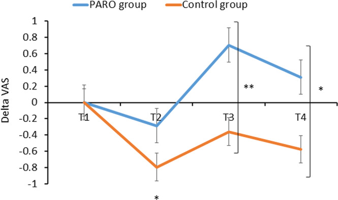
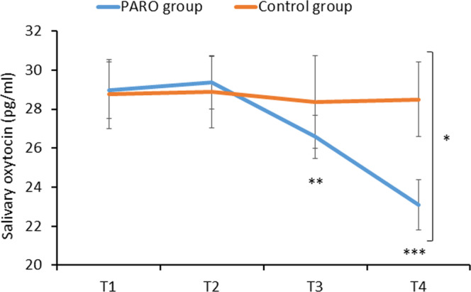
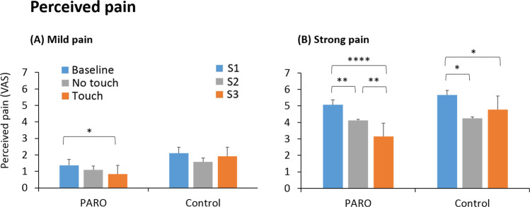
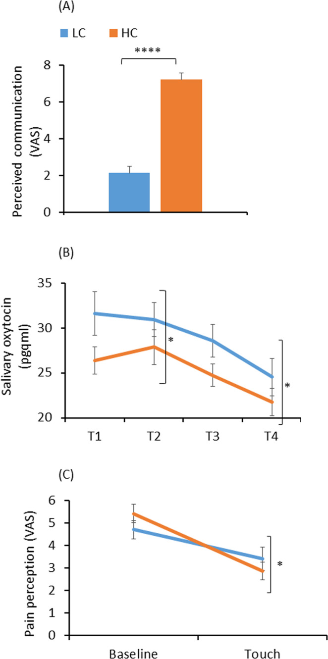
Human-human social touch improves mood and alleviates pain. No studies have so far tested the effect of human-robot emotional touch on experimentally induced pain ratings, on mood and on oxytocin levels in healthy young adults. Here, we assessed the effect of touching the robot PARO on pain perception, on mood and on salivary oxytocin levels, in 83 young adults. We measured their perceived pain, happiness state, and salivary oxytocin. For the 63 participants in the PARO group, pain was assessed in three conditions: Baseline, Touch (touching PARO) and No-Touch (PARO present). The control group (20 participants) underwent the same measurements without ever encountering PARO. There was a decrease in pain ratings and in oxytocin levels and an increase in happiness ratings compared to baseline only in the PARO group. The Touch condition yielded a larger decrease in pain ratings compared to No-Touch. These effects correlated with the participants’ positive perceptions of the interaction with PARO. Participants with higher perceived ability to communicate with PARO experienced a greater hypoalgesic effect when touching PARO. We show that human-robot social touch is effective in reducing pain ratings, improving mood and - surprisingly - reducing salivary oxytocin levels in adults.
Overview
The claims and statistics of this paper haven’t been checked yet.
Run the check — free, no account needed. It reads the open-access text and checks the sources it cites.
Integrity signals
Registry
Cites
Statistics
Figures
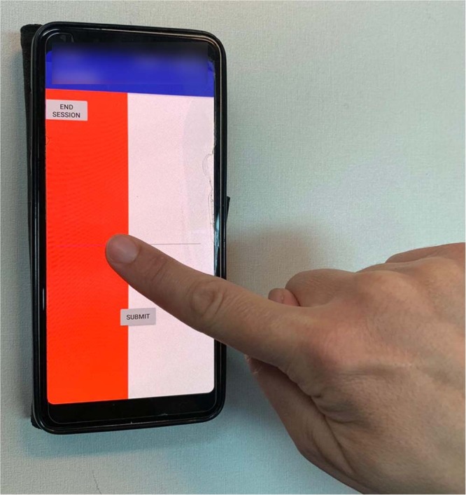
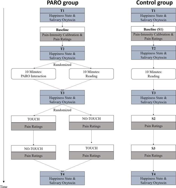
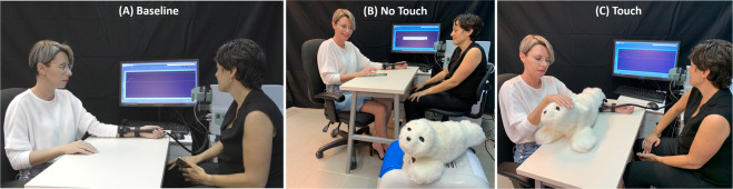
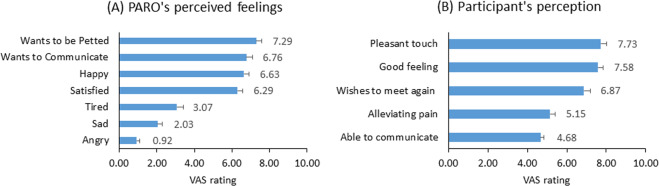
Verification
Claims the paper makes
The claims and statistics of this paper haven’t been checked yet.
Run the check — free, no account needed. It reads the open-access text and checks the sources it cites.
Claims other papers make about this one
Each is checked against this paper’s own text: does it say what the citing paper says it does?
The source backs part of the claim, not all of it — or backs it for a different population, condition or comparison.
In the citing paper
“Earlier experimental work similarly suggested that touching PARO, compared with its mere presence, was associated with reduced pain perception and increased happiness, with stronger hypoallergic effects observed among participants who reported more positive experiences and a greater perceived ability to communicate with the robot [24,54].”
From The Effectiveness of Artificial Intelligence-Based Pet Therapy in Improving the Care of Patients: A Systematic Review (2026) · cites this paper as [54]
Experimental — accuracy not yet measured
Self-check
Percentages, sums and reported differences re-computed from the paper’s own numbers.
The self-check needs the paper’s full text.
Citations
Cites
Faul, F., Erdfelder, E., Lang, A.-G., & Buchner, A. (2007) · Behavior Research Methods · doi:10.3758/bf03193146 · Cited by 65,971
Measuring individual differences in empathy: Evidence for a multidimensional approach.
Davis, M. H. (1983) · Journal of Personality and Social Psychology · doi:10.1037/0022-3514.44.1.113 · Cited by 10,272
Social Relationships and Health.
Cohen, S. (2004) · American Psychologist · doi:10.1037/0003-066x.59.8.676 · Cited by 4,852
The validation of visual analogue scales as ratio scale measures for chronic and experimental pain
Price, D. D., McGrath, P. A., Rafii, A., & Buckingham, B. (1983) · Pain · doi:10.1016/0304-3959(83)90126-4 · Cited by 3,921
Social relationships and health a flashpoint for health policy
Umberson, D., & Karas Montez, J. (2010) · Journal of Health and Social Behavior · doi:10.1177/0022146510383501 · Cited by 2,639
Neuroendocrine perspectives on social attachment and love
Sue Carter, C. (1998) · Psychoneuroendocrinology · doi:10.1016/s0306-4530(98)00055-9 · Cited by 1,425
Lending a Hand: Social Regulation of the Neural Response to Threat
Coan, J. A., Schaefer, H. S., & Davidson, R. J. (2006) · Psychological Science · doi:10.1111/j.1467-9280.2006.01832.x · Cited by 1,218
Advancing social connection as a public health priority in the United States.
Holt-Lunstad, J., Robles, T. F., & Sbarra, D. A. (2017) · American Psychologist · doi:10.1037/amp0000103 · Cited by 978
The Social Salience Hypothesis of Oxytocin
Shamay-Tsoory, S. G., & Abu-Akel, A. (2016) · Biological Psychiatry · doi:10.1016/j.biopsych.2015.07.020 · Cited by 904
Beetz, A., Uvnäs-Moberg, K., Julius, H., & Kotrschal, K. (2012) · Frontiers in Psychology · doi:10.3389/fpsyg.2012.00234 · Cited by 904
Research works printed but not linked: 91
- Berscheid, E. The human’s greatest strength: Other humans., 37–47 (American Psychological Association, 2003). · DOI
- Frank, D. A., Klass, P. E., Earls, F. & Eisenberg, L. Infants and young children in orphanages: One view from pediatrics and child psychiatry. Pediatrics 97, 569–578 (1996).
- Rutter, M. & O’connor, T. G. Are there biological programming effects for psychological development? Findings from a study of Romanian adoptees. Developmental psychology 40, 81 (2004). · DOI
- Thompson, R. A. Social support and child protection: Lessons learned and learning. Child Abuse & Neglect 41, 19–29 (2015). · DOI · Paper page
- DeWall, C. N. & Bushman, B. J. Social acceptance and rejection: The sweet and the bitter. Current Directions in Psychological Science 20, 256–260 (2011). · DOI · Paper page
- Galovski, T. & Lyons, J. A. Psychological sequelae of combat violence: A review of the impact of PTSD on the veteran’s family and possible interventions. Aggression and violent behavior 9, 477–501 (2004). · DOI
- Rintala, D. H., Hart, K. A. & Priebe, M. M. Predicting consistency of pain over a 10-year period in persons with spinal cord injury. Journal of Rehabilitation Research & Development 41 (2004). · DOI
- Åslund, C., Larm, P., Starrin, B. & Nilsson, K. W. The buffering effect of tangible social support on financial stress: influence on psychological well-being and psychosomatic symptoms in a large sample of the adult general population. International journal for equity in health 13, 85 (2014). · DOI
- Divney, A. A. et al. Depression during pregnancy among young couples: the effect of personal and partner experiences of stressors and the buffering effects of social relationships. Journal of pediatric and adolescent gynecology 25, 201–207 (2012). · DOI
- Koopman, C., Hermanson, K., Diamond, S., Angell, K. & Spiegel, D. Social support, life stress, pain and emotional adjustment to advanced breast cancer. Psycho‐Oncology: Journal of the Psychological, Social and Behavioral Dimensions of Cancer 7, 101–111 (1998). · DOI
- Turner, J. B. & Turner, R. J. In Handbook of the sociology of mental health 341–356 (Springer, 2013). · DOI
- Gliga, T., Farroni, T. & Cascio, C. J. Social touch: A new vista for developmental cognitive neuroscience? Developmental cognitive neuroscience 35, 1 (2019). · DOI
- Prescott, T. J., Diamond, M. E. & Wing, A. M. Active touch sensing. Philosophical Transactions of the Royal Society B 366, 2989–2995 (2011). · DOI
- Cohen, S., Janicki-Deverts, D., Turner, R. B. & Doyle, W. J. Does hugging provide stress-buffering social support? A study of susceptibility to upper respiratory infection and illness. Psychological science 26, 135–147 (2015). · DOI · Paper page
- Bufalari, I. & Ionta, S. The social and personality neuroscience of empathy for pain and touch. Frontiers in human neuroscience 7, 393 (2013). · DOI
- Feldman, R., Gordon, I., Schneiderman, I., Weisman, O. & Zagoory-Sharon, O. Natural variations in maternal and paternal care are associated with systematic changes in oxytocin following parent–infant contact. Psychoneuroendocrinology 35, 1133–1141 (2010). · DOI · Paper page
- Moberg, K. U. & Moberg, K. The oxytocin factor: Tapping the hormone of calm, love, and healing. (Da Capo Press, 2003).
- Uvnäs-Moberg, K., Arn, I. & Magnusson, D. The psychobiology of emotion: the role of the oxytocinergic system. International journal of behavioral medicine 12, 59–65 (2005). · DOI · Paper page
- Agren, G., Lundeberg, T., Uvnäs-Moberg, K. & Sato, A. The oxytocin antagonist 1-deamino-2-D-Tyr-(Oet)-4-Thr-8-Orn-oxytocin reverses the increase in the withdrawal response latency to thermal, but not mechanical nociceptive stimuli following oxytocin administration or massage-like stroking in rats. Neuroscience letters 187, 49–52 (1995). · DOI
- Krahé, C., Drabek, M. M., Paloyelis, Y. & Fotopoulou, A. Affective touch and attachment style modulate pain: a laser-evoked potentials study. Philosophical Transactions of the Royal Society B: Biological Sciences 371, 20160009 (2016). · DOI
- Master, S. L. et al. A picture’s worth: Partner photographs reduce experimentally induced pain. Psychological Science 20, 1316–1318 (2009). · DOI · Paper page
- Feingold Polak, R. et al. Differences between young and old users when interacting with a humanoid robot: a qualitative usability study. Paladyn, Journal of Behavioral Robotics 9, 183–192 (2018). · DOI
- Feingold Polak, R. & Levy-Tzedek, S. A Social Robot for Rehabilitation: Expert Clinicians and Post-Stroke Patients’ Evaluation Following a Long-Term Intervention. In Proceedings of the 2020 ACM/IEEE International Conference on Human-Robot Interaction (pp. 151–160). · DOI
- Eizicovits, D., Edan, Y., Tabak, I. & Levy-Tzedek, S. Robotic gaming prototype for upper limb exercise: Effects of age and embodiment on user preferences and movement. Restorative neurology and neuroscience 36, 261–274 (2018). · DOI
- Kashi, S. & Levy-Tzedek, S. Smooth leader or sharp follower? Playing the mirror game with a robot. Restorative neurology and neuroscience 36, 147–159 (2018). · DOI
- Li, J. & Chignell, M. Communication of emotion in social robots through simple head and arm movements. International Journal of Social Robotics 3, 125–142 (2011). · DOI
- Hoffman, G. & Weinberg, G. Interactive improvisation with a robotic marimba player. Autonomous Robots 31, 133–153 (2011). · DOI
- Clabaugh, C., Tsiakas, K. & Mataric, M. In Proceedings of the Synergies between Learning and Interaction Workshop, IROS, Vancouver, BC, Canada. 24–28.
- Kellmeyer, P., Mueller, O., Feingold-Polak, R. & Levy-Tzedek, S. Social robots in rehabilitation: A question of trust. Sci. Robot. 3, eaat1587 (2018). · DOI
- Langer, A., Feingold-Polak, R., Mueller, O., Kellmeyer, P. & Levy-Tzedek, S. Trust in Socially Assistive Robots: Considerations for use in Rehabilitation. Neuroscience & Biobehavioral Reviews 104, 231–239 (2019). · DOI
- Shibata, T. Integration of therapeutic robot, paro, into welfare systems. Proceedings of the 28th Annual European Conference on Cognitive Ergonomics 3 (2010). · DOI
- Moyle, W. et al. Social robots helping people with dementia: Assessing efficacy of social robots in the nursing home environment. 6th International Conference on Human System Interactions (HSI). 608–613 (2013). · DOI
- Wada, K. & Shibata, T. Living with seal robots—its sociopsychological and physiological influences on the elderly at a care house. IEEE transactions on robotics 23, 972–980 (2007). · DOI · Paper page
- Shibata, T. et al. Mental commit robot and its application to therapy of children. IEEE/ASME International Conference on Advanced Intelligent Mechatronics. 1053–1058 (2001). · DOI
- Eskander, R., Tewari, K., Osann, K. & Shibata, T. Pilot study of the PARO therapeutic robot demonstrates decreased pain, fatigue, and anxiety among patients with recurrent ovarian carcinoma. Gynecologic Oncology 130, e144–e145 (2013). · DOI
- Beran, T. N., Ramirez-Serrano, A., Vanderkooi, O. G. & Kuhn, S. Humanoid robotics in health care: An exploration of children’s and parents’ emotional reactions. Journal of health psychology 20, 984–989 (2015). · DOI
- Trost, M. J., Ford, A. R., Kysh, L., Gold, J. I. & Matarić, M. Socially Assistive Robots for Helping Pediatric Distress and Pain. The Clinical Journal of Pain 35, 451–458 (2019). · DOI
- Koyama, Y., Koyama, T., Kroncke, A. P. & Coghill, R. C. Effects of stimulus duration on heat induced pain: the relationship between real-time and post-stimulus pain ratings. Pain 107, 256–266 (2004). · DOI
- Sue Carter, C. et al. Oxytocin: Behavioral Associations and Potential as a Salivary Biomarker. Annals of the New York Academy of Sciences 1098, 312–322 (2007). · DOI · Paper page
- Wada, K., Shibata, T., Saito, T. & Tanie, K. Effects of robot assisted activity for elderly people at day service center and analysis of its factors. Proceedings of the 4th World Congress on Intelligent Control and Automation 1301–1305 (2002). · DOI
- Calo, C. J., Hunt-Bull, N., Lewis, L. & Metzler, T. Ethical implications of using the paro robot, with a focus on dementia patient care. Workshops at the Twenty-Fifth AAAI Conference on Artificial Intelligence (2011).
- Veenhoven, R. Informed pursuit of happiness: What we should know, do know and can get to know. Journal of Happiness Studies 16, 1035–1071 (2015). · DOI
- Steptoe, A. Happiness and health. Annual review of public health 40, 339–359 (2019). · DOI · Paper page
- Hooghe, M. & Vanhoutte, B. Subjective well-being and social capital in Belgian communities. The impact of community characteristics on subjective well-being indicators in Belgium. Social Indicators Research 100, 17–36 (2011). · DOI
- Hsu, H.-C. & Chang, W.-C. Social connections and happiness among the elder population of Taiwan. Aging & mental health 19, 1131–1137 (2015). · DOI
- Oerlemans, W. G., Bakker, A. B. & Veenhoven, R. Finding the key to happy aging: A day reconstruction study of happiness. Journals of Gerontology Series B: Psychological Sciences and Social Sciences 66, 665–674 (2011). · DOI
- Requena, F. Welfare systems, support networks and subjective well-being among retired persons. Social Indicators Research 99, 511–529 (2010). · DOI
- Bao, K. J. & Schreer, G. Pets and happiness: Examining the association between pet ownership and wellbeing. Anthrozoös 29, 283–296 (2016). · DOI
- Walsh, F. Human‐animal bonds I: The relational significance of companion animals. Family process 48, 462–480 (2009). · DOI · Paper page
- Moerman, C. J., van der Heide, L. & Heerink, M. Social robots to support children’s well-being under medical treatment: A systematic state-of-the-art review. Journal of Child Health Care 23, 596–612 (2019). · DOI · Paper page
- Fiocco, A. & Hunse, A. The buffer effect of therapy dog exposure on stress reactivity in undergraduate students. International journal of environmental research and public health 14, 707 (2017). · DOI
- Kerns, K. A., Stuart‐Parrigon, K. L., Coifman, K. G., van Dulmen, M. H. & Koehn, A. Pet dogs: Does their presence influence preadolescents’ emotional responses to a social stressor? Social Development 27, 34–44 (2018). · DOI
- Crossman, M. K., Kazdin, A. E. & Kitt, E. R. The influence of a socially assistive robot on mood, anxiety, and arousal in children. Professional Psychology: Research and Practice 49, 48 (2018). · DOI
- Bernhard, A. et al. Adolescent oxytocin response to stress and its behavioral and endocrine correlates. Hormones and behavior 105, 157–165 (2018). · DOI
- Jurek, B. & Neumann, I. D. The oxytocin receptor: from intracellular signaling to behavior. Physiological reviews 98, 1805–1908 (2018). · DOI · Paper page
- Pierrehumbert, B. et al. Oxytocin response to an experimental psychosocial challenge in adults exposed to traumatic experiences during childhood or adolescence. Neuroscience 166, 168–177 (2010). · DOI · Paper page
- Taylor, S. E. et al. Relation of oxytocin to psychological stress responses and hypothalamic-pituitary-adrenocortical axis activity in older women. Psychosomatic medicine 68, 238–245 (2006). · DOI · Paper page
- Brown, C. A., Cardoso, C. & Ellenbogen, M. A. A meta-analytic review of the correlation between peripheral oxytocin and cortisol concentrations. Frontiers in neuroendocrinology 43, 19–27 (2016). · DOI
- Feldman, R., Gordon, I. & Zagoory‐Sharon, O. Maternal and paternal plasma, salivary, and urinary oxytocin and parent–infant synchrony: considering stress and affiliation components of human bonding. Developmental science 14, 752–761 (2011). · DOI · Paper page
- Schneiderman, I., Kanat-Maymon, Y., Zagoory-Sharon, O. & Feldman, R. Mutual influences between partners’ hormones shape conflict dialog and relationship duration at the initiation of romantic love. Social Neuroscience 9, 337–351 (2014). · DOI
- Crockford, C. et al. Urinary oxytocin and social bonding in related and unrelated wild chimpanzees. Proceedings of the Royal Society B: Biological Sciences 280, 20122765 (2013). · DOI · Paper page
- Campbell, P., Ophir, A. G. & Phelps, S. M. Central vasopressin and oxytocin receptor distributions in two species of singing mice. Journal of Comparative Neurology 516, 321–333 (2009). · DOI
- De Dreu, C. K. et al. The neuropeptide oxytocin regulates parochial altruism in intergroup conflict among humans. Science 328, 1408–1411 (2010). · DOI · Paper page
- Dębiec, J. Peptides of love and fear: vasopressin and oxytocin modulate the integration of information in the amygdala. Bioessays 27, 869–873 (2005). · DOI
- Pedersen, C. A. Biological aspects of social bonding and the roots of human violence. Annals of the New York Academy of Sciences 1036, 106–127 (2004). · DOI
- Pfundmair, M., Reinelt, A., DeWall, C. N. & Feldmann, L. Oxytocin strengthens the link between provocation and aggression among low anxiety people. Psychoneuroendocrinology 93, 124–132 (2018). · DOI
- Romney, C., Hahn-Holbrook, J., Norman, G. J., Moore, A. & Holt-Lunstad, J. Where is the love? A double-blind, randomized study of the effects of intranasal oxytocin on stress regulation and aggression. International journal of psychophysiology 136, 15–21 (2019). · DOI
- Edwards, C., Edwards, A., Stoll, B., Lin, X. & Massey, N. Evaluations of an artificial intelligence instructor’s voice: Social Identity Theory in human-robot interactions. Computers in Human Behavior 90, 357–362 (2019). · DOI
- Quintana, D. S. & Guastella, A. An Allostatic Theory of Oxytocin Signaling. (2019). · DOI
- Okita, S. Y. Self–Other’s Perspective Taking: The Use of Therapeutic Robot Companions as Social Agents for Reducing Pain and Anxiety in Pediatric Patients. Cyberpsychology, Behavior, and Social Networking 16, 436–441 (2013). · DOI
- Stinson, J., Jibb, L., Nathan, P., Beran, T. & Hum, V. Using a humanoid robot to reduce procedural pain in children with cancer: a pilot randomized controlled trial. Pediatric Blood & Cancer. S54–S55 (Wiley-Blackwell, NJ USA).
- Nie, J., Park, M., Marin, A. L. & Sundar, S. S. Can you hold my hand? Physical warmth in human-robot interaction. 7th ACM/IEEE International Conference on Human-Robot Interaction (HRI). 201–202 (2012). · DOI
- Willemse, C. J. & van Erp, J. B. Social Touch in Human–Robot Interaction: Robot-Initiated Touches can Induce Positive Responses without Extensive Prior Bonding. International journal of social robotics, 1–20 (2018). · DOI
- Robinson, H., MacDonald, B. & Broadbent, E. Physiological effects of a companion robot on blood pressure of older people in residential care facility: A pilot study. Australasian journal on ageing 34, 27–32 (2015). · DOI
- Geva, N., Pruessner, J. & Defrin, R. Acute psychosocial stress reduces pain modulation capabilities in healthy men. PAIN 155, 2418–2425 (2014). · DOI
- Geva, N., Pruessner, J. & Defrin, R. Triathletes lose their advantageous pain modulation under acute psychosocial stress. Medicine & Science in Sports & Exercise 49, 333–341 (2017). · DOI
- Dunford, E. & Thompson, M. Relaxation and mindfulness in pain: A review. Reviews in pain 4, 18–22 (2010). · DOI
- Kwekkeboom, K. L. & Gretarsdottir, E. Systematic review of relaxation interventions for pain. Journal of nursing scholarship 38, 269–277 (2006). · DOI · Paper page
- Smith, C. A. et al. Relaxation techniques for pain management in labour. Cochrane Database of Systematic Reviews 3 (2018). · DOI
- Chayadi, E. & McConnell, B. L. Gaining insights on the influence of attention, anxiety, and anticipation on pain perception. Journal of Pain Research 12, 851 (2019). · DOI
- DeMore, M. & Cohen, L. L. Distraction for pediatric immunization pain: A critical review. Journal of Clinical Psychology in Medical Settings 12, 281–291 (2005). · DOI
- Johnson, M. H., Breakwell, G., Douglas, W. & Humphries, S. The effects of imagery and sensory detection distractors on different measures of pain: how does distraction work? British Journal of Clinical Psychology 37, 141–154 (1998). · DOI
- Mancini, F., Beaumont, A.-L., Hu, L., Haggard, P. & Iannetti, G. D. D. Touch inhibits subcortical and cortical nociceptive responses. Pain 156, 1936 (2015). · DOI · Paper page
- Mancini, F., Nash, T., Iannetti, G. D. & Haggard, P. Pain relief by touch: a quantitative approach. PAIN® 155, 635–642 (2014). · DOI
- Bourne, S., Machado, A. G. & Nagel, S. J. Basic anatomy and physiology of pain pathways. Neurosurgery Clinics 25, 629–638 (2014).
- Melzack, R. From the gate to the neuromatrix. Pain 82, S121–S126 (1999). · DOI · Paper page
- Barnett, A. G., Van Der Pols, J. C. & Dobson, A. J. Regression to the mean: what it is and how to deal with it. International journal of epidemiology 34, 215–220 (2004). · DOI · Paper page
- De Vignemont, F. & Singer, T. The empathic brain: how, when and why? Trends in cognitive sciences 10, 435–441 (2006). · DOI
- Akert, R. M. & Panter, A. T. Extraversion and the ability to decode nonverbal communication. Personality and Individual Differences 9, 965–972 (1988). · DOI
- Rahbar, F. et al. In International Conference on Social Robotics. 543–553 (Springer).
- Erk, S. M., Toet, A. & Van Erp, J. B. Effects of mediated social touch on affective experiences and trust. PeerJ 3, e1297 (2015). · DOI
Cited by
Sorted by citations
Trajbarič, T., Muršec, D., Svenšek, A., Štiglic, G., & Gosak, L. (2026) · Applied Sciences · doi:10.3390/app16104683
Metadata
- Authors
- Geva, N., Uzefovsky, F., & Levy-Tzedek, S.
- Venue
- Scientific Reports
- Published
- 2020-06-17
- Volume
- 10
- Issue
- 1
- ISSN
- 2045-2322
- PMC
- PMC7299999
- Cited by
- 111
- References
- 25
- Language
- en
- Funders
- Helmsley Charitable Trust, Marcus Endowment Fund, the Borten Family Foundation grants, the Israel Pain Association, the Rosetrees Trust, Consolidated Anti-Aging Foundation
About these checks
Registry, retraction, Cites and statistics results are computed, not judged, with one exception: a printed reference that no index lists is read once by a model before it counts as a citation error. Claims are extracted by a model and kept only when their quote appears verbatim in the text; each cited source is then checked, with a model’s help, for whether it says what the claim relies on. Those verdicts can be wrong — read the claim’s quote and the cited source before relying on one.
Statistics and arithmetic are recomputed, not judged.
Confidence bands are shown only for fields calibrated on our annotated test set. None is calibrated yet, so no band is shown.
What counts as a claim ›How we measure confidence ›How we check this ›
Are you an author, or do you see a problem with this page? Request a review
