

The DOI matches its Crossref record; 68 of the 68 references it cites were found in a registry or an index.
Enhanced osteogenic differentiation of stem cells by 3D printed PCL scaffolds coated with collagen and hydroxyapatite
Open accessAbstract
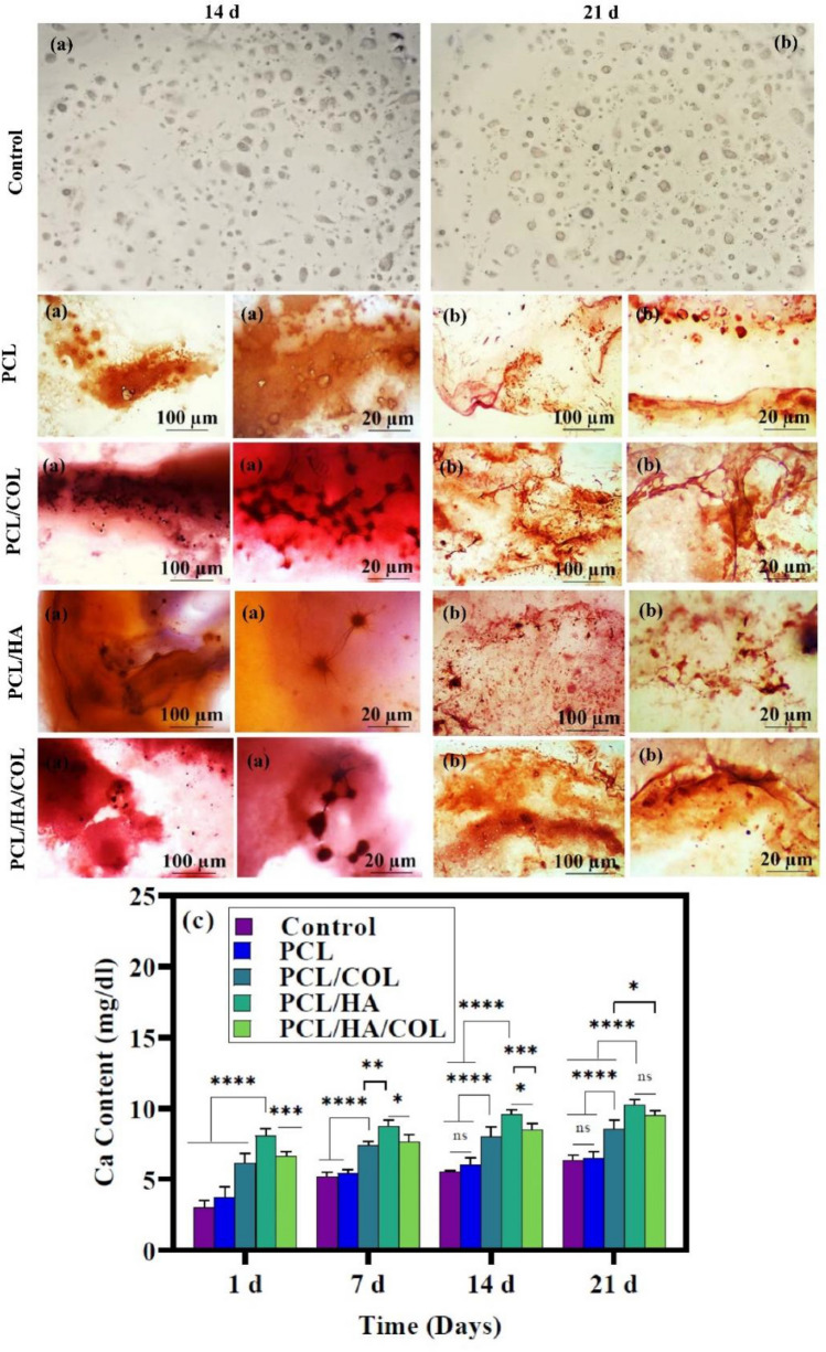
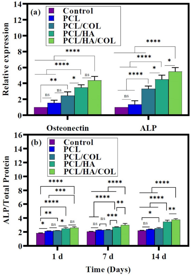
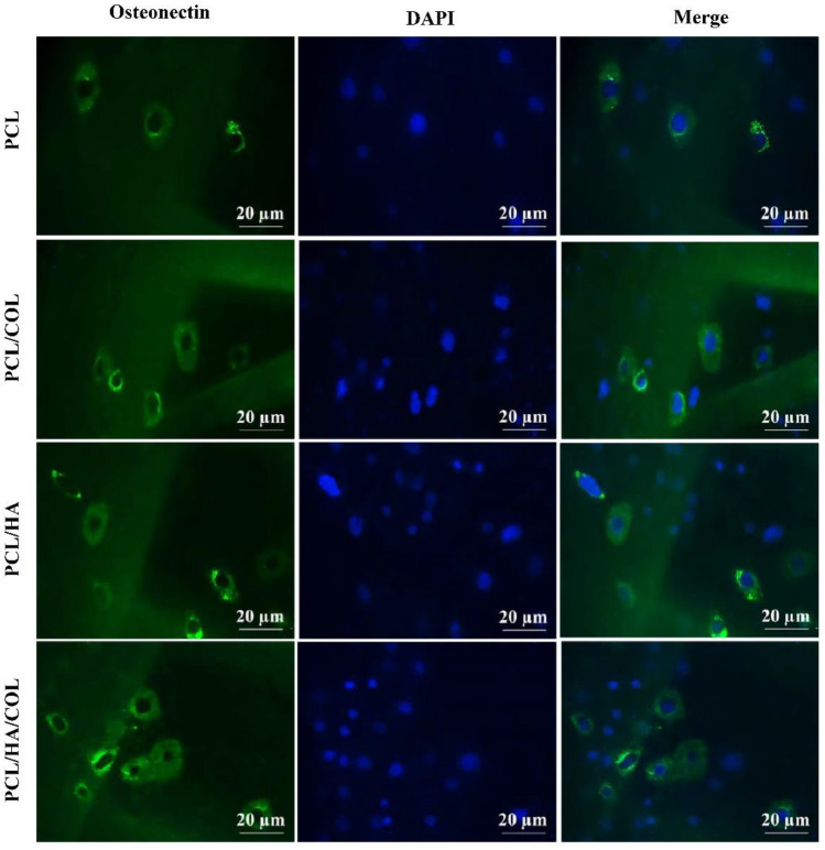
Bone tissue engineering uses various methods and materials to find suitable scaffolds that regenerate lost bone due to disease or injury. Poly(ε-caprolactone) (PCL) can be used in 3D printing for producing biodegradable scaffolds by fused deposition modeling (FDM). However, the hydrophobic surfaces of PCL and its non-osteogenic nature reduces adhesion and cell bioactivity at the time of implantation. This work aims to enhance bone formation, osteogenic differentiation, and in vitro biocompatibility via PCL scaffolds modification with Hydroxyapatite (HA) and Collagen type I (COL). This study evaluated the osteosupportive capacity, biological behavior, and physicochemical properties of 3D-printed PCL, PCL/HA, PCL/COL, and PCL/HA/COL scaffolds. Biocompatibility and cells proliferation were investigated by seeding human adipose tissue-derived mesenchymal stem cells (hADSCs) onto the scaffolds, which were analyzed by 3-(4,5-dimethylthiazol-2-yl)-2,5-diphenyl tetrazolium bromide (MTT) assay, and 6-diamidino-2-phenylindole (DAPI) staining. In addition, the bone differentiation potential of the hADSCs was assessed using calcium deposition, alkaline phosphatase (ALP) activity, and bone-related protein and genes. Although all constructed scaffolds support hADSCs proliferation and differentiation, the results showed that scaffold coating with HA and COL can boost these capacities in a synergistic manner. According to the findings, the tricomponent 3D-printed scaffold can be considered as a promising choice for bone tissue regeneration and rebuilding.
Overview
The claims and statistics of this paper haven’t been checked yet.
Run the check — free, no account needed. It reads the open-access text and checks the sources it cites.
Integrity signals
Registry
Cites
Statistics
Figures
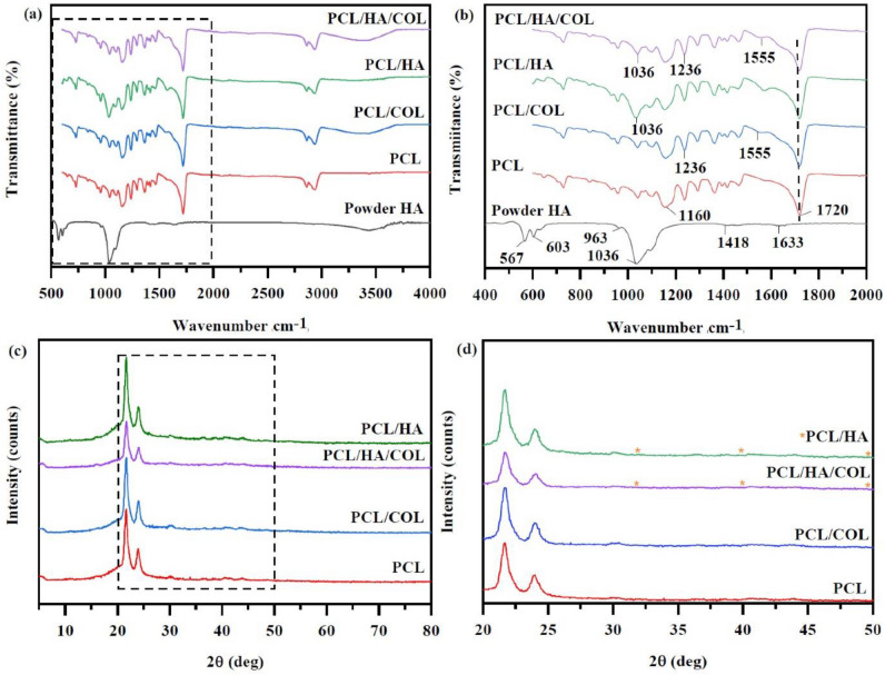
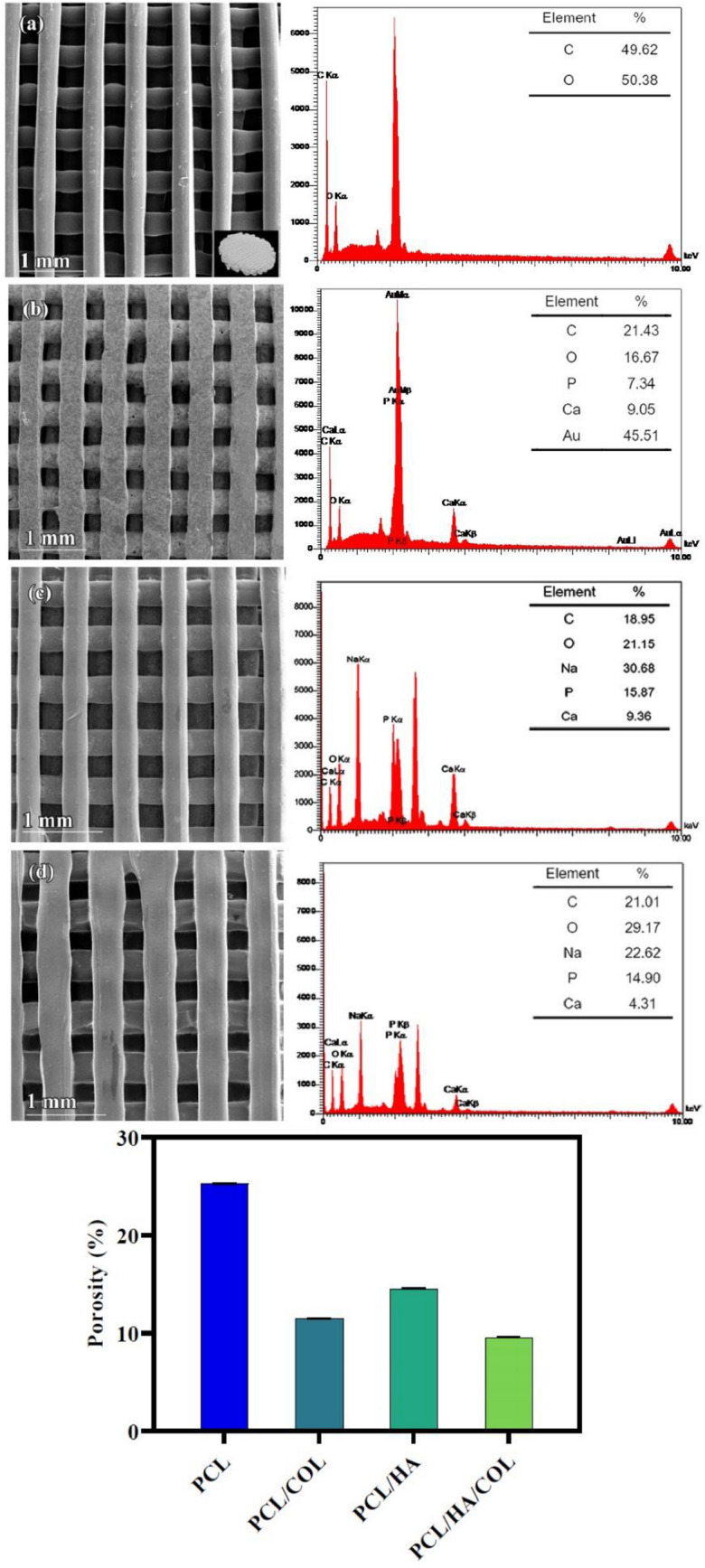
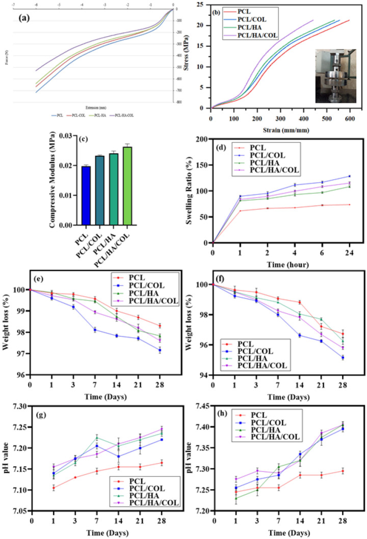
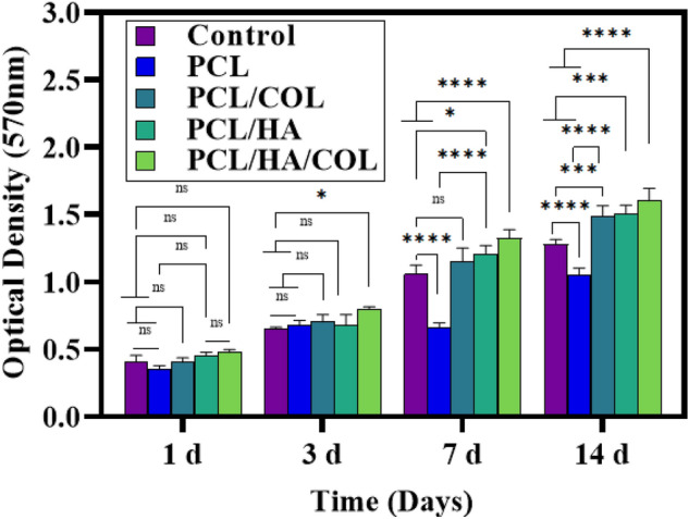
Verification
Claims the paper makes
The claims and statistics of this paper haven’t been checked yet.
Run the check — free, no account needed. It reads the open-access text and checks the sources it cites.
Claims other papers make about this one
Each is checked against this paper’s own text: does it say what the citing paper says it does?
The source backs part of the claim, not all of it — or backs it for a different population, condition or comparison.
In the citing paper
“PCL scaffold coated with collagen and HA showed enhanced proliferation and differentiation of human adipose tissue-derived mesenchymal stem cells through a synergistic effect.”
From Nano-hydroxyapatite/collagen composite as scaffold material for bone regeneration. (2023) · cites this paper as [95]
Experimental — accuracy not yet measuredThe cited source says what the claim attributes to it.
In the citing paper
“These results also substantiated that the coating of collagen and HA can improve the overall bioactivity of the scaffold.”
From Nano-hydroxyapatite/collagen composite as scaffold material for bone regeneration. (2023) · cites this paper as [95]
Self-check
Percentages, sums and reported differences re-computed from the paper’s own numbers.
The self-check needs the paper’s full text.
Citations
Cites
Polymers for 3D Printing and Customized Additive Manufacturing
Ligon, S. C., Liska, R., Stampfl, J., Gurr, M., & Mülhaupt, R. (2017) · Chemical Reviews · doi:10.1021/acs.chemrev.7b00074 · Cited by 3,942
3D bioactive composite scaffolds for bone tissue engineering
Turnbull, G. S., Clarke, J., Picard, F., Riches, P. E., Jia, L., Han, F., Li, B., & Shu, W. (2017) · Bioactive Materials · doi:10.1016/j.bioactmat.2017.10.001 · Cited by 1,388
Designing Biomaterials for 3D Printing
Güvendiren, M., Molde, J., Soares, R. M. D., & Kohn, J. (2016) · ACS Biomaterials Science & Engineering · doi:10.1021/acsbiomaterials.6b00121 · Cited by 761
3D printing of bone tissue engineering scaffolds
Wang, C., Huang, W., Zhou, Y., He, L., He, Z., Chen, Z., He, X., Tian, S., Liao, J., Lu, B., et al. (2020) · Bioactive Materials · doi:10.1016/j.bioactmat.2020.01.004 · Cited by 661
Kafle, A., Luis, E., Silwal, R., Pan, H. M., Shrestha, P. L., & Bastola, A. K. (2021) · Polymers · doi:10.3390/polym13183101 · Cited by 587
PCL-Based Composite Scaffold Matrices for Tissue Engineering Applications
Siddiqui, N., Asawa, S., Birru, B., Baadhe, R. R., & Rao, S. (2018) · Molecular Biotechnology · doi:10.1007/s12033-018-0084-5 · Cited by 461
The development of collagen based composite scaffolds for bone regeneration
Zhang, D., Wu, X., Chen, J., & Lin, K. (2017) · Bioactive Materials · doi:10.1016/j.bioactmat.2017.08.004 · Cited by 442
Characterization of printed PLA scaffolds for bone tissue engineering
Grémare, A., Gudurić, V., Bareille, R., Héroguez, V., Latour, S., L’Heureux, N., Fricain, J., Catros, S., & Nihouannen, D. L. (2017) · Journal of Biomedical Materials Research Part A · doi:10.1002/jbm.a.36289 · Cited by 363
Hassanajili, S., Karami-Pour, A., Oryan, A., & Talaei‐Khozani, T. (2019) · Materials Science and Engineering C · doi:10.1016/j.msec.2019.109960 · Cited by 346
Strategies for Bone Regeneration: From Graft to Tissue Engineering
Battafarano, G., Rossi, M., Martino, V. D., Marampon, F., Borro, L., Secinaro, A., & Fattore, A. D. (2021) · International Journal of Molecular Sciences · doi:10.3390/ijms22031128 · Cited by 299
Review of 3D printable hydrogels and constructs
Li, H., Tan, C., & Li, L. (2018) · Materials & Design · doi:10.1016/j.matdes.2018.08.023 · Cited by 253
Holländer, J., Genina, N., Jukarainen, H., Khajeheian, M., Rosling, A., Mäkilä, E. M., & Sandler, N. (2016) · Journal of Pharmaceutical Sciences · doi:10.1016/j.xphs.2015.12.012 · Cited by 252
Chitosan and gelatin-based electrospun fibers for bone tissue engineering
Sruthi, R., Balagangadharan, K., & Selvamurugan, N. (2019) · International Journal of Biological Macromolecules · doi:10.1016/j.ijbiomac.2019.04.115 · Cited by 236
Gonçalves, E. M., Oliveira, F. J., Silva, R. F., Neto, M. A., Fernandes, M. H., Amaral, M., Vallet‐Regí, M., & Vila, M. (2015) · Journal of Biomedical Materials Research Part B Applied Biomaterials · doi:10.1002/jbm.b.33432 · Cited by 222
Klimek, K., & Ginalska, G. (2020) · Polymers · doi:10.3390/polym12040844 · Cited by 220
(2015) · Johnson Matthey Technology Review · doi:10.1595/205651315x688406 · Cited by 205
Mondal, S., Nguyen, T. P., Pham, V. H., Hoang, G., Manivasagan, P., Kim, M. H., Nam, S. Y., & Oh, J. (2019) · Ceramics International · doi:10.1016/j.ceramint.2019.10.057 · Cited by 204
3D printing of HA / PCL composite tissue engineering scaffolds
Jiao, Z., Luo, B., Xiang, S., Ma, H., Yu, Y., & Yang, W. (2019) · Advanced Industrial and Engineering Polymer Research · doi:10.1016/j.aiepr.2019.09.003 · Cited by 147
Rajzer, I., Menaszek, E., Kwiatkowski, R., Planell, J. A., & Castaño, Ó. (2014) · Materials Science and Engineering C · doi:10.1016/j.msec.2014.08.017 · Cited by 146
Trakoolwannachai, V., Kheolamai, P., & Ummartyotin, S. (2019) · Composites Part B Engineering · doi:10.1016/j.compositesb.2019.106974 · Cited by 142
Babilotte, J., Gudurić, V., Nihouannen, D. L., Naveau, A., Fricain, J., & Catros, S. (2019) · Journal of Biomedical Materials Research Part B Applied Biomaterials · doi:10.1002/jbm.b.34348 · Cited by 134
Bose, S., Sarkar, N., & Banerjee, D. (2018) · Materials Today Chemistry · doi:10.1016/j.mtchem.2018.03.005 · Cited by 132
Materials-Directed Differentiation of Mesenchymal Stem Cells for Tissue Engineering and Regeneration
Leach, J. K., & Whitehead, J. (2017) · ACS Biomaterials Science & Engineering · doi:10.1021/acsbiomaterials.6b00741 · Cited by 129
Zhang, W., Ullah, I., Shi, L., Zhang, Y., Ou, H., Zhou, J., Ullah, M. W., Zhang, X., & Li, W. (2019) · Materials & Design · doi:10.1016/j.matdes.2019.107946 · Cited by 126
Alksnė, M., Kalvaitytė, M., Šimoliūnas, E., Rinkūnaitė, I., Gendvilienė, I., Ločs, J., Rutkūnas, V., & Bukelskienė, V. (2020) · Journal of the mechanical behavior of biomedical materials/Journal of mechanical behavior of biomedical materials · doi:10.1016/j.jmbbm.2020.103641 · Cited by 103
Cestari, F., Petretta, M., Yang, Y., Motta, A., Grigolo, B., & Sglavo, V. M. (2021) · Sustainable materials and technologies · doi:10.1016/j.susmat.2021.e00318 · Cited by 103
Rashad, A., Mohamed‐Ahmed, S., Ojansivu, M., Berstad, K., Yassin, M. A., Kivijärvi, T., Heggset, E. B., Syverud, K., & Mustafa, K. (2018) · Biomacromolecules · doi:10.1021/acs.biomac.8b01194 · Cited by 101
Aidun, A., Firoozabady, A. S., Moharrami, M., Ahmadi, A., Haghighipour, N., Bonakdar, S., & Faghihi, S. (2019) · Artificial Organs · doi:10.1111/aor.13474 · Cited by 97
Mineralization of Biomaterials for Bone Tissue Engineering
Wu, X., Walsh, K., Hoff, B. L., & Camci‐Unal, G. (2020) · Bioengineering · doi:10.3390/bioengineering7040132 · Cited by 97
Zou, L., Zhang, Y., Liu, X., Chen, J., & Zhang, Q. (2019) · Colloids and Surfaces B Biointerfaces · doi:10.1016/j.colsurfb.2019.03.004 · Cited by 96
Huang, B., Vyas, C., Byun, J. J., El-Newehy, M. H., Huang, Z., & Bártolo, P. (2019) · Materials Science and Engineering C · doi:10.1016/j.msec.2019.110374 · Cited by 96
3D Printing for the Clinic: Examining Contemporary Polymeric Biomaterials and Their Clinical Utility
Weems, A. C., Pérez‐Madrigal, M. M., Arno, M. C., & Dove, A. P. (2020) · Biomacromolecules · doi:10.1021/acs.biomac.9b01539 · Cited by 93
Formation of porous HPCL/LPCL/HA scaffolds with supercritical CO2 gas foaming method
Moghadam, M., Hassanajili, S., Esmaeilzadeh, F., Ayatollahi, M., & Ahmadi, M. (2017) · Journal of the mechanical behavior of biomedical materials/Journal of mechanical behavior of biomedical materials · doi:10.1016/j.jmbbm.2016.12.014 · Cited by 91
Hassan, M. I., & Sultana, N. (2017) · 3 Biotech · doi:10.1007/s13205-017-0889-0 · Cited by 90
New Materials for 3D-Printing Based on Polycaprolactone with Gum Rosin and Beeswax as Additives
Pavón, C., Aldás, M., López, J., & Bou, S. F. (2020) · Polymers · doi:10.3390/polym12020334 · Cited by 88
Puyana, V. M. P., Wieringa, P. A., Yuste, Y., de la Portilla, F., Guererro, A., Romero, A., & Moroni, L. (2021) · Journal of Biomedical Materials Research Part A · doi:10.1002/jbm.a.37156 · Cited by 87
Enzymatic degradation of graphene/polycaprolactone materials for tissue engineering
Murray, E., Thompson, B. C., Sayyar, S., & Wallace, G. G. (2014) · Polymer Degradation and Stability · doi:10.1016/j.polymdegradstab.2014.10.010 · Cited by 86
Kim, C. G., Han, K. S., Lee, S., Kim, M. C., Kim, S. Y., & Nah, J. (2021) · Applied Sciences · doi:10.3390/app11146351 · Cited by 81
Brugmans, M. C. P., Söntjens, S. H. M., Cox, M. A. J., Nandakumar, A., Bosman, A. W., Mes, T., Janssen, H. M., Bouten, C. V. C., Baaijens, F. F., & Driessen‐Mol, A. (2015) · Acta Biomaterialia · doi:10.1016/j.actbio.2015.08.034 · Cited by 80
Li, Q., Lei, X., Wang, X., Cai, Z., Lyu, P., & Zhang, G. (2019) · Tissue Engineering Part A · doi:10.1089/ten.tea.2018.0201 · Cited by 74
Wang, Q., Ma, Z., Wang, Y., Zhong, L., & Xie, W. (2020) · Bio-Design and Manufacturing · doi:10.1007/s42242-020-00095-3 · Cited by 71
Yu, J., Xu, Y., Li, S. C., Seifert, G. V., & Becker, M. L. (2017) · Biomacromolecules · doi:10.1021/acs.biomac.7b01222 · Cited by 67
Kim, Y., Lim, J. Y., Yang, G. H., Seo, J.-H., Ryu, H., & Kim, G. (2019) · Journal of Industrial and Engineering Chemistry · doi:10.1016/j.jiec.2019.06.027 · Cited by 66
A 3D-Printed PLCL Scaffold Coated with Collagen Type I and Its Biocompatibility
He, Y., Liu, W., Guan, L., Chen, J., Duan, L., Jia, Z., Huang, J., Li, W., Liu, J., Xiong, J., et al. (2018) · BioMed Research International · doi:10.1155/2018/5147156 · Cited by 63
Patil, V. A., & Masters, K. S. B. (2020) · Bioengineering · doi:10.3390/bioengineering7040163 · Cited by 63
Raina, D. B., Larsson, D., Mrkonjic, F., Isaksson, H., Kumar, A., Lidgren, L., & Tägil, M. (2018) · Journal of Controlled Release · doi:10.1016/j.jconrel.2018.01.006 · Cited by 62
Kim, M. H., Yun, C., Chalisserry, E. P., Lee, Y. W., Kang, H. W., Park, S., Jung, W., Oh, J., & Nam, S. Y. (2018) · Materials Letters · doi:10.1016/j.matlet.2018.03.025 · Cited by 62
Goodarzi, H., Hashemi‐Najafabadi, S., Baheiraei, N., & Bagheri, F. (2019) · Tissue Engineering and Regenerative Medicine · doi:10.1007/s13770-019-00184-0 · Cited by 58
Boron Nitride Based Nanobiocomposites: Design by 3D Printing for Bone Tissue Engineering
Belaid, H., Nagarajan, S., Barou, C., Huon, V., Barés, J., Balme, S., Miele, P., Cornu, D., Cavaillès, V., Teyssier, C., et al. (2020) · ACS Applied Bio Materials · doi:10.1021/acsabm.9b00965 · Cited by 53
Mystiridou, E., Patsidis, A. C., & Bouropoulos, N. (2021) · Applied Sciences · doi:10.3390/app11094253 · Cited by 52
Park, S., Kim, J. E., Han, J., Jeong, S. J., Lim, J. W., Lee, M. C., Son, H., Kim, H. B., Choung, Y., Seonwoo, H., et al. (2021) · Polymers · doi:10.3390/polym13020257 · Cited by 49
Lynch, C. R., Kondiah, P. P. D., & Choonara, Y. E. (2021) · Molecules · doi:10.3390/molecules26092518 · Cited by 49
Non-Cytotoxic Agarose/Hydroxyapatite Composite Scaffolds for Drug Release
Witzler, M., Ottensmeyer, P. F., Gericke, M., Heinze, T. J., Tobiasch, E., & Schulze, M. (2019) · International Journal of Molecular Sciences · doi:10.3390/ijms20143565 · Cited by 44
Karimi, Z., Seyedjafari, E., Mahdavi, F. S., Hashemi, S. M., Khojasteh, A., Kazemi, B., & Mohammadi‐Yeganeh, S. (2019) · Journal of Biomedical Materials Research Part A · doi:10.1002/jbm.a.36638 · Cited by 39
Self-citationMashhadikhan, M., Soleimani, M., Parivar, K., & Yaghmaei, P. (2015) · PubMed Central · Cited by 34
Arab‐Ahmadi, S., Irani, S., Bakhshi, H., Atyabi, F., & Ghalandari, B. (2020) · Polymers for Advanced Technologies · doi:10.1002/pat.5128 · Cited by 34
Self-citationYu, J., Lee, S., Choi, S., Kim, K. K., Ryu, B., Kim, C., Jung, C., Min, B., Xin, Y.-Z., Park, S. A., et al. (2020) · Polymers · doi:10.3390/polym12102203 · Cited by 31
Shapourzadeh, A., Atyabi, S.-M., Irani, S., & Bakhshi, H. (2020) · International Journal of Biological Macromolecules · doi:10.1016/j.ijbiomac.2020.02.036 · Cited by 28
Self-citationLee, C.-F., Hsu, Y., Lin, Y., Nguyen, T.-T., Chen, H.-W., Nabilla, S. C., Hou, S., Chang, F., & Chung, R. (2021) · Polymers · doi:10.3390/polym13183123 · Cited by 27
3D-printed poly-ε-caprolactone-CaCO3-biocomposite-scaffolds for hard tissue regeneration
Neumann, R., Neunzehn, J., Hinüber, C., Flath, T., Schulze, F. P., & Wiesmann, H.-P. (2018) · eXPRESS Polymer Letters · doi:10.3144/expresspolymlett.2019.2 · Cited by 22
Hashemi, S., Amirabad, L. M., Farzad‐Mohajeri, S., Rad, M. R., Fahimipour, F., Ardeshirylajimi, A., Dashtimoghadam, E., Salehi, M., Soleimani, M., Dehghan, M. M., et al. (2021) · Cell and Tissue Research · doi:10.1007/s00441-020-03374-8 · Cited by 22
Self-citationRosales‐Ibáñez, R., Cubo-Mateo, N., Rodríguez-Navarrete, A., González‐González, A. M., Villamar-Duque, T. E., Flores-Sánchez, L. O., & Rodríguez-Lorenzo, L. M. (2021) · Polymers · doi:10.3390/polym13071154 · Cited by 21
Coating of Laponite on PLA Nanofibrous for Bone Tissue Engineering Application
Orafa, Z., Irani, S., Zamanian, A., Bakhshi, H., Nikukar, H., & Ghalandari, B. (2021) · Macromolecular Research · doi:10.1007/s13233-021-9028-1 · Cited by 20
Self-citationLi, B., Ren, K., Wang, Y., Qi, Y., Chen, X., & Huang, Y. (2016) · ACS Applied Materials & Interfaces · doi:10.1021/acsami.6b11287 · Cited by 19
Song, Y., Mou, R., Li, Y., & Yang, T. (2020) · Medical Science Monitor · doi:10.12659/msm.919309 · Cited by 17
Karimzadeh, L., Seyedjafari, E., Hossein, G., Nabiuni, M., Ara, M. H. M., & Salber, J. (2021) · International Journal of Molecular Sciences · doi:10.3390/ijms221910332 · Cited by 16
Self-citationModulating the chemo-mechanical response of structured DNA assemblies through binding molecules
Lee, C., Kim, Y., Kim, K. S., Lee, J. Y., & Kim, D. (2021) · Nucleic Acids Research · doi:10.1093/nar/gkab1119 · Cited by 15
Marouf, N., Nojehdehian, H., & Ghorbani, F. (2019) · Polymers and Polymer Composites · doi:10.1177/0967391119874972 · Cited by 5
Research works printed but not linked: 1
- Jayatissa, N. U., Unagolla, J. & Bhaduri, S. 3D printed polymer scaffolds for bone tissue engineering. Ohio J. Sci. 118, A15–A16 (2018).
Cited by
Sorted by citations
Structural and Functional Adaptive Artificial Bone: Materials, Fabrications, and Properties
Feng, P., Zhao, R., Tang, W., Yang, F. Y., Tian, H., Peng, S., Pan, H., & Shuai, C. (2023) · Advanced Functional Materials · doi:10.1002/adfm.202214726 · Cited by 251
Huang, H., Qiang, L., Fan, M., Liu, Y., Yang, A., Chang, D., Li, J., Sun, T. S., Wang, Y., Guo, R., et al. (2023) · Bioactive Materials · doi:10.1016/j.bioactmat.2023.07.004 · Cited by 87
Collagen as a bio-ink for 3D printing: a critical review
Debnath, S., Agrawal, A., Jain, N., Chatterjee, K., & Player, D. J. (2025) · Journal of Materials Chemistry B · doi:10.1039/d4tb01060d · Cited by 71
Integrated and Bifunctional Bilayer 3D Printing Scaffold for Osteochondral Defect Repair
Li, C., Zhang, W., Nie, Y., Jiang, D., Jia, J., Zhang, W., Li, L., Yao, Z., Qin, L., & Lai, Y. (2023) · Advanced Functional Materials · doi:10.1002/adfm.202214158 · Cited by 59
Nano-hydroxyapatite/collagen composite as scaffold material for bone regeneration.
Kavitha Sri, A., Arthi, C., Neya, N. R., & Hikku, G. S. (2023) · Biomedical Materials · doi:10.1088/1748-605x/acc99e · Cited by 57
Metadata
- Authors
- Ebrahimi, Z., Irani, S., Ardeshirylajimi, A., & Seyedjafari, E.
- Affiliations
- Islamic Azad University, Tehran (IR)
- Shahid Beheshti University (IR)
- University of Tehran (IR)
- Corresponding author
- Seyedjafari, Ehsan
- Venue
- Scientific Reports
- Published
- 2022-07-20
- Volume
- 12
- Issue
- 1
- Pages
- 12359-12359
- ISSN
- 2045-2322 (ISSN-L)
- PMC
- PMC9300684
- OpenAlex
- W4286504968
- Cited by
- 141
- References
- 68
- Citations by year
- 2026: 39 · 2025: 49 · 2024: 31 · 2023: 18 · 2022: 4
- FWCI
- 7.41
- Type
- journal article
- Language
- en
- Open access
- gold
- doi.org
- Locations
- Scientific Reports (publishedVersion, cc-by)
- PubMed Central (publishedVersion, cc-by)
- PubMed (publishedVersion)
- DOAJ (DOAJ: Directory of Open Access Journals) (submittedVersion, cc-by-sa)
- Topics
- Physical Sciences › Engineering › Biomedical Engineering › Bone Tissue Engineering Materials
- Physical Sciences › Engineering › Biomedical Engineering › 3D Printing in Biomedical Research
- Physical Sciences › Materials Science › Biomaterials › biodegradable polymer synthesis and properties
- Keywords
- polycaprolactone, 3D-printed scaffolds, osteogenic differentiation, hydroxyapatite coating, human adipose-derived mesenchymal stem cells, bone tissue engineering, biocompatibility, cell proliferation, alkaline phosphatase activity, calcium deposition, fused deposition modeling
- MeSH terms
- Collagen, Humans, Osteogenesis, Polyesters, Stem Cells, Durapatite, Tissue Scaffolds, Printing, Three-Dimensional
About these checks
Registry, retraction, Cites and statistics results are computed, not judged, with one exception: a printed reference that no index lists is read once by a model before it counts as a citation error. Claims are extracted by a model and kept only when their quote appears verbatim in the text; each cited source is then checked, with a model’s help, for whether it says what the claim relies on. Those verdicts can be wrong — read the claim’s quote and the cited source before relying on one.
Statistics and arithmetic are recomputed, not judged.
Confidence bands are shown only for fields calibrated on our annotated test set. None is calibrated yet, so no band is shown.
What counts as a claim ›How we measure confidence ›How we check this ›
Are you an author, or do you see a problem with this page? Request a review
