

The DOI matches its Crossref record; 4 of the 4 references it cites were found in a registry or an index.
Synaptic vesicles contain small ribonucleic acids (sRNAs) including transfer RNA fragments (trfRNA) and microRNAs (miRNA)
Abstract
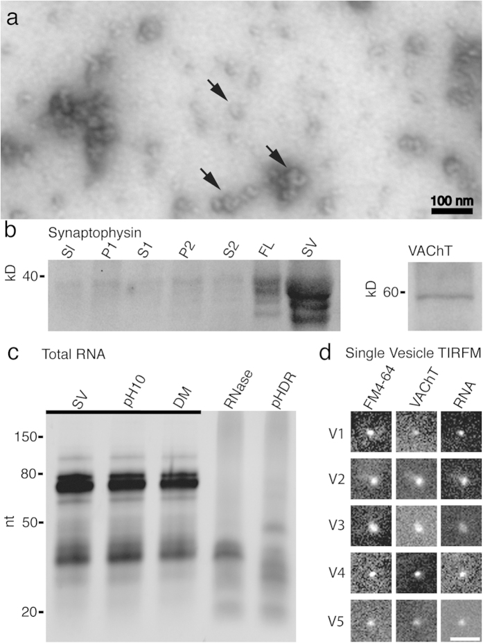
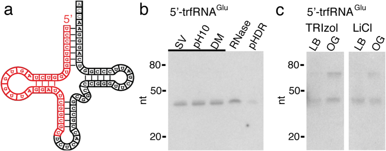
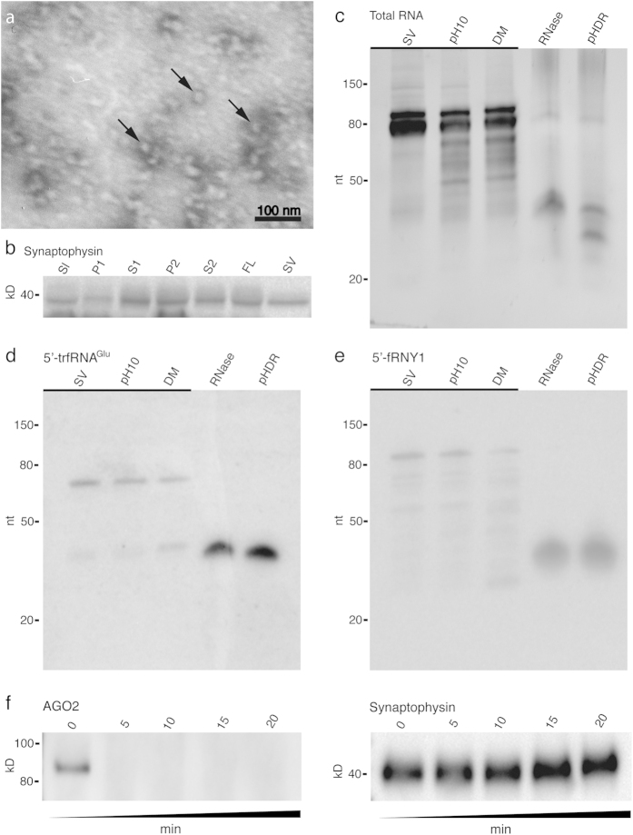
Synaptic vesicles (SVs) are neuronal presynaptic organelles that load and release neurotransmitter at chemical synapses. In addition to classic neurotransmitters, we have found that synaptic vesicles isolated from the electric organ of Torpedo californica,a model cholinergic synapse, contain small ribonucleic acids (sRNAs), primarily the 5′ ends of transfer RNAs (tRNAs) termed tRNA fragments (trfRNAs). To test the evolutionary conservation of SV sRNAs we examined isolated SVs from the mouse central nervous system (CNS). We found abundant levels of sRNAs in mouse SVs, including trfRNAs and micro RNAs (miRNAs) known to be involved in transcriptional and translational regulation. This discovery suggests that, in addition to inducing changes in local dendritic excitability through the release of neurotransmitters, SVs may, through the release of specific trfRNAs and miRNAs, directly regulate local protein synthesis. We believe these findings have broad implications for the study of chemical synaptic transmission.
Overview
The claims and statistics of this paper haven’t been checked yet.
Run the check — free, no account needed. It reads the open-access text and checks the sources it cites.
Integrity signals
Registry
Cites
Statistics
Figures
Verification
Claims the paper makes
The claims and statistics of this paper haven’t been checked yet.
Run the check — free, no account needed. It reads the open-access text and checks the sources it cites.
Claims other papers make about this one
Each is checked against this paper’s own text: does it say what the citing paper says it does?
In the citing paper
“Neurons secrete miRNAs complexed with proteins, such as Argonaute2 and high-density lipoproteins [78–80]”
From Neurodegenerative disease-associated microRNAs acting as signaling molecules modulate CNS neuron structure and viability (2025) · cites this paper as [78]
Self-check
Percentages, sums and reported differences re-computed from the paper’s own numbers.
The self-check needs the paper’s full text.
Citations
Cites
Molecular anatomy of a trafficking organelle
Takamori, S., Holt, M., Stenius, K., Lemke, E. A., Grønborg, M., Riedel, D., Urlaub, H., Schenck, S., Brügger, B., Ringler, P., et al. (2006) · Cell · doi:10.1016/j.cell.2006.10.030 · Cited by 2,373
Mutch, S. A., Kensel-Hammes, P., Gadd, J. C., Fujimoto, B. S., Allen, R. W., Schiro, P. G., Lorenz, R. M., Kuyper, C. L., Kuo, J. S., Bajjalieh, S. M., et al. (2011) · The Journal of Neuroscience · doi:10.1523/jneurosci.3805-10.2011 · Cited by 185
MicroRNA-99 Family Targets AKT/mTOR Signaling Pathway in Dermal Wound Healing
Jin, Y., Tymen, S. D., Chen, D., Fang, Z. J., Zhao, Y., Dragas, D., Dai, Y., Marucha, P. T., & Zhou, X. (2013) · PLoS ONE · doi:10.1371/journal.pone.0064434 · Cited by 172
Current advances in local protein synthesis and synaptic plasticity
Pfeiffer, B. E., & Huber, K. M. (2006) · The Journal of Neuroscience · doi:10.1523/jneurosci.1797-06.2006 · Cited by 168
Research works printed but not linked: 49
- Dale, H. H. The action of certain esters and ethers of choline and their relation to muscarine. J. Pharmacol. Exp. Ther. 6, 147–190 (1914). · DOI
- Loewi, O. Humoral transferability of the heart nerve effect. I. Announcement. Pflug Arch Ges Phys 189, 239–242, 10.1007/Bf01738910 (1921). · DOI
- Fatt, P. & Katz, B. Spontaneous Subthreshold Activity at Motor Nerve Endings. J Physiol-London 117, 109–128 (1952). · DOI
- Bliss, T. V. P. & Lomo, T. Long-Lasting Potentiation of Synaptic Transmission in Dentate Area of Anesthetized Rabbit Following Stimulation of Perforant Path. J Physiol-London 232, 331–356 (1973). · DOI · Paper page
- Davis, H. P. & Squire, L. R. Protein-Synthesis and Memory—a Review. Psychol. Bull. 96, 518–559, 10.1037/0033-2909.96.3.518 (1984). · DOI
- Steward, O. & Levy, W. B. Preferential localization of polyribosomes under the base of dendritic spines in granule cells of the dentate gyrus. J. Neurosci. 2, 284–291 (1982). · DOI · Paper page
- Hawkins, R. D., Kandel, E. R. & Bailey, C. H. Molecular mechanisms of memory storage in Aplysia. Biol. Bull. 210, 174–191 (2006). · DOI
- Holt, C. E. & Schuman, E. M. The central dogma decentralized: new perspectives on RNA function and local translation in neurons. Neuron 80, 648–657, 10.1016/j.neuron.2013.10.036 (2013). · DOI · Paper page
- Smalheiser, N. R. The RNA-centred view of the synapse: non-coding RNAs and synaptic plasticity. Philos. Trans. R. Soc. Lond. B. Biol. Sci. 369, 10.1098/rstb.2013.0504 (2014). · DOI
- Bramham, C. R. & Wells, D. G. Dendritic mRNA: transport, translation and function. Nat. Rev. Neurosci. 8, 776–789, 10.1038/Nrn2150 (2007). · DOI
- Pichardo-Casas, I. et al. Expression profiling of synaptic microRNAs from the adult rat brain identifies regional differences and seizure-induced dynamic modulation. Brain Res. 1436, 20–33, 10.1016/j.brainres.2011.12.001 (2012). · DOI
- Xu, J., Chen, Q., Zen, K., Zhang, C. & Zhang, Q. Synaptosomes secrete and uptake functionally active microRNAs via exocytosis and endocytosis pathways. J. Neurochem. 124, 15–25, 10.1111/jnc.12057 (2013). · DOI · Paper page
- Whittaker, V. P. The structure and function of cholinergic synaptic vesicles. The Third Thudichum Lecture. Biochem. Soc. Trans. 12, 561–576 (1984). · DOI
- Perreault, J., Perreault, J. P. & Boire, G. Ro-associated Y RNAs in metazoans: evolution and diversification. Mol. Biol. Evol. 24, 1678–1689, 10.1093/molbev/msm084 (2007). · DOI
- Whittaker, V. P., Essman, W. B. & Dowe, G. H. The isolation of pure cholinergic synaptic vesicles from the electric organs of elasmobranch fish of the family Torpedinidae. Biochem. J. 128, 833–845 (1972). · DOI
- Hell, J. W., Maycox, P. R., Stadler, H. & Jahn, R. Uptake of GABA by rat brain synaptic vesicles isolated by a new procedure. EMBO J. 7, 3023–3029 (1988). · DOI
- Lamparski, H. G. et al. Production and characterization of clinical grade exosomes derived from dendritic cells. J. Immunol. Methods 270, 211–226 (2002). · DOI
- Witwer, K. W. et al. Standardization of sample collection, isolation and analysis methods in extracellular vesicle research. Journal of extracellular vesicles 2, 10.3402/jev.v2i0.20360 (2013). · DOI
- Montecalvo, A. et al. Mechanism of transfer of functional microRNAs between mouse dendritic cells via exosomes. Blood 119, 756–766, 10.1182/Blood-2011-02-338004 (2012). · DOI · Paper page
- Valadi, H. et al. Exosome-mediated transfer of mRNAs and microRNAs is a novel mechanism of genetic exchange between cells. Nat. Cell Biol. 9, 654–659, 10.1038/ncb1596 (2007). · DOI · Paper page
- Fallon, J. R., Nitkin, R. M., Reist, N. E., Wallace, B. G. & McMahan, U. J. Acetycholine receptor-aggregating factor is similar to molecules concentrated at neuromuscular junctions. Nature 315, 571–574 (1985). · DOI
- Nagwaney, S. et al. Macromolecular connections of active zone material to docked synaptic vesicles and presynaptic membrane at neuromuscular junctions of mouse. J. Comp. Neurol. 513, 457–468 (2009). · DOI
- Tashiro, T. & Stadler, H. Chemical composition of cholinergic synaptic vesicles from Torpedo marmorata based on improved purification. Eur. J. Biochem. 90, 479–487 (1978). · DOI
- Wiedenmann, B. & Franke, W. W. Identification and localization of synaptophysin, an integral membrane glycoprotein of Mr 38,000 characteristic of presynaptic vesicles. Cell 41, 1017–1028 (1985). · DOI
- Erickson, J. D. et al. Functional identification of a vesicular acetylcholine transporter and its expression from a “cholinergic” gene locus. J. Biol. Chem. 269, 21929–21932 (1994). · DOI
- Kao, T. H. & Crothers, D. M. A Proton-Coupled Conformational Switch of Escherichia-Coli 5s Ribosomal-Rna. P Natl Acad Sci-Biol 77, 3360–3364, 10.1073/Pnas.77.6.3360 (1980). · DOI
- Vanaken, T., Foxallvanaken, S., Castleman, S. & Fergusonmiller, S. Alkyl Glycoside Detergents - Synthesis and Applications to the Study of Membrane-Proteins. Methods Enzymol. 125, 27–35 (1986). · DOI
- Li, H. & Harlow, M. L. Individual synaptic vesicles from the electroplaque of Torpedo californica, a classic cholinergic synapse, also contain transporters for glutamate and ATP. Physiological reports 2, e00206, 10.1002/phy2.206 (2014). · DOI
- Takamori, S., Riedel, D. & Jahn, R. Immunoisolation of GABA-specific synaptic vesicles defines a functionally distinct subset of synaptic vesicles. J. Neurosci. 20, 4904–4911 (2000). · DOI
- Fu, H. et al. Stress induces tRNA cleavage by angiogenin in mammalian cells. FEBS Lett. 583, 437–442, 10.1016/j.febslet.2008.12.043 (2009). · DOI
- Yamasaki, S., Ivanov, P., Hu, G. F. & Anderson, P. Angiogenin cleaves tRNA and promotes stress-induced translational repression. J. Cell Biol. 185, 35–42, 10.1083/jcb.200811106 (2009). · DOI
- Wang, Q. et al. Identification and functional characterization of tRNA-derived RNA fragments (tRFs) in respiratory syncytial virus infection. Molecular therapy: the journal of the American Society of Gene Therapy 21, 368–379, 10.1038/mt.2012.237 (2013). · DOI · Paper page
- Varshney, U., Lee, C. P. & RajBhandary, U. L. Direct analysis of aminoacylation levels of tRNAs in vivo. Application to studying recognition of Escherichia coli initiator tRNA mutants by glutaminyl-tRNA synthetase. J. Biol. Chem. 266, 24712–24718 (1991). · DOI
- Anger, A. M. et al. Structures of the human and Drosophila 80S ribosome. Nature 497, 80–85, 10.1038/nature12104 (2013). · DOI
- Hammond, S. M., Bernstein, E., Beach, D. & Hannon, G. J. An RNA-directed nuclease mediates post-transcriptional gene silencing in Drosophila cells. Nature 404, 293–296, 10.1038/35005107 (2000). · DOI
- Gruber, A. R., Lorenz, R., Bernhart, S. H., Neubock, R. & Hofacker, I. L. The Vienna RNA websuite. Nucleic Acids Res. 36, W70–74, 10.1093/nar/gkn188 (2008). · DOI
- Popenda, M. et al. Automated 3D structure composition for large RNAs. Nucleic Acids Res. 40, e112, 10.1093/nar/gks339 (2012). · DOI
- Hanada, T. et al. CLP1 links tRNA metabolism to progressive motor-neuron loss. Nature 495, 474–480, 10.1038/nature11923 (2013). · DOI
- Thompson, D. M. & Parker, R. Stressing out over tRNA cleavage. Cell 138, 215–219, 10.1016/j.cell.2009.07.001 (2009). · DOI
- Peng, H. et al. A novel class of tRNA-derived small RNAs extremely enriched in mature mouse sperm. Cell research 22, 1609–1612, 10.1038/cr.2012.141 (2012). · DOI
- Tan, C. L. et al. MicroRNA-128 governs neuronal excitability and motor behavior in mice. Science 342, 1254–1258, 10.1126/science.1244193 (2013). · DOI · Paper page
- Jovicic, A., Jolissaint, J. F. Z., Moser, R., Santos, M. D. S. & Luthi-Carter, R. MicroRNA-22 (miR-22) Overexpression Is Neuroprotective via General Anti-Apoptotic Effects and May also Target Specific Huntington’s Disease-Related Mechanisms. PloS one 8, ARTN e54222, 10.1371/journal.pone.0054222 (2013). · DOI
- Gardiner, E. et al. Imprinted DLK1-DIO3 region of 14q32 defines a schizophrenia-associated miRNA signature in peripheral blood mononuclear cells. Molecular psychiatry 17, 827–840, 10.1038/mp.2011.78 (2012). · DOI
- Goodarzi, H. et al. Endogenous tRNA-Derived Fragments Suppress Breast Cancer Progression via YBX1 Displacement. Cell 161, 790–802, 10.1016/j.cell.2015.02.053 (2015). · DOI · Paper page
- Ivanov, P. et al. G-quadruplex structures contribute to the neuroprotective effects of angiogenin-induced tRNA fragments. Proc Natl Acad Sci USA 111, 18201–18206, 10.1073/pnas.1407361111 (2014). · DOI
- Ohsawa, K., Dowe, G. H., Morris, S. J. & Whittaker, V. P. The lipid and protein content of cholinergic synaptic vesicles from the electric organ of Torpedo marmorata purified to constant composition: implications for vesicle structure. Brain Res. 161, 447–457 (1979). · DOI
- Mutch, S. A. et al. Determining the number of specific proteins in cellular compartments by quantitative microscopy. Nature protocols 6, 1953–1968, 10.1038/nprot.2011.414 (2011). · DOI
- Gaffield, M. A. & Betz, W. J. Imaging synaptic vesicle exocytosis and endocytosis with FM dyes. Nature protocols 1, 2916–2921, 10.1038/nprot.2006.476 (2006). · DOI
- Lagendijk, A. K., Moulton, J. D. & Bakkers, J. Revealing details: whole mount microRNA in situ hybridization protocol for zebrafish embryos and adult tissues. Biology open 1, 566–569, 10.1242/bio.2012810 (2012). · DOI
Cited by
Sorted by citations
Epigenetic inheritance of acquired traits through sperm RNAs and sperm RNA modifications
Chen, Q., Yan, W., & Duan, E. (2016) · Nature Reviews Genetics · doi:10.1038/nrg.2016.106 · Cited by 576
Kumbol, V., Ivanov, A., McGurran, H., Schüler, J., Zhai, Y., Ludwik, K., Hinkelmann, L., Brehm, M., Krüger, C., Küchler, J., et al. (2025) · Cell Communication and Signaling · doi:10.1186/s12964-025-02199-8 · Cited by 8
Metadata
- Authors
- Li, H., Wu, C., Aramayo, R., Sachs, M. S., & Harlow, M. L.
- Venue
- Scientific Reports
- Published
- 2015-10-08
- Volume
- 5
- Issue
- 1
- ISSN
- 2045-2322
- PMC
- PMC4597359
- Cited by
- 31
- References
- 4
- Language
- en
About these checks
Registry, retraction, Cites and statistics results are computed, not judged, with one exception: a printed reference that no index lists is read once by a model before it counts as a citation error. Claims are extracted by a model and kept only when their quote appears verbatim in the text; each cited source is then checked, with a model’s help, for whether it says what the claim relies on. Those verdicts can be wrong — read the claim’s quote and the cited source before relying on one.
Statistics and arithmetic are recomputed, not judged.
Confidence bands are shown only for fields calibrated on our annotated test set. None is calibrated yet, so no band is shown.
What counts as a claim ›How we measure confidence ›How we check this ›
Are you an author, or do you see a problem with this page? Request a review
