

Phosphatidylserine binding directly regulates TIM-3 function
Open accessThe DOI matches its Crossref record; 88 of the 88 references it cites were found in a registry or an index.
Abstract
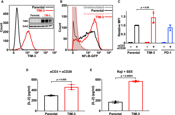
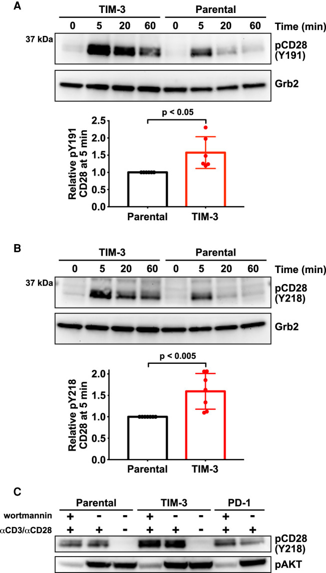
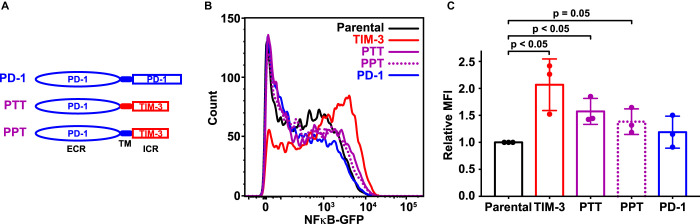
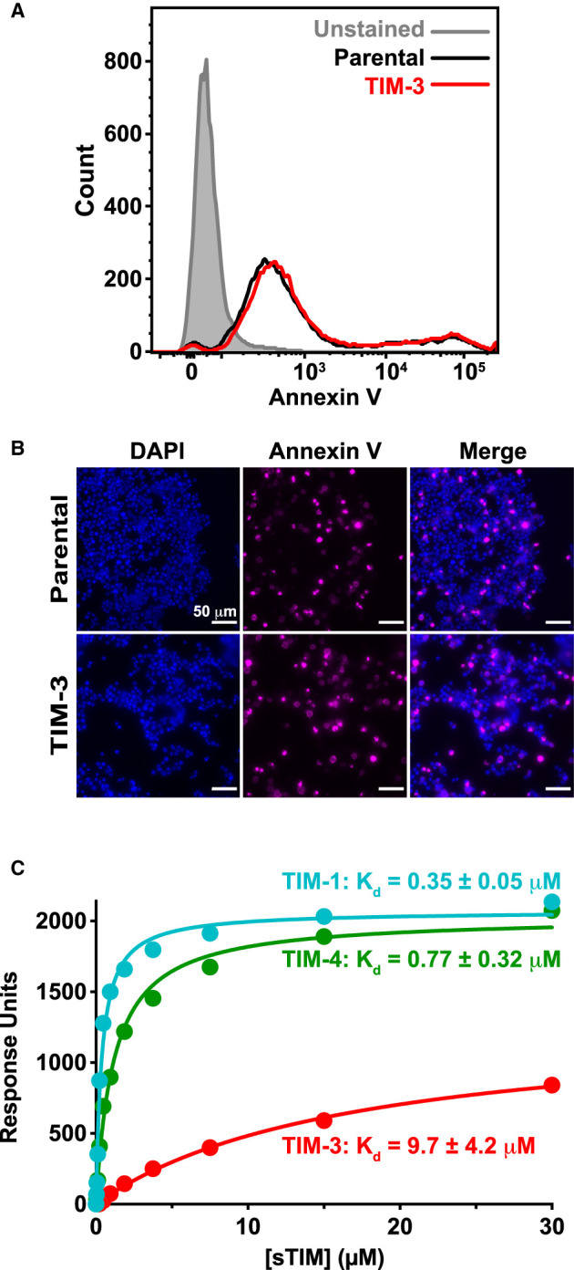
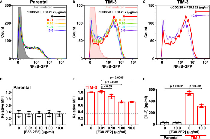
Co-signaling receptors for the T cell receptor (TCR) are important therapeutic targets, with blockade of co-inhibitory receptors such as PD-1 now central in immuno-oncology. Advancing additional therapeutic immune modulation approaches requires understanding ligand regulation of other co-signaling receptors. One poorly understood potential therapeutic target is TIM-3 (T cell immunoglobulin and mucin domain containing-3). Which of TIM-3's several proposed regulatory ligands is/are relevant for signaling is unclear, and different studies have reported TIM-3 as a co-inhibitory or co-stimulatory receptor in T cells. Here, we show that TIM-3 promotes NF-κB signaling and IL-2 secretion following TCR stimulation in Jurkat cells, and that this activity is regulated by binding to phosphatidylserine (PS). TIM-3 signaling is stimulated by PS exposed constitutively in cultured Jurkat cells, and can be blocked by mutating the PS-binding site or by occluding this site with an antibody. We also find that TIM-3 signaling alters CD28 phosphorylation. Our findings clarify the importance of PS as a functional TIM-3 ligand, and may inform the future exploitation of TIM-3 as a therapeutic target.
Overview
The claims and statistics of this paper haven’t been checked yet.
Free: it reads the open-access text and checks the first 5 claims that cite a source we can check. Create a free account to check every claim.
Integrity signals
Registry
Cites
Statistics
Figures

Verification
Claims the paper makes
The claims and statistics of this paper haven’t been checked yet.
Free: it reads the open-access text and checks the first 5 claims that cite a source we can check. Create a free account to check every claim.
Claims other papers make about this one
Each is checked against this paper’s own text: does it say what the citing paper says it does?
The source backs part of the claim, not all of it — or backs it for a different population, condition or comparison.
In the citing paper
“Another data revealed that the blockade activity of the TIM‐3 or galectin 9 axis is due to the action of galectin 3 by clustering TIM‐3 to inhibit the binding to PS [314].”
From Tumor microenvironment signaling and therapeutics in cancer progression (2023) · cites this paper as [314]
Experimental — accuracy not yet measured
Self-check
Percentages, sums and reported differences re-computed from the paper’s own numbers.
The self-check needs the paper’s full text.
Citations
Cites
Molecular mechanisms of T cell co-stimulation and co-inhibition
Chen, L., & Flies, D. B. (2013) · Nature Reviews Immunology · doi:10.1038/nri3405 · Cited by 3,114 · cited 1×
“The functional outcome when an antigen engages the T cell receptor (TCR) depends on the activity of a wide range of ‘ co-signaling receptors ’ in T cells [1], which can be stimulatory (like CD28) or inhibitory (like CTLA-4 and PD-1).”
Targeting Tim-3 and PD-1 pathways to reverse T cell exhaustion and restore anti-tumor immunity
Sakuishi, K., Apetoh, L., Sullivan, J. M., Blazar, B. R., Kuchroo, V. K., & Anderson, A. C. (2010) · Journal of Experimental Medicine · doi:10.1084/jem.20100643 · Cited by 2,072 · cited 1×
“Nonetheless, pre-clinical studies have indicated that antibody blockade of TIM-3, in combination with PD-1 blockade, may be a promising therapeutic approach in cancer [9,10].”
The Tim-3 ligand galectin-9 negatively regulates T helper type 1 immunity
Zhu, C., Anderson, A. C., Schubart, A., Xiong, H., Imitola, J., Khoury, S. J., Zheng, X. X., Strom, T. B., & Kuchroo, V. K. (2005) · Nature Immunology · doi:10.1038/ni1271 · Cited by 2,057 · cited 2×
“The fi rst was the lectin family member galectin-9 [26], which has two β -galactoside-binding carbohydrate-recognition domains.”
Monney, L., Sabatos, C. A., Gaglia, J. L., Ryu, A., Waldner, H., Chernova, T., Manning, S., Greenfield, E. A., Coyle, A. J., Sobel, R. A., et al. (2002) · Nature · doi:10.1038/415536a · Cited by 1,472
Membrane recognition by phospholipid-binding domains
Lemmon, M. A. (2008) · Nature Reviews Molecular Cell Biology · doi:10.1038/nrm2328 · Cited by 1,419 · cited 1×
“…values are in the same range as those measured for other PS-binding proteins, pleckstrin homology domains and other peripheral membrane proteins [45,61,62], and indeed, for recognition of the prototypical T cell co-receptors CD28 and PD-1 for their respective ligands, CD80/86 [63] and PD-L1 [64].”
Self-citationFourcade, J., Sun, Z., Benallaoua, M., Guillaume, P., Luescher, I. F., Sander, C., Kirkwood, J. M., Kuchroo, V., & Zarour, H. M. (2010) · Journal of Experimental Medicine · doi:10.1084/jem.20100637 · Cited by 1,307 · cited 1×
“Nonetheless, pre-clinical studies have indicated that antibody blockade of TIM-3, in combination with PD-1 blockade, may be a promising therapeutic approach in cancer [9,10].”
Identification of Tim4 as a phosphatidylserine receptor
Miyanishi, M., Tada, K., Koike, M., Uchiyama, Y., Kitamura, T., & Nagata, S. (2007) · Nature · doi:10.1038/nature06307 · Cited by 1,180 · cited 2×
“PS was initially suggested as a TIM-3 ligand based on homology between TIM-3 and the known PS receptor TIM-4 [33].”
Hayden, M. S., West, A. P., & Ghosh, S. (2006) · Oncogene · doi:10.1038/sj.onc.1209943 · Cited by 1,178
Membrane Phosphatidylserine Regulates Surface Charge and Protein Localization
Yeung, T., Gilbert, G. E., Shi, J., Silvius, J., Kapus, A., & Grinstein, S. (2008) · Science · doi:10.1126/science.1152066 · Cited by 1,107 · cited 1×
“…values are in the same range as those measured for other PS-binding proteins, pleckstrin homology domains and other peripheral membrane proteins [45,61,62], and indeed, for recognition of the prototypical T cell co-receptors CD28 and PD-1 for their respective ligands, CD80/86 [63] and PD-L1 [64].”
Potentiating the antitumour response of CD8+ T cells by modulating cholesterol metabolism
Yang, W., Bai, Y., Xiong, Y., Zhang, J., Chen, S., Zheng, X., Meng, X., Li, L., Wang, J., Xu, C., et al. (2016) · Nature · doi:10.1038/nature17412 · Cited by 1,010 · cited 1×
“…adding annexin V elevated TCR-driven NF-κ B signaling independently of TIM-3 expression (Supplementary Figure S5C,D) — likely because of its reported effects on membrane structure [57], which are likely to alter TCR clustering and activity as seen when cholesterol levels are changed [58].”
Cited by
Tumor microenvironment signaling and therapeutics in cancer progression
Goenka, A., Khan, F., Verma, B., Sinha, P., Dmello, C. C., Jogalekar, M. P., Gangadaran, P., & Ahn, B. (2023) · Cancer Communications · doi:10.1002/cac2.12416 · Cited by 323
Immune checkpoint proteins: Signaling mechanisms and molecular interactions in cancer immunotherapy
Gaikwad, S. R., Agrawal, M. Y., Kaushik, I., Ramachandran, S., & Srivastava, S. (2022) · Seminars in Cancer Biology · doi:10.1016/j.semcancer.2022.03.014 · Cited by 172
TIM-3 as a promising target for cancer immunotherapy in a wide range of tumors
Sauer, N. J., Janicka, N., Szlasa, W., Skinderowicz, B., Kołodzińska, K., Dwernicka, W., Oślizło, M., Kulbacka, J., Novickij, V., & Karłowicz-Bodalska, K. (2023) · Cancer Immunology Immunotherapy · doi:10.1007/s00262-023-03516-1 · Cited by 158
“Furthermore, phosphatidylserine stimulates NF-κB signaling and IL-2 secretion [17].”
Backgroundİmani, S., Farghadani, R., Roozitalab, G., Maghsoudloo, M., Emadi, M., Moradi, A., Abedi, B., & Kaboli, P. J. (2025) · Journal of Experimental & Clinical Cancer Research · doi:10.1186/s13046-025-03394-8 · Cited by 132
Immune Checkpoint Receptors Signaling in T Cells
Baldanzi, G. (2022) · International Journal of Molecular Sciences · doi:10.3390/ijms23073529 · Cited by 80
“TIM-3 is a promiscuous receptor suggested to bind heterologous ligands, such phosphatidylserine, from apoptotic cell [69], carcin embryonic antigen cell adhesion molecule 1 (CEACAM1, [70]), the alarmin protein high mobility group B1 (HMGB1), and the carbohydrate receptor galectin-9 [71].”
BackgroundResult
Metadata
- Authors
- Smith, C. M., Li, A., Krishnamurthy, N., & Lemmon, M. A.
- Affiliations
- Yale University (US)
- Corresponding author
- Smith, Courtney M.
- Lemmon, Mark A.
- Venue
- Biochemical Journal
- Publisher
- Portland Press Ltd.
- Published
- 2021-09-14
- Volume
- 478
- Issue
- 17
- Pages
- 3331-3349
- ISSN
- 0264-6021 (ISSN-L)
- 1470-8728
- PMC
- PMC8454703
- OpenAlex
- W3193547547
- Cited by
- 61
- References
- 87
- Citations by year
- 2026: 15 · 2025: 19 · 2024: 9 · 2023: 10 · 2022: 6 · 2021: 2
- FWCI
- 2.51
- Type
- journal article
- Language
- en
- Open access
- hybrid
- doi.org
- Licence
- Locations
- Biochemical Journal (publishedVersion, cc-by-nc-nd)
- Europe PMC (PubMed Central) (publishedVersion, other-oa)
- PubMed Central (publishedVersion, cc-by-nc-nd)
- PubMed (publishedVersion)
- Funders
- National Institute of General Medical Sciences (R35GM122485)
- Topics
- Life Sciences › Immunology and Microbiology › Immunology › T-cell and B-cell Immunology
- Health Sciences › Medicine › Oncology › Cancer Immunotherapy and Biomarkers
- Life Sciences › Immunology and Microbiology › Immunology › Phagocytosis and Immune Regulation
- Keywords
- TIM-3, phosphatidylserine, NF-κB signaling, IL-2 secretion, T cell receptor signaling, immunotherapy targets, Jurkat cells
- MeSH terms
- Hepatitis A Virus Cellular Receptor 2, Antibodies, Binding Sites, Humans, Interleukin-2, Ligands, Macrophages, Phosphatidylserines, Phosphorylation, Receptors, Antigen, T-Cell, T-Lymphocytes, Transfection, Signal Transduction, NF-kappa B, Apoptosis, CD28 Antigens, Jurkat Cells, HEK293 Cells
About these checks
Registry, retraction, Cites and statistics results are computed, not judged, with one exception: a printed reference that no index lists is read once by a model before it counts as a citation error. Claims are extracted by a model and kept only when their quote appears verbatim in the text; each cited source is then checked, with a model’s help, for whether it says what the claim relies on. Those verdicts can be wrong — read the claim’s quote and the cited source before relying on one.
Statistics and arithmetic are recomputed, not judged.
Confidence bands are shown only for fields calibrated on our annotated test set. None is calibrated yet, so no band is shown.
What counts as a claim ›How we measure confidence ›How we check this ›
Are you an author, or do you see a problem with this page? Request a review
How to cite
Cite CoChat for the checks on this page, and the paper for its own findings.
- This page
- CoChat. (2026). Citation check: Phosphatidylserine binding directly regulates TIM-3 function. https://app.cochat.ai/papers/10.1042/bcj20210425
- The paper
- Smith, C. M., Li, A., Krishnamurthy, N., & Lemmon, M. A. (2021). Phosphatidylserine binding directly regulates TIM-3 function. Biochemical Journal. https://doi.org/10.1042/bcj20210425
Data sources
Bibliographic data: Semantic Scholar, OpenAlex, Crossref.
Contains information from the Semantic Scholar Academic Graph Datasets, made available under the ODC Attribution License (ODC-BY 1.0).
Semantic Scholar’s data is described in: Kinney, R., et al. (2023). The Semantic Scholar Open Data Platform. arXiv:2301.10140.
