

Phosphatidylserine binding directly regulates TIM-3 function
Open accessThe DOI matches its Crossref record; 88 of the 88 references it cites were found in a registry or an index.
Abstract
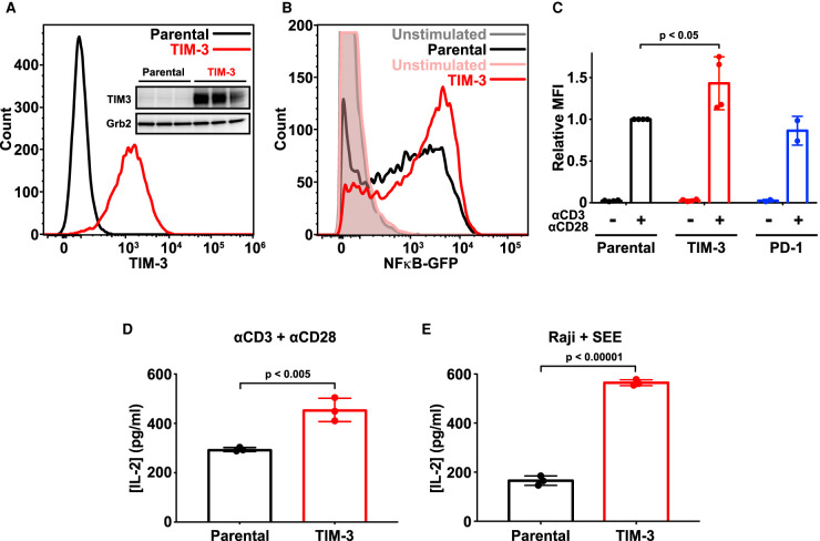
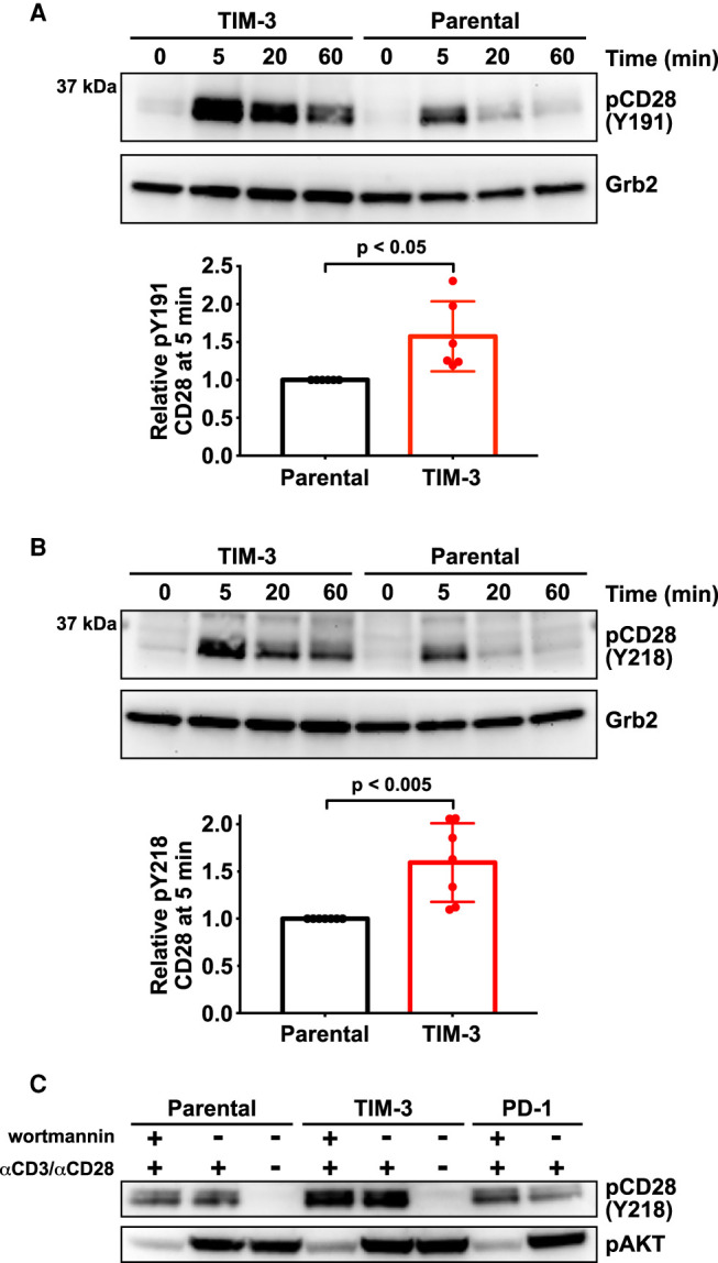
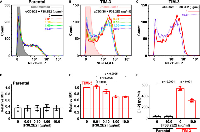
Co-signaling receptors for the T cell receptor (TCR) are important therapeutic targets, with blockade of co-inhibitory receptors such as PD-1 now central in immuno-oncology. Advancing additional therapeutic immune modulation approaches requires understanding ligand regulation of other co-signaling receptors. One poorly understood potential therapeutic target is TIM-3 (T cell immunoglobulin and mucin domain containing-3). Which of TIM-3's several proposed regulatory ligands is/are relevant for signaling is unclear, and different studies have reported TIM-3 as a co-inhibitory or co-stimulatory receptor in T cells. Here, we show that TIM-3 promotes NF-κB signaling and IL-2 secretion following TCR stimulation in Jurkat cells, and that this activity is regulated by binding to phosphatidylserine (PS). TIM-3 signaling is stimulated by PS exposed constitutively in cultured Jurkat cells, and can be blocked by mutating the PS-binding site or by occluding this site with an antibody. We also find that TIM-3 signaling alters CD28 phosphorylation. Our findings clarify the importance of PS as a functional TIM-3 ligand, and may inform the future exploitation of TIM-3 as a therapeutic target.
Overview
The claims and statistics of this paper haven’t been checked yet.
Free: it reads the open-access text and checks the first 5 claims that cite a source we can check. Create a free account to check every claim.
Integrity signals
Registry
Cites
Statistics
Figures

Verification
Claims the paper makes
The claims and statistics of this paper haven’t been checked yet.
Free: it reads the open-access text and checks the first 5 claims that cite a source we can check. Create a free account to check every claim.
Claims other papers make about this one
Each is checked against this paper’s own text: does it say what the citing paper says it does?
The source backs part of the claim, not all of it — or backs it for a different population, condition or comparison.
In the citing paper
“Another data revealed that the blockade activity of the TIM‐3 or galectin 9 axis is due to the action of galectin 3 by clustering TIM‐3 to inhibit the binding to PS [314].”
From Tumor microenvironment signaling and therapeutics in cancer progression (2023) · cites this paper as [314]
Experimental — accuracy not yet measured
Self-check
Percentages, sums and reported differences re-computed from the paper’s own numbers.
The self-check needs the paper’s full text.
Citations
Cites
Molecular mechanisms of T cell co-stimulation and co-inhibition
Chen, L., & Flies, D. B. (2013) · Nature Reviews Immunology · doi:10.1038/nri3405 · Cited by 3,114 · cited 1×
“The functional outcome when an antigen engages the T cell receptor (TCR) depends on the activity of a wide range of ‘ co-signaling receptors ’ in T cells [1], which can be stimulatory (like CD28) or inhibitory (like CTLA-4 and PD-1).”
Targeting Tim-3 and PD-1 pathways to reverse T cell exhaustion and restore anti-tumor immunity
Sakuishi, K., Apetoh, L., Sullivan, J. M., Blazar, B. R., Kuchroo, V. K., & Anderson, A. C. (2010) · Journal of Experimental Medicine · doi:10.1084/jem.20100643 · Cited by 2,072 · cited 1×
“Nonetheless, pre-clinical studies have indicated that antibody blockade of TIM-3, in combination with PD-1 blockade, may be a promising therapeutic approach in cancer [9,10].”
The Tim-3 ligand galectin-9 negatively regulates T helper type 1 immunity
Zhu, C., Anderson, A. C., Schubart, A., Xiong, H., Imitola, J., Khoury, S. J., Zheng, X. X., Strom, T. B., & Kuchroo, V. K. (2005) · Nature Immunology · doi:10.1038/ni1271 · Cited by 2,057 · cited 2×
“The fi rst was the lectin family member galectin-9 [26], which has two β -galactoside-binding carbohydrate-recognition domains.”
Monney, L., Sabatos, C. A., Gaglia, J. L., Ryu, A., Waldner, H., Chernova, T., Manning, S., Greenfield, E. A., Coyle, A. J., Sobel, R. A., et al. (2002) · Nature · doi:10.1038/415536a · Cited by 1,472
Membrane recognition by phospholipid-binding domains
Lemmon, M. A. (2008) · Nature Reviews Molecular Cell Biology · doi:10.1038/nrm2328 · Cited by 1,419 · cited 1×
“…values are in the same range as those measured for other PS-binding proteins, pleckstrin homology domains and other peripheral membrane proteins [45,61,62], and indeed, for recognition of the prototypical T cell co-receptors CD28 and PD-1 for their respective ligands, CD80/86 [63] and PD-L1 [64].”
Self-citationFourcade, J., Sun, Z., Benallaoua, M., Guillaume, P., Luescher, I. F., Sander, C., Kirkwood, J. M., Kuchroo, V., & Zarour, H. M. (2010) · Journal of Experimental Medicine · doi:10.1084/jem.20100637 · Cited by 1,307 · cited 1×
“Nonetheless, pre-clinical studies have indicated that antibody blockade of TIM-3, in combination with PD-1 blockade, may be a promising therapeutic approach in cancer [9,10].”
Identification of Tim4 as a phosphatidylserine receptor
Miyanishi, M., Tada, K., Koike, M., Uchiyama, Y., Kitamura, T., & Nagata, S. (2007) · Nature · doi:10.1038/nature06307 · Cited by 1,180 · cited 2×
“PS was initially suggested as a TIM-3 ligand based on homology between TIM-3 and the known PS receptor TIM-4 [33].”
Hayden, M. S., West, A. P., & Ghosh, S. (2006) · Oncogene · doi:10.1038/sj.onc.1209943 · Cited by 1,178
Membrane Phosphatidylserine Regulates Surface Charge and Protein Localization
Yeung, T., Gilbert, G. E., Shi, J., Silvius, J., Kapus, A., & Grinstein, S. (2008) · Science · doi:10.1126/science.1152066 · Cited by 1,107 · cited 1×
“…values are in the same range as those measured for other PS-binding proteins, pleckstrin homology domains and other peripheral membrane proteins [45,61,62], and indeed, for recognition of the prototypical T cell co-receptors CD28 and PD-1 for their respective ligands, CD80/86 [63] and PD-L1 [64].”
Potentiating the antitumour response of CD8+ T cells by modulating cholesterol metabolism
Yang, W., Bai, Y., Xiong, Y., Zhang, J., Chen, S., Zheng, X., Meng, X., Li, L., Wang, J., Xu, C., et al. (2016) · Nature · doi:10.1038/nature17412 · Cited by 1,010 · cited 1×
“…adding annexin V elevated TCR-driven NF-κ B signaling independently of TIM-3 expression (Supplementary Figure S5C,D) — likely because of its reported effects on membrane structure [57], which are likely to alter TCR clustering and activity as seen when cholesterol levels are changed [58].”
Neoadjuvant checkpoint blockade for cancer immunotherapy
Topalian, S. L., Taube, J. M., & Pardoll, D. M. (2020) · Science · doi:10.1126/science.aax0182 · Cited by 1,001
The Distribution and Function of Phosphatidylserine in Cellular Membranes
Leventis, P. A., & Grinstein, S. (2010) · Annual Review of Biophysics · doi:10.1146/annurev.biophys.093008.131234 · Cited by 988
TIM3 comes of age as an inhibitory receptor
Wolf, Y., Anderson, A. C., & Kuchroo, V. K. (2020) · Nature Reviews Immunology · doi:10.1038/s41577-019-0224-6 · Cited by 959
Cooperation of Tim-3 and PD-1 in CD8 T-cell exhaustion during chronic viral infection
Jin, H.-T., Anderson, A. C., Tan, W. G., West, E. E., Ha, S.-J., Araki, K., Freeman, G. J., Kuchroo, V. K., & Ahmed, R. (2010) · Proceedings of the National Academy of Sciences · doi:10.1073/pnas.1009731107 · Cited by 858 · cited 1×
“TIM-3 was fi rst identi fi ed as a marker of CD4 T helper 1 (T H 1) cells and CD8 cytotoxic T (T C 1) cells [12], and was later identi fi ed on exhausted T cells in chronic viral infection and cancer [13 – 15].”
Jones, R. B., Ndhlovu, L. C., Barbour, J. D., Sheth, P. M., Jha, A. R., Long, B. R., Wong, J. C., Satkunarajah, M., Schweneker, M., Chapman, J. M., et al. (2008) · The Journal of Experimental Medicine · doi:10.1084/jem.20081398 · Cited by 753
Enzymatic Assembly of Overlapping DNA Fragments
Gibson, D. G. (2011) · Methods in Enzymology · doi:10.1016/b978-0-12-385120-8.00015-2 · Cited by 736 · cited 1×
“1 (+) using Gibson assembly [38].”
Sánchez-Fueyo, A., Tian, J., Picarella, D., Domenig, C., Zheng, X. X., Sabatos, C. A., Manlongat, N., Bender, O., Kamradt, T., Kuchroo, V. K., et al. (2003) · Nature Immunology · doi:10.1038/ni987 · Cited by 712 · cited 4×
“Early in vivo studies led to the suggestion of a co-inhibitory signaling role for TIM-3 in T cells [16,17].”
InfluentialPromotion of Tissue Inflammation by the Immune Receptor Tim-3 Expressed on Innate Immune Cells
Anderson, A. C., Anderson, D. E., Bregoli, L., Hastings, W. D., Kassam, N., Lei, C., Chandwaskar, R., Karman, J., Su, E. W., Hirashima, M., et al. (2007) · Science · doi:10.1126/science.1148536 · Cited by 701
Zhou, Q., Munger, M. E., Veenstra, R. G., Weigel, B. J., Hirashima, M., Munn, D. H., Murphy, W. J., Azuma, M., Anderson, A. C., Kuchroo, V. K., et al. (2011) · Blood · doi:10.1182/blood-2010-10-310425 · Cited by 675
Human Immunodeficiency Virus Type 1 Spinoculation Enhances Infection through Virus Binding
O'Doherty, U., Swiggard, W. J., & Malim, M. H. (2000) · Journal of Virology · doi:10.1128/jvi.74.21.10074-10080.2000 · Cited by 664 · cited 1×
“Filtered virus suspension was then added to Jurkat cells (2 × 10 5 cells), and cells were transduced with lentivirus by spinfection (spinning at 800× g for 30 min at 32°C) [41] in sealed aerosol containment lids.”
Sabatos, C. A., Chakravarti, S., Cha, E., Schubart, A., Sánchez-Fueyo, A., Zheng, X. X., Coyle, A. J., Strom, T. B., Freeman, G. J., & Kuchroo, V. K. (2003) · Nature Immunology · doi:10.1038/ni988 · Cited by 628 · cited 4×
“Early in vivo studies led to the suggestion of a co-inhibitory signaling role for TIM-3 in T cells [16,17].”
InfluentialFreeman, G. J., Casasnovas, J. M., Umetsu, D. T., & DeKruyff, R. H. (2010) · Immunological Reviews · doi:10.1111/j.0105-2896.2010.00903.x · Cited by 614 · cited 1×
“The role of PS binding in regulating signaling effects of TIM-3 has remained largely unexplored [8,70,71].”
Jurkat T cells and development of the T-cell receptor signalling paradigm
Abraham, R. T., & Weiss, A. (2004) · Nature Reviews Immunology · doi:10.1038/nri1330 · Cited by 598
Inhibitory receptors and ligands beyond PD-1, PD-L1 and CTLA-4: breakthroughs or backups
Andrews, L. P., Yano, H., & Vignali, D. A. A. (2019) · Nature Immunology · doi:10.1038/s41590-019-0512-0 · Cited by 543
Tim-3 mediates phagocytosis of apoptotic cells and cross-presentation
Nakayama, M., Akiba, H., Takeda, K., Kojima, Y., Hashiguchi, M., Azuma, M., Yagita, H., & Okumura, K. (2009) · Blood · doi:10.1182/blood-2008-10-185884 · Cited by 432 · cited 2×
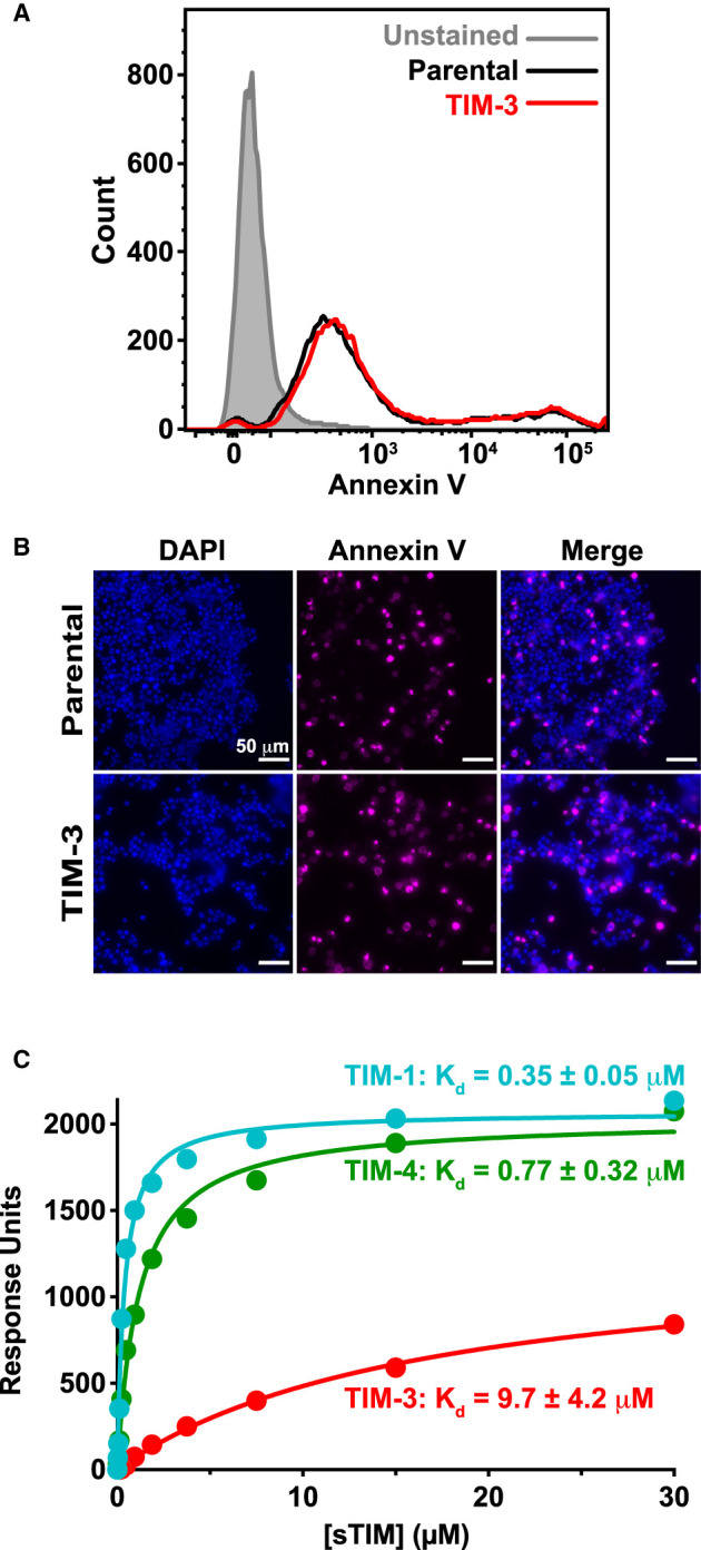
“The relative af fi nities that we observed here re fl ect what has been reported for murine TIM proteins using ELISA-type assays [34,35] and other approaches [36], with sTIM-3 binding PS membranes 12 – 27-fold more weakly than sTIM-1 or sTIM-4.”
Binding of phosphatidyl-inositol-3-OH kinase to CD28 is required for T-cell signalling
Pagès, F., Ragueneau, M., Rottapel, R., Truneh, A., Nunes, J., Imbert, J., & Olive, D. (1994) · Nature · doi:10.1038/369327a0 · Cited by 398
Tim-3 finds its place in the cancer immunotherapy landscape
Acharya, N., Sabatos-Peyton, C., & Anderson, A. C. (2020) · Journal for ImmunoTherapy of Cancer · doi:10.1136/jitc-2020-000911 · Cited by 394 · cited 3×
“Several TIM-3 antibodies are now in clinical trials [11], underlining the need to understand this co-receptor mechanistically.”
InfluentialStructural Biology of the Immune Checkpoint Receptor PD-1 and Its Ligands PD-L1/PD-L2
Zak, K. M., Grudnik, P., Magiera, K., Dömling, A., Dubin, G., & Holak, T. A. (2017) · Structure · doi:10.1016/j.str.2017.06.011 · Cited by 388 · cited 1×
“…values are in the same range as those measured for other PS-binding proteins, pleckstrin homology domains and other peripheral membrane proteins [45,61,62], and indeed, for recognition of the prototypical T cell co-receptors CD28 and PD-1 for their respective ligands, CD80/86 [63] and PD-L1 [64].”
Gleason, M. K., Lenvik, T. R., McCullar, V., Felices, M., O'Brien, M. S., Cooley, S. A., Verneris, M. R., Cichocki, F., Holman, C. J., Panoskaltsis-Mortari, A., et al. (2012) · Blood · doi:10.1182/blood-2011-06-360321 · Cited by 373 · cited 1×
“TIM-3 has also been reported to enhance DC and natural killer (NK) cell activation, as well as Fc ε R1 signaling in mast cells [19,79,80].”
TIM-3 restrains anti-tumour immunity by regulating inflammasome activation
Dixon, K. O., Tabaka, M., Schramm, M. A., Xiao, S., Tang, R., Dionne, D., Anderson, A. C., Rozenblatt-Rosen, O., Regev, A., & Kuchroo, V. K. (2021) · Nature · doi:10.1038/s41586-021-03626-9 · Cited by 372 · cited 2×
“However, as mentioned in the Introduction, very recent studies have suggested that the inhibitory effect of TIM-3 on anti-tumor immunity actually originates from its effects on NLRP3 in fl ammasome activation in dendritic cells [18], and not its effects in T cells.”
Immunoreceptor tyrosine‐based inhibition motifs: a quest in the past and future
Daëron, M., Jaeger, S., Du Pasquier, L., & Vivier, E. (2008) · Immunological Reviews · doi:10.1111/j.1600-065x.2008.00666.x · Cited by 370 · cited 1×
“TIM-3 does not have a de fi nable intracellular ITIM (immunoreceptor tyrosine-based inhibitory motif) or ITSM (immunoreceptor tyrosine-based switch motif), motifs that normally characterize co-inhibitory receptors and recruit SH2 domain-containing phosphatases to reduce T cell signaling [25].”
CD28, CTLA‐4 and their ligands: who does what and to whom?
Sansom, D. M. (2000) · Immunology · doi:10.1046/j.1365-2567.2000.00121.x · Cited by 362 · cited 1×
“…values are in the same range as those measured for other PS-binding proteins, pleckstrin homology domains and other peripheral membrane proteins [45,61,62], and indeed, for recognition of the prototypical T cell co-receptors CD28 and PD-1 for their respective ligands, CD80/86 [63] and PD-L1 [64].”
Shan, X., Czar, M. J., Bunnell, S. C., Liu, P., Liu, Y., Schwartzberg, P. L., & Wange, R. L. (2000) · Molecular and Cellular Biology · doi:10.1128/mcb.20.18.6945-6957.2000 · Cited by 350 · cited 1×
“Phosphorylation of Y191 in CD28 promotes recruitment of phosphoinositide 3-kinase (PI3K) to CD28 [53,54], and the consequences of altering its phosphorylation are likely blunted in Jurkat cells, which lack PTEN [55].”
Galectin-9-CD44 Interaction Enhances Stability and Function of Adaptive Regulatory T Cells
Wu, C., Thalhamer, T., Franca, R. F., Xiao, S., Wang, C., Hotta, C., Zhu, C., Hirashima, M., Anderson, A. C., & Kuchroo, V. K. (2014) · Immunity · doi:10.1016/j.immuni.2014.06.011 · Cited by 349
DeKruyff, R. H., Bu, X., Ballesteros, A., Santiago, C., Chim, Y.-L. E., Lee, H.-H., Karisola, P., Pichavant, M., Kaplan, G. G., Umetsu, D. T., et al. (2010) · The Journal of Immunology · doi:10.4049/jimmunol.0903059 · Cited by 330 · cited 7×
“( A ) Details of the PS-binding site in murine TIM-3 (PDB: 3KAA — blue) overlaid on human TIM-3 (PDB: 6TXZ — red), revealing key interacting residues [34,82].”
InfluentialPrasad, K. V., Cai, Y. C., Raab, M., Duckworth, B., Cantley, L., Shoelson, S. E., & Rudd, C. E. (1994) · Proceedings of the National Academy of Sciences · doi:10.1073/pnas.91.7.2834 · Cited by 295
Phosphotyrosine-Dependent Coupling of Tim-3 to T-Cell Receptor Signaling Pathways
Lee, J., Su, E. W., Zhu, C., Hainline, S., Phuah, J., Moroco, J. A., Smithgall, T. E., Kuchroo, V. K., & Kane, L. P. (2011) · Molecular and Cellular Biology · doi:10.1128/mcb.05297-11 · Cited by 285 · cited 9×
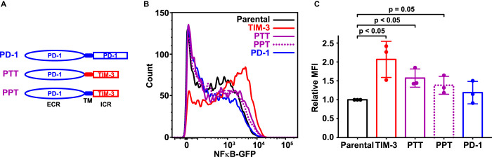
“Lee et al. [22] previously used a similar strategy to investigate signaling requirements of the TIM-3 cytoplasmic tail.”
InfluentialAn Enigmatic Tail of CD28 Signaling
Boomer, J. S., & Green, J. M. (2010) · Cold Spring Harbor Perspectives in Biology · doi:10.1101/cshperspect.a002436 · Cited by 283 · cited 1×
“As expected [50], elevated CD28 phosphorylation was seen at both sites following α CD3/ α CD28 stimulation of parental cells, but TIM-3 expression further increased the magnitude of this response by ∼ 50% — with no apparent elevation of baseline CD28 phosphorylation.”
Bi, S., Hong, P. W., Lee, B., & Baum, L. G. (2011) · Proceedings of the National Academy of Sciences · doi:10.1073/pnas.1017954108 · Cited by 248
Exposure of Phosphatidylethanolamine on the Surface of Apoptotic Cells
Emoto, K., Toyama-Sorimachi, N., Karasuyama, H., Inoue, K., & Umeda, M. (1997) · Experimental Cell Research · doi:10.1006/excr.1997.3521 · Cited by 248 · cited 1×
“We also asked whether the sTIM proteins can bind other phospholipids known to become exposed on the cell surface during cell death, namely phosphatidic acid (PA) and phosphatidylethanolamine (PE) [65,66]. sTIM-1, sTIM-3, and sTIM-4 all bound to 20% PA-containing membranes (Supplementary Figure S7C –”
Cao, E., Zang, X., Ramagopal, U. A., Mukhopadhaya, A., Fedorov, A., Fedorov, E., Zencheck, W. D., Lary, J. W., Cole, J. L., Deng, H., et al. (2007) · Immunity · doi:10.1016/j.immuni.2007.01.016 · Cited by 228 · cited 3×
“E. coli -derived recombinant murine TIM-3 IgV domain was previously shown to bind T cells, fi broblasts and macrophages from several species [56] — consistent with the recognition of a conserved component of these cells, such as PS.”
Targeting co-stimulatory molecules in autoimmune disease
Edner, N. M., Carlesso, G., Rush, J. S., & Walker, L. S. K. (2020) · Nature Reviews Drug Discovery · doi:10.1038/s41573-020-0081-9 · Cited by 228 · cited 1×
“Both classes of co-receptor offer important opportunities in immunotherapy, including suppression of co-stimulatory receptor signaling in autoimmunity [2] and suppression of co-inhibitory receptors — or immune checkpoint blockade (ICB) — in cancer [3,4].”
Membrane phosphatidylserine distribution as a non-apoptotic signalling mechanism in lymphocytes
Elliott, J. I., Surprenant, A., Marelli-Berg, F. M., Cooper, J. C., Cassady-Cain, R. L., Wooding, C., Linton, K., Alexander, D. R., & Higgins, C. F. (2005) · Nature Cell Biology · doi:10.1038/ncb1279 · Cited by 227 · cited 2×
“Another major TIM-3 ligand is the membrane phospholipid phosphatidylserine (PS), exposed on the surface of cells undergoing apoptosis and other processes [29,30], including T cell activation [31,32].”
DNASU plasmid and PSI: biology-materials repositories: resources to accelerate biological research
Seiler, C. Y., Park, J. G., Sharma, A., Hunter, P., Surapaneni, P., Sedillo, C., Field, J., Algar, R., Price, A., Steel, J., et al. (2014) · Nucleic Acids Research · doi:10.1093/nar/gkt1060 · Cited by 224
Loke, P., MacDonald, A. S., Robb, A., Maizels, R. M., & Allen, J. E. (2000) · European Journal of Immunology · doi:10.1002/1521-4141(200009)30:9<2669::aid-immu2669>3.0.co;2-1 · Cited by 211
Flipping the dogma – phosphatidylserine in non-apoptotic cell death
Shlomovitz, I., Speir, M., & Gerlic, M. (2019) · Cell Communication and Signaling · doi:10.1186/s12964-019-0437-0 · Cited by 189 · cited 1×
“Another major TIM-3 ligand is the membrane phospholipid phosphatidylserine (PS), exposed on the surface of cells undergoing apoptosis and other processes [29,30], including T cell activation [31,32].”
Survival of pancreatic cancer cells lacking KRAS function
Muzumdar, M. D., Chen, P.-Y., Dorans, K. J., Chung, K. M., Bhutkar, A., Hong, E., Noll, E. M., Sprick, M. R., Trumpp, A., & Jacks, T. (2017) · Nature Communications · doi:10.1038/s41467-017-00942-5 · Cited by 177 · cited 1×
“…transfer plasmid was provided by the laboratory of Mandar Muzumdar at Yale University and contained AmpR, PuroR under the SV40 promoter, and the gene of interest under the PGK promoter [40]. cDNA fragments encoding human TIM-3 and human PD-1 were sub-cloned into the lentivirus transfer plasmid.”
Too Much of a Good Thing? Tim-3 and TCR Signaling in T Cell Exhaustion
Ferris, R. L., Lu, B., & Kane, L. P. (2014) · The Journal of Immunology · doi:10.4049/jimmunol.1400557 · Cited by 173
Niki, T., Tsutsui, S., Hirose, S., Aradono, S., Sugimoto, Y., Takeshita, K., Nishi, N., & Hirashima, M. (2009) · Journal of Biological Chemistry · doi:10.1074/jbc.m109.035196 · Cited by 170 · cited 1×
“However, as a carbohydrate-binding protein, galectin-9 is known to bind numerous other targets [74 – 77], and several studies have argued that key effects of galectin-9 on T cells are TIM-3-independent [28,77,78] — or alternatively that galectin-9 does not bind TIM-3 at all [27].”
TIM-3 and Its Regulatory Role in Immune Responses
Zhu, C. Y., Anderson, A. C., & Kuchroo, V. K. (2010) · Current topics in microbiology and immunology · doi:10.1007/82_2010_84 · Cited by 169 · cited 1×
“Nonetheless, the prevailing hypothesis in the fi eld is that galectin-9 is the key TIM-3 ligand and binds TIM-3 to trigger co-inhibitory signaling and thus T cell death [8,26,73].”
Moravcevic, K., Mendrola, J. M., Schmitz, K. R., Wang, Y.-H., Slochower, D., Janmey, P. A., & Lemmon, M. A. (2010) · Cell · doi:10.1016/j.cell.2010.11.028 · Cited by 169
Self-citationAntigen recognition induces phosphatidylserine exposure on the cell surface of human CD8+ T cells
Fischer, K., Voelkl, S., Berger, J., Andreesen, R., Pomorski, T., & Mackensen, A. (2006) · Blood · doi:10.1182/blood-2006-03-011742 · Cited by 169 · cited 4×
“Another major TIM-3 ligand is the membrane phospholipid phosphatidylserine (PS), exposed on the surface of cells undergoing apoptosis and other processes [29,30], including T cell activation [31,32].”
InfluentialAvery, L., Filderman, J., Szymczak-Workman, A. L., & Kane, L. P. (2018) · Proceedings of the National Academy of Sciences · doi:10.1073/pnas.1712107115 · Cited by 169
Harding, J. J., Moreno, V., Bang, Y.-J., Hong, M. H., Patnaik, A., Trigo, J., Szpurka, A. M., Yamamoto, N., Doi, T., Fu, S., et al. (2021) · Clinical Cancer Research · doi:10.1158/1078-0432.ccr-20-4405 · Cited by 161 · cited 1×
“Most TIM-3 antibodies under investigation have been shown to recognize the PS-binding site of TIM-3 [37,82], including Sym023 [83], BGB-A425 [84], ICAGN02390 [85], IBI104 [86], and LY3321367 [87].”
Determining selectivity of phosphoinositide-binding domains
Narayan, K., & Lemmon, M. A. (2006) · Methods · doi:10.1016/j.ymeth.2006.05.006 · Cited by 150 · cited 2×
“The actual K d, app values that we report are substantially weaker than those suggested using ELISA methods (which were in the nM range), as is typical for multivalent systems when comparing results from direct binding studies such as SPR with those from ELISA assays [46,60].”
Self-citationTan, S., Xu, Y., Wang, Z., Wang, T., Du, X., Song, X., Guo, X., Peng, J., Zhang, J., Liang, Y., et al. (2020) · Cancer Research · doi:10.1158/0008-5472.can-19-2332 · Cited by 147 · cited 1×
“Moreover, stimulation of TIM-3 with PS was recently reported to promote TIM-3 phosphorylation in NK cells, leading to inhibition of cytokine secretion [81].”
Richard, A. S., Zhang, A., Park, S.-J., Farzan, M., Zong, M., & Choe, H. (2015) · Proceedings of the National Academy of Sciences · doi:10.1073/pnas.1508095112 · Cited by 144 · cited 1×
“This is consistent with a previous report [67] that also indicated that PE on the surface of apoptotic cells promotes their TIM-1-mediated phagocytosis.”
Clayton, K. L., Haaland, M. S., Douglas-Vail, M. B., Mujib, S., Chew, G. M., Ndhlovu, L. C., & Ostrowski, M. A. (2014) · The Journal of Immunology · doi:10.4049/jimmunol.1302663 · Cited by 134
Galectin-9 suppresses Th17 cell development in an IL-2-dependent but Tim-3-independent manner
Oomizu, S., Arikawa, T., Niki, T., Kadowaki, T., Ueno, M., Nishi, N., Yamauchi, A., & Hirashima, M. (2012) · Clinical Immunology · doi:10.1016/j.clim.2012.01.004 · Cited by 131 · cited 1×
“However, as a carbohydrate-binding protein, galectin-9 is known to bind numerous other targets [74 – 77], and several studies have argued that key effects of galectin-9 on T cells are TIM-3-independent [28,77,78] — or alternatively that galectin-9 does not bind TIM-3 at all [27].”
PD-1 and BTLA regulate T cell signaling differentially and only partially through SHP1 and SHP2
Xu, X., Hou, B., Fulzele, A., Masubuchi, T., Zhao, Y., Wu, Z., Hu, Y., Jiang, Y., Ma, Y., Wang, H., et al. (2020) · Journal of Cell Biology · doi:10.1083/jcb.201905085 · Cited by 130 · cited 1×
“After starvation, 2 × 10 5 Jurkat cells were mixed with 10 5 SEE-loaded Raji B cells per well in a 96-well U-bottom plate, as described [43].”
Phosphatidylserine Is the Signal for TAM Receptors and Their Ligands
Lemke, G. (2017) · Trends in Biochemical Sciences · doi:10.1016/j.tibs.2017.06.004 · Cited by 128 · cited 1×
“…a PS probe did prove useful for assessing the exposure of this phospholipid on our Jurkat cells, revealing that PS is indeed exposed on their surface in our cultures — presumably as cells undergo apoptosis and/or activation, two processes known to lead to PS exposure on the cell surface [29,31,59].”
Sabatos-Peyton, C. A., Nevin, J. T., Brock, A., Venable, J. D., Tan, D. J., Kassam, N., Xu, F., Taraszka, J. A., Wesemann, L. D., Pertel, T., et al. (2017) · OncoImmunology · doi:10.1080/2162402x.2017.1385690 · Cited by 124 · cited 4×
“Most TIM-3 antibodies under investigation have been shown to recognize the PS-binding site of TIM-3 [37,82], including Sym023 [83], BGB-A425 [84], ICAGN02390 [85], IBI104 [86], and LY3321367 [87].”
InfluentialAnnexin-V stabilizes membrane defects by inducing lipid phase transition
Lin, Y., Chipot, C. J., & Scheuring, S. (2020) · Nature Communications · doi:10.1038/s41467-019-14045-w · Cited by 115 · cited 1×
“…adding annexin V elevated TCR-driven NF-κ B signaling independently of TIM-3 expression (Supplementary Figure S5C,D) — likely because of its reported effects on membrane structure [57], which are likely to alter TCR clustering and activity as seen when cholesterol levels are changed [58].”
Galectin-9 regulates T helper cell function independently of Tim-3
Su, E. W., Bi, S., & Kane, L. P. (2011) · Glycobiology · doi:10.1093/glycob/cwq214 · Cited by 111 · cited 2×
“However, as a carbohydrate-binding protein, galectin-9 is known to bind numerous other targets [74 – 77], and several studies have argued that key effects of galectin-9 on T cells are TIM-3-independent [28,77,78] — or alternatively that galectin-9 does not bind TIM-3 at all [27].”
Tim-3 enhances FcεRI-proximal signaling to modulate mast cell activation
Phong, B. L., Avery, L., Sumpter, T. L., Gorman, J. V., Watkins, S. C., Colgan, J. D., & Kane, L. P. (2015) · Journal of Experimental Medicine · doi:10.1084/jem.20150388 · Cited by 110 · cited 1×
“TIM-3 has also been reported to enhance DC and natural killer (NK) cell activation, as well as Fc ε R1 signaling in mast cells [19,79,80].”
Galectin-9 binds IgM-BCR to regulate B cell signaling
Cao, A., Alluqmani, N., Buhari, F. H. M., Wasim, L., Smith, L. K., Quaile, A. T., Shannon, M., Hakim, Z., Furmli, H., Owen, D. M., et al. (2018) · Nature Communications · doi:10.1038/s41467-018-05771-8 · Cited by 105
Mujib, S., Jones, R. B., Lo, C., Aidarus, N., Clayton, K., Sakhdari, A., Benko, E., Kovacs, C., & Ostrowski, M. A. (2012) · The Journal of Immunology · doi:10.4049/jimmunol.1102609 · Cited by 101 · cited 1×
“Cells were maintained between 0.25 and 0.5 × 10 6 cells per cm 2 with α CD3/ α CD28 stimulation for 7 days to promote expression of TIM-3 [42].”
InfluentialHeinrich, L., Tissot, N., Hartmann, D. J., & Cohen, R. (2009) · Journal of Immunological Methods · doi:10.1016/j.jim.2009.10.002 · Cited by 96
Tim-3 Directly Enhances CD8 T Cell Responses to Acute Listeria monocytogenes Infection
Gorman, J. V., Starbeck-Miller, G., Pham, N.-L. L., Traver, G. L., Rothman, P. B., Harty, J. T., & Colgan, J. D. (2014) · The Journal of Immunology · doi:10.4049/jimmunol.1302290 · Cited by 92
TIM-3 Does Not Act as a Receptor for Galectin-9
Leitner, J., Rieger, A., Pickl, W. F., Zlabinger, G., Grabmeier-Pfistershammer, K., & Steinberger, P. (2013) · PLoS Pathogens · doi:10.1371/journal.ppat.1003253 · Cited by 90 · cited 2×
“However, as a carbohydrate-binding protein, galectin-9 is known to bind numerous other targets [74 – 77], and several studies have argued that key effects of galectin-9 on T cells are TIM-3-independent [28,77,78] — or alternatively that galectin-9 does not bind TIM-3 at all [27].”
Chen, D., Iijima, H., Nagaishi, T., Nakajima, A., Russell, S., Raychowdhury, R., Morales, V., Rudd, C. E., Utku, N., & Blumberg, R. S. (2004) · The Journal of Immunology · doi:10.4049/jimmunol.172.6.3535 · Cited by 89 · cited 1×
“As CEACAM1 is not endogenously expressed in Jurkat cells [68,69], any observed effects from F38.”
Tietjen, G. T., Gong, Z., Chen, C.-H., Vargas, E., Crooks, J. E., Cao, K. D., Heffern, C. T. R., Henderson, J. M., Meron, M., Lin, B., et al. (2014) · Proceedings of the National Academy of Sciences · doi:10.1073/pnas.1320174111 · Cited by 83 · cited 4×
“Crystallographic and binding studies have since con fi rmed that TIM-3 binds PS [34], and TIM-3 can also facilitate binding to and engulfment of apoptotic cells (efferocytosis) by macrophages like its relatives TIM-1 and TIM-4 [34 – 36].”
InfluentialTomkowicz, B., Walsh, E., Cotty, A., Verona, R., Sabins, N., Kaplan, F., Santulli-Marotto, S., Chin, C.-N., Mooney, J., Lingham, R. B., et al. (2015) · PLOS ONE · doi:10.1371/journal.pone.0140694 · Cited by 83
Locally inducible CD66a (CEACAM1) as an amplifier of the human intestinal T cell response
Donda, A., Mori, L., Shamshiev, A. T., Carena, I., Mottet, C., Heim, M. H., Beglinger, C., Grunert, F., Rochlitz, C. F., Terracciano, L., et al. (2000) · European Journal of Immunology · Cited by 59
Kataoka, S., Manandhar, P., Lee, J., Workman, C. J., Banerjee, H., Szymczak-Workman, A. L., Kvorjak, M., Lohmueller, J. J., & Kane, L. (2021) · Science Signaling · doi:10.1126/scisignal.aba0717 · Cited by 54 · cited 3×
“TIM-3 appears to be recruited to the IS [49], and was recently reported to be recruited to the IS upon Raji B cell activation of Jurkat T cells in this way [21].”
TIMs, TAMs, and PS- antibody targeting: implications for cancer immunotherapy
Dayoub, A. S., & Brekken, R. A. (2020) · Cell Communication and Signaling · doi:10.1186/s12964-020-0521-5 · Cited by 51 · cited 1×
“The role of PS binding in regulating signaling effects of TIM-3 has remained largely unexplored [8,70,71].”
Identification and characterization of M6903, an antagonistic anti–TIM-3 monoclonal antibody
Zhang, D., Jiang, F., Zaynagetdinov, R., Huang, H., Sood, V. D., Wang, H., Zhao, X., Jenkins, M. H., Ji, Q., Wang, Y., et al. (2020) · OncoImmunology · doi:10.1080/2162402x.2020.1744921 · Cited by 49 · cited 2×
“Most TIM-3 antibodies under investigation have been shown to recognize the PS-binding site of TIM-3 [37,82], including Sym023 [83], BGB-A425 [84], ICAGN02390 [85], IBI104 [86], and LY3321367 [87].”
Baxter, A. A., Hulett, M. D., & Poon, I. K. H. (2015) · Cell Death and Differentiation · doi:10.1038/cdd.2015.122 · Cited by 44 · cited 1×
“We also asked whether the sTIM proteins can bind other phospholipids known to become exposed on the cell surface during cell death, namely phosphatidic acid (PA) and phosphatidylethanolamine (PE) [65,66]. sTIM-1, sTIM-3, and sTIM-4 all bound to 20% PA-containing membranes (Supplementary Figure S7C –”
Frontline Science: Tim-3-mediated dysfunctional engulfment of apoptotic cells in SLE
Zhao, D., Guo, M., Liu, B., Lin, Q., Xie, T., Zhang, Q., Jia, X., Shu, Q., Liang, X., Gao, L., et al. (2017) · Journal of Leukocyte Biology · doi:10.1189/jlb.3hi0117-005rr · Cited by 41 · cited 1×
“PS binding to TIM-3 on macrophages, dendritic cells and fi broblasts has been shown to mediate their phagocytosis of apoptotic cells [34,35,72], whereas the same interaction only allows T cells to form conjugates with apoptotic cells — with no engulfment [34].”
Lee, M. J., Woo, M.-Y., Chwae, Y., Kwon, M., Kim, K. H., & Park, S. (2012) · Immunobiology · doi:10.1016/j.imbio.2012.01.012 · Cited by 32 · cited 3×
“The GFP expression signal was signi fi cantly increased by TIM-3 expression only after T cell stimulation (Figure 1B,C), suggesting a co-stimulatory role for TIM-3 in Jurkat cell activation consistent with the majority of previous studies [6,19 – 22] — but differing from two other reports [23,24].”
CD28-dependent HIV-1 Transcription Is Associated with Vav, Rac, and NF-κB Activation
Cook, J. A., Albacker, L. A., August, A., & Henderson, A. J. (2003) · Journal of Biological Chemistry · doi:10.1074/jbc.m302878200 · Cited by 31
Teng, J. M. C., King, P. D., Sadra, A., Liu, X., Han, A. S., Selvakumar, A., August, A., & Dupont, B. (1996) · Tissue Antigens · doi:10.1111/j.1399-0039.1996.tb02643.x · Cited by 23 · cited 1×
“Moreover, phosphorylation of all three distal tyrosines in the CD28 tail (including Y218) appears to be required for stimulation of IL-2 secretion by T cells [52].”
Signal Transduction Via Co-stimulatory and Co-inhibitory Receptors
Ogawa, S., & Abe, R. (2019) · Advances in experimental medicine and biology · doi:10.1007/978-981-32-9717-3_4 · Cited by 23
A novel antibody targeting TIM-3 resulting in receptor internalization for cancer immunotherapy
Kuang, Z., Li, L., Zhang, P., Chen, B., Wu, M., Ni, H., Yi, S., Zou, J., & Liu, J. (2020) · Antibody Therapeutics · doi:10.1093/abt/tbaa022 · Cited by 22 · cited 1×
“Most TIM-3 antibodies under investigation have been shown to recognize the PS-binding site of TIM-3 [37,82], including Sym023 [83], BGB-A425 [84], ICAGN02390 [85], IBI104 [86], and LY3321367 [87].”
Waight, J. D., Iyer, P., Breous-Nystrom, E., Riordan, C., Findeis, M. A., Underwood, D. J., Connolly, J., Sanicola-Nadel, M., Nastri, H. G., Scherle, P., et al. (2018) · Cancer Research · doi:10.1158/1538-7445.am2018-3825 · Cited by 19 · cited 1×
“Most TIM-3 antibodies under investigation have been shown to recognize the PS-binding site of TIM-3 [37,82], including Sym023 [83], BGB-A425 [84], ICAGN02390 [85], IBI104 [86], and LY3321367 [87].”
Multistrip Western Blotting: A Tool for Comparative Quantitative Analysis of Multiple Proteins
Aksamitiene, E., Hoek, J. B., & Kiyatkin, A. B. (2015) · Methods in molecular biology · doi:10.1007/978-1-4939-2694-7_23 · Cited by 18 · cited 1×
“To probe multiple proteins simultaneously without requiring stripping and reprobing, the multistrip Western blotting procedure was used [44], in which horizontal strips are excised from the nitrocellulose membrane corresponding to molecular mass guides to allow probing of one blot with several…”
Lindsted, T., Gad, M., Grandal, M. V., Frölich, C., Bhatia, V. K., Gjetting, T., Lantto, J., Horak, I. D., Kragh, M., Koefoed, K., et al. (2018) · Cancer Research · doi:10.1158/1538-7445.am2018-5629 · Cited by 18 · cited 1×
“Most TIM-3 antibodies under investigation have been shown to recognize the PS-binding site of TIM-3 [37,82], including Sym023 [83], BGB-A425 [84], ICAGN02390 [85], IBI104 [86], and LY3321367 [87].”
Zhang, T., Liu, X., Zhang, J., Liu, Q., Ma, J., Zhang, Y., Shi, Y., Hou, H., Peng, H. F., Liu, N., et al. (2017) · Cancer Research · doi:10.1158/1538-7445.am2017-2628 · Cited by 10
Cited by
Tumor microenvironment signaling and therapeutics in cancer progression
Goenka, A., Khan, F., Verma, B., Sinha, P., Dmello, C. C., Jogalekar, M. P., Gangadaran, P., & Ahn, B. (2023) · Cancer Communications · doi:10.1002/cac2.12416 · Cited by 323
Immune checkpoint proteins: Signaling mechanisms and molecular interactions in cancer immunotherapy
Gaikwad, S. R., Agrawal, M. Y., Kaushik, I., Ramachandran, S., & Srivastava, S. (2022) · Seminars in Cancer Biology · doi:10.1016/j.semcancer.2022.03.014 · Cited by 172
TIM-3 as a promising target for cancer immunotherapy in a wide range of tumors
Sauer, N. J., Janicka, N., Szlasa, W., Skinderowicz, B., Kołodzińska, K., Dwernicka, W., Oślizło, M., Kulbacka, J., Novickij, V., & Karłowicz-Bodalska, K. (2023) · Cancer Immunology Immunotherapy · doi:10.1007/s00262-023-03516-1 · Cited by 158
“Furthermore, phosphatidylserine stimulates NF-κB signaling and IL-2 secretion [17].”
Backgroundİmani, S., Farghadani, R., Roozitalab, G., Maghsoudloo, M., Emadi, M., Moradi, A., Abedi, B., & Kaboli, P. J. (2025) · Journal of Experimental & Clinical Cancer Research · doi:10.1186/s13046-025-03394-8 · Cited by 132
Immune Checkpoint Receptors Signaling in T Cells
Baldanzi, G. (2022) · International Journal of Molecular Sciences · doi:10.3390/ijms23073529 · Cited by 80
“TIM-3 is a promiscuous receptor suggested to bind heterologous ligands, such phosphatidylserine, from apoptotic cell [69], carcin embryonic antigen cell adhesion molecule 1 (CEACAM1, [70]), the alarmin protein high mobility group B1 (HMGB1), and the carbohydrate receptor galectin-9 [71].”
BackgroundResult
Metadata
- Authors
- Smith, C. M., Li, A., Krishnamurthy, N., & Lemmon, M. A.
- Affiliations
- Yale University (US)
- Corresponding author
- Smith, Courtney M.
- Lemmon, Mark A.
- Venue
- Biochemical Journal
- Publisher
- Portland Press Ltd.
- Published
- 2021-09-14
- Volume
- 478
- Issue
- 17
- Pages
- 3331-3349
- ISSN
- 0264-6021 (ISSN-L)
- 1470-8728
- PMC
- PMC8454703
- OpenAlex
- W3193547547
- Cited by
- 61
- References
- 87
- Citations by year
- 2026: 15 · 2025: 19 · 2024: 9 · 2023: 10 · 2022: 6 · 2021: 2
- FWCI
- 2.51
- Type
- journal article
- Language
- en
- Open access
- hybrid
- doi.org
- Licence
- Locations
- Biochemical Journal (publishedVersion, cc-by-nc-nd)
- Europe PMC (PubMed Central) (publishedVersion, other-oa)
- PubMed Central (publishedVersion, cc-by-nc-nd)
- PubMed (publishedVersion)
- Funders
- National Institute of General Medical Sciences (R35GM122485)
- Topics
- Life Sciences › Immunology and Microbiology › Immunology › T-cell and B-cell Immunology
- Health Sciences › Medicine › Oncology › Cancer Immunotherapy and Biomarkers
- Life Sciences › Immunology and Microbiology › Immunology › Phagocytosis and Immune Regulation
- Keywords
- TIM-3, phosphatidylserine, NF-κB signaling, IL-2 secretion, T cell receptor signaling, immunotherapy targets, Jurkat cells
- MeSH terms
- Hepatitis A Virus Cellular Receptor 2, Antibodies, Binding Sites, Humans, Interleukin-2, Ligands, Macrophages, Phosphatidylserines, Phosphorylation, Receptors, Antigen, T-Cell, T-Lymphocytes, Transfection, Signal Transduction, NF-kappa B, Apoptosis, CD28 Antigens, Jurkat Cells, HEK293 Cells
About these checks
Registry, retraction, Cites and statistics results are computed, not judged, with one exception: a printed reference that no index lists is read once by a model before it counts as a citation error. Claims are extracted by a model and kept only when their quote appears verbatim in the text; each cited source is then checked, with a model’s help, for whether it says what the claim relies on. Those verdicts can be wrong — read the claim’s quote and the cited source before relying on one.
Statistics and arithmetic are recomputed, not judged.
Confidence bands are shown only for fields calibrated on our annotated test set. None is calibrated yet, so no band is shown.
What counts as a claim ›How we measure confidence ›How we check this ›
Are you an author, or do you see a problem with this page? Request a review
Data sources
Bibliographic data: Semantic Scholar, OpenAlex, Crossref.
Contains information from the Semantic Scholar Academic Graph Datasets, made available under the ODC Attribution License (ODC-BY 1.0).
Semantic Scholar’s data is described in: Kinney, R., et al. (2023). The Semantic Scholar Open Data Platform. arXiv:2301.10140.
