

The DOI matches its Crossref record; 1 of the 1 references it cites were found in a registry or an index.
Receptor-associated protein (RAP) has two high-affinity binding sites for the low-density lipoprotein receptor-related protein (LRP): consequences for the chaperone functions of RAP
Abstract
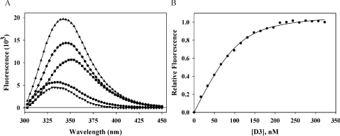
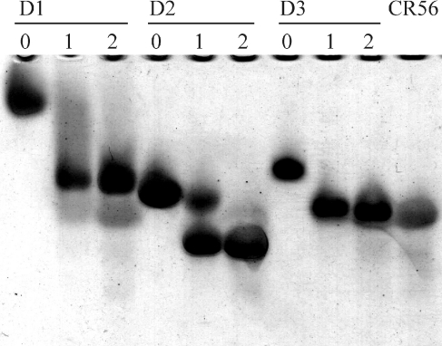
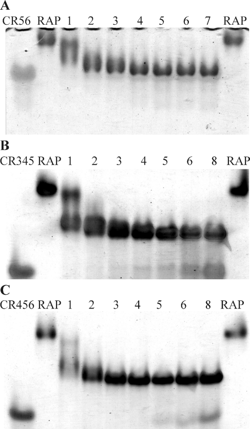
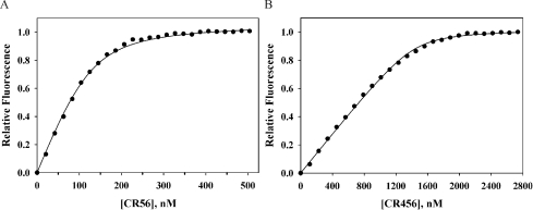
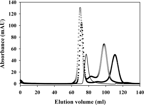
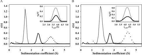
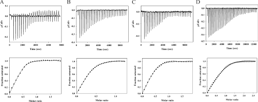
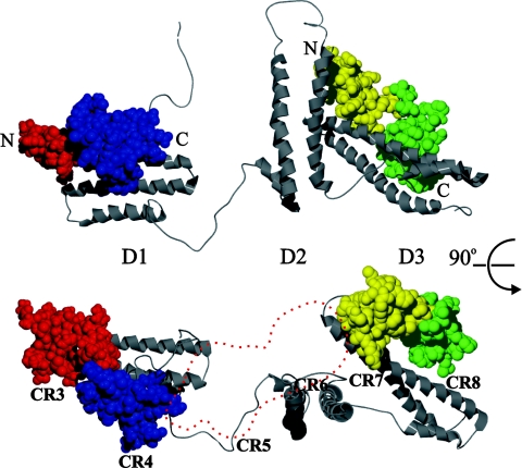
RAP (receptor-associated protein) is a three domain 38 kDa ER (endoplasmic reticulum)-resident protein that is a chaperone for the LRP (low-density lipoprotein receptor-related protein). Whereas RAP is known to compete for binding of all known LRP ligands, neither the location, the number of binding sites on LRP, nor the domains of RAP involved in binding is known with certainty. We have systematically examined the binding of each of the three RAP domains (D1, D2 and D3) to tandem and triple CRs (complement-like repeats) that span the principal ligand-binding region, cluster II, of LRP. We found that D3 binds with low nanomolar affinity to all (CR)2 species examined. Addition of a third CR domain increases the affinity for D3 slightly. A pH change from 7.4 to 5.5 gave only a 6-fold increase in Kd for D3 at 37 °C, whereas temperature change from 22 °C to 37 °C has a similar small effect on affinity, raising questions about the recently proposed D3-destabilization mechanism of RAP release from LRP. Surprisingly, and in contrast to literature suggestions, D1 and D2 also bind to most (CR)2 and (CR)3 constructs with nanomolar affinity. Although this suggested that there might be three high-affinity binding sites in RAP for LRP, studies with intact RAP showed that only two binding sites are available in the intact chaperone. These findings suggest a new model for RAP to function as a folding chaperone and also for the involvement of YWTD domains in RAP release from LRP in the Golgi.
Overview
The claims and statistics of this paper haven’t been checked yet.
Run the check — free, no account needed. It reads the open-access text and checks the sources it cites.
Integrity signals
Registry
Cites
Statistics
Figures
Verification
Claims the paper makes
The claims and statistics of this paper haven’t been checked yet.
Run the check — free, no account needed. It reads the open-access text and checks the sources it cites.
Claims other papers make about this one
Each is checked against this paper’s own text: does it say what the citing paper says it does?
In the citing paper
“while LRPAP1 can bind domains II, III and IV in competition with all known ligands (46–50).”
From LRPAP1 is released from activated microglia and inhibits microglial phagocytosis and amyloid beta aggregation (2023) · cites this paper as [49]
Self-check
Percentages, sums and reported differences re-computed from the paper’s own numbers.
The self-check needs the paper’s full text.
Citations
Cites
Willnow, T. E., Rohlmann, A., Horton, J., Otani, H., Braun, J. R., Hammer, R. E., & Herz, J. (1996) · The EMBO Journal · doi:10.1002/j.1460-2075.1996.tb00623.x · Cited by 276
Research works printed but not linked: 35
- Hussain, 1999, The mammalian low-density lipoprotein receptor family, Annu. Rev. Nutr., 19, 141, 10.1146/annurev.nutr.19.1.141 · DOI
- Herz, 2001, LRP: a multifunctional scavenger and signaling receptor, J. Clin. Invest., 108, 779, 10.1172/JCI200113992 · DOI · Paper page
- Willnow, 1995, Functional expression of low density lipoprotein receptor-related protein is controlled by receptor-associated protein in vivo, Proc. Natl. Acad. Sci. U.S.A., 92, 4537, 10.1073/pnas.92.10.4537 · DOI · Paper page
- Orlando, 2004, The low-density lipoprotein receptor-related protein associates with calnexin, calreticulin, and protein disulfide isomerase in receptor-associated-protein- deficient fibroblasts, Exp. Cell Res., 294, 244, 10.1016/j.yexcr.2003.11.004 · DOI
- Bu, 1996, Receptor-associated protein is a folding chaperone for low density lipoprotein receptor-related protein, J. Biol. Chem., 271, 22218, 10.1074/jbc.271.36.22218 · DOI · Paper page
- Herz, 2003, Coaxing the LDL receptor family into the fold, Cell, 112, 289, 10.1016/S0092-8674(03)00073-4 · DOI
- Bu, 2000, Role of RAP in the biogenesis of lipoprotein receptors, Trends Cardiovasc. Med., 10, 148, 10.1016/S1050-1738(00)00045-1 · DOI
- Obermoeller, 1997, Differential functions of triplicated repeats suggest two independent roles for the receptor-associated protein as a molecular chaperone, J. Biol. Chem., 272, 10761, 10.1074/jbc.272.16.10761 · DOI · Paper page
- Lazic, 2003, Structural organization of the receptor associated protein, Biochemistry, 42, 14913, 10.1021/bi035779e · DOI
- Bu, 1997, ERD2 proteins mediate ER retention of the HNEL signal of LRP's receptor associated protein (RAP), J. Cell Sci., 110, 65, 10.1242/jcs.110.1.65 · DOI
- Bu, 1995, 39 kDa receptor-associated protein is an ER resident protein and molecular chaperone for LDL receptor-related protein, EMBO J., 14, 2269, 10.1002/j.1460-2075.1995.tb07221.x · DOI · Paper page
- Orlando, 1994, Functional domains of the receptor-associated protein (RAP), Proc. Natl. Acad. Sci. U.S.A., 91, 3161, 10.1073/pnas.91.8.3161 · DOI
- Andersen, 2001, Dominant thermodynamic role of the third independent receptor binding site in the receptor-associated protein, Biochemistry, 40, 15408, 10.1021/bi0110692 · DOI
- Jensen, 2006, Binding site structure of one LRP-RAP complex: implications for a common ligand-receptor binding motif, J. Mol. Biol., 362, 700, 10.1016/j.jmb.2006.07.013 · DOI
- Dolmer, 2006, Three complement-like repeats compose the complete α2-macroglobulin binding site in the second ligand binding cluster of the low density lipoprotein receptor-related protein, J. Biol. Chem., 281, 34189, 10.1074/jbc.M604389200 · DOI
- Schuck, 2000, Size-distribution analysis of macromolecules by sedimentation velocity ultracentrifugation and Lamm equation modeling, Biophys. J., 78, 1606, 10.1016/S0006-3495(00)76713-0 · DOI
- Balbo, 2005, Studying multiprotein complexes by multisignal sedimentation velocity analytical centrifugation, Proc. Natl. Acad. Sci. U.S.A., 102, 81, 10.1073/pnas.0408399102 · DOI
- Andersen, 2000, Identification of the minimal functional unit in the low density lipoprotein receptor-related protein for binding the receptor-associated protein (RAP), J. Biol. Chem., 275, 21017, 10.1074/jbc.M000507200 · DOI
- Medved, 1999, Domain organization of the 39-kDa receptor-associated protein, J. Biol. Chem., 274, 717, 10.1074/jbc.274.2.717 · DOI
- Fisher, 2006, Structure of an LDLR-RAP complex reveals a general mode for ligand recognition by lipoprotein receptors, Mol. Cell, 22, 277, 10.1016/j.molcel.2006.02.021 · DOI · Paper page
- Lee, 2006, RAP uses a histidine switch to regulate its interaction with LRP in the ER and Golgi, Mol. Cell, 22, 423, 10.1016/j.molcel.2006.04.011 · DOI
- Estrada, 2008, Unfolding of the RAP-D3 helical bundle facilitates dissociation of RAP-receptor complexes, Biochemistry, 47, 1532, 10.1021/bi702076y · DOI
- Herz, 1988, Surface location and high calcium affinity of a 500kD liver membrane protein closely related to the LDL-receptor suggest a physiological role as lipoprotein receptor, EMBO J., 7, 4119, 10.1002/j.1460-2075.1988.tb03306.x · DOI
- Moestrup, 1990, The human α2-macroglobulin receptor contains high affinity calcium binding sites important for receptor conformation and ligand recognition, J. Biol. Chem., 265, 12623, 10.1016/S0021-9258(19)38389-9 · DOI
- Simonovic, 2001, Calcium coordination and pH dependence of the calcium affinity of ligand-binding repeat CR7 from the LRP. Comparison with related domains from the LRP and the LDL receptor, Biochemistry, 40, 15127, 10.1021/bi015688m · DOI
- Wang, 2001, Role of calcium in protein folding and function of Tva, the receptor of subgroup A avian sarcoma and leukosis virus, J. Virol., 75, 2051, 10.1128/JVI.75.5.2051-2058.2001 · DOI
- Lazic, 2006, Dissection of RAP-LRP interactions. Binding of RAP and RAP fragments to complement-like repeats 7 and 8 from cluster II of LRP, Arch. Biochem. Biophys., 450, 167, 10.1016/j.abb.2006.04.007 · DOI
- Lee, 2007, The structure of receptor-associated protein (RAP), Protein Sci., 16, 1628, 10.1110/ps.072865407 · DOI
- Rao, 1998, A trivalent system from vancomycin.D-Ala-D-Ala with higher affinity than avidin.biotin, Science, 280, 708, 10.1126/science.280.5364.708 · DOI
- Zhou, 2006, Quantitative relation between intermolecular and intramolecular binding of Pro-rich peptides to SH3 domains, Biophys. J., 91, 3170, 10.1529/biophysj.106.090258 · DOI
- Tolkatchev, 2005, Transforming bivalent ligands into retractable enzyme inhibitors through polypeptide-protein interactions, Bioorg. Med. Chem. Lett., 15, 5120, 10.1016/j.bmcl.2005.08.085 · DOI
- Springer, 1998, An extracellular β-propellor module predicted in lipoprotein and scavenger receptors, tyrosine kinases, epidermal growth factor precursor, and extracellular matrix components, J. Mol. Biol., 283, 837, 10.1006/jmbi.1998.2115 · DOI
- Rudenko, 2002, Structure of the LDL receptor extracellular domain at endosomal pH, Science, 298, 2353, 10.1126/science.1078124 · DOI · Paper page
- Zhao, 2008, The epidermal growth factor homology domain of the LDL receptor drives lipoprotein release through an allosteric mechanism involving H190, H562, and H586, J. Biol. Chem., 283, 26528, 10.1074/jbc.M804624200 · DOI
- Obermoeller-McCormick, 2001, Dissection of receptor folding and ligand-binding property with functional minireceptors of LDL receptor-related protein, J. Cell Sci., 114, 899, 10.1242/jcs.114.5.899 · DOI · Paper page
Cited by
Sorted by citations
Reid, K. M., & Brown, G. C. (2023) · Frontiers in Immunology · doi:10.3389/fimmu.2023.1286474 · Cited by 13
Metadata
- Authors
- Jensen, J. K., Dolmer, K., Schar, C., & Gettins, P. G. W.
- Affiliations
- Department of Biochemistry and Molecular Genetics, University of Illinois at Chicago, 900 S. Ashland, M/C 669, Chicago, IL 60607, U.S.A.
- Venue
- Biochemical Journal
- Publisher
- Portland Press Ltd.
- Published
- 2009-06-26
- Volume
- 421
- Issue
- 2
- Pages
- 273-282
- ISSN
- 0264-6021
- 1470-8728
- PMC
- PMC2708935
- Cited by
- 39
- References
- 1
- Language
- en
About these checks
Registry, retraction, Cites and statistics results are computed, not judged, with one exception: a printed reference that no index lists is read once by a model before it counts as a citation error. Claims are extracted by a model and kept only when their quote appears verbatim in the text; each cited source is then checked, with a model’s help, for whether it says what the claim relies on. Those verdicts can be wrong — read the claim’s quote and the cited source before relying on one.
Statistics and arithmetic are recomputed, not judged.
Confidence bands are shown only for fields calibrated on our annotated test set. None is calibrated yet, so no band is shown.
What counts as a claim ›How we measure confidence ›How we check this ›
Are you an author, or do you see a problem with this page? Request a review
How to cite
Cite CoChat for the checks on this page, and the paper for its own findings.
- This page
- CoChat. (2026). Citation check: Receptor-associated protein (RAP) has two high-affinity binding sites for the low-density lipoprotein receptor-related protein (LRP): consequences for the chaperone functions of RAP. https://app.cochat.ai/papers/10.1042/bj20090175
- The paper
- Jensen, J. K., Dolmer, K., Schar, C., & Gettins, P. G. W. (2009). Receptor-associated protein (RAP) has two high-affinity binding sites for the low-density lipoprotein receptor-related protein (LRP): consequences for the chaperone functions of RAP. Biochemical Journal. https://doi.org/10.1042/bj20090175
