

Spatial transcriptome profiling by MERFISH reveals subcellular RNA compartmentalization and cell cycle-dependent gene expression
Open accessThe DOI matches its Crossref record; 50 of the 50 references it cites were found in a registry or an index.
Abstract
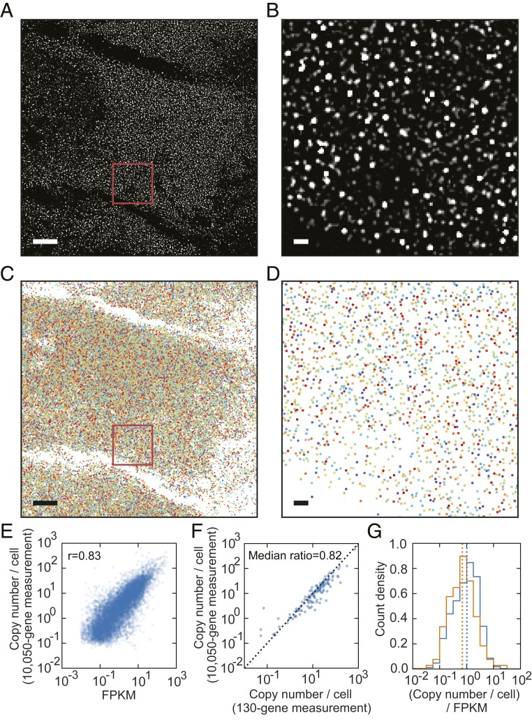
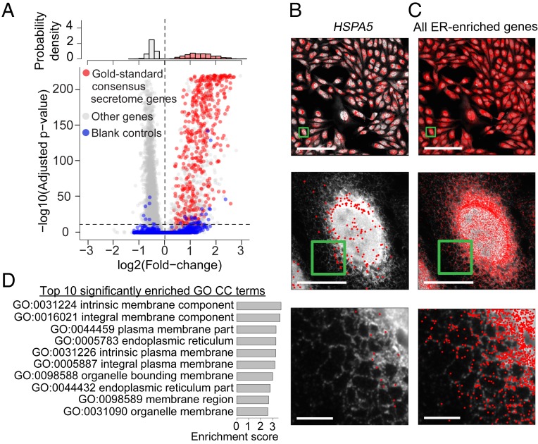
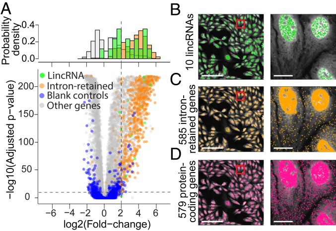
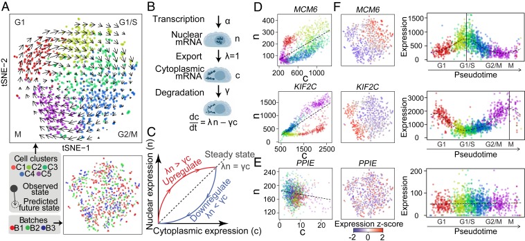
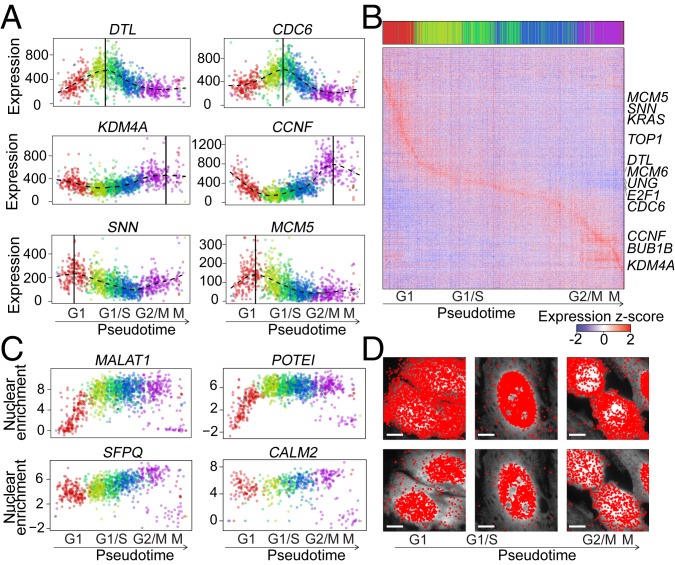
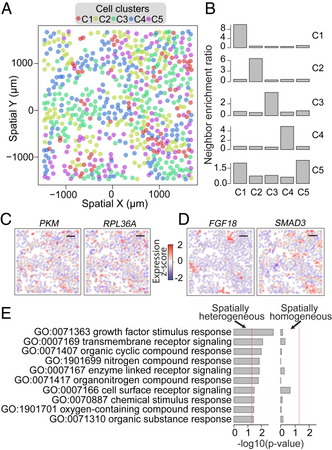
The expression profiles and spatial distributions of RNAs regulate many cellular functions. Image-based transcriptomic approaches provide powerful means to measure both expression and spatial information of RNAs in individual cells within their native environment. Among these approaches, multiplexed error-robust fluorescence in situ hybridization (MERFISH) has achieved spatially resolved RNA quantification at transcriptome scale by massively multiplexing single-molecule FISH measurements. Here, we increased the gene throughput of MERFISH and demonstrated simultaneous measurements of RNA transcripts from ∼10,000 genes in individual cells with ∼80% detection efficiency and ∼4% misidentification rate. We combined MERFISH with cellular structure imaging to determine subcellular compartmentalization of RNAs. We validated this approach by showing enrichment of secretome transcripts at the endoplasmic reticulum, and further revealed enrichment of long noncoding RNAs, RNAs with retained introns, and a subgroup of protein-coding mRNAs in the cell nucleus. Leveraging spatially resolved RNA profiling, we developed an approach to determine RNA velocity in situ using the balance of nuclear versus cytoplasmic RNA counts. We applied this approach to infer pseudotime ordering of cells and identified cells at different cell-cycle states, revealing ∼1,600 genes with putative cell cycle-dependent expression and a gradual transcription profile change as cells progress through cell-cycle stages. Our analysis further revealed cell cycle-dependent and cell cycle-independent spatial heterogeneity of transcriptionally distinct cells. We envision that the ability to perform spatially resolved, genome-wide RNA profiling with high detection efficiency and accuracy by MERFISH could help address a wide array of questions ranging from the regulation of gene expression in cells to the development of cell fate and organization in tissues.
Overview
The claims and statistics of this paper haven’t been checked yet.
Free: it reads the open-access text and checks the first 5 claims that cite a source we can check. Create a free account to check every claim.
Integrity signals
Registry
Cites
Statistics
Figures
Verification
Claims the paper makes
The claims and statistics of this paper haven’t been checked yet.
Free: it reads the open-access text and checks the first 5 claims that cite a source we can check. Create a free account to check every claim.
Claims other papers make about this one
Each is checked against this paper’s own text: does it say what the citing paper says it does?
In the citing paper
“Finer spatial resolution can be achieved by super-resolution imaging (Lubeck and Cai, 2012) or isotropic physical expansion of tissue prior to imaging (Chen et al., 2015a; Xia et al., 2019).”
From Single-Cell Sequencing of Brain Cell Transcriptomes and Epigenomes (2021) · cites this paper as Xia et al., 2019
Self-check
Percentages, sums and reported differences re-computed from the paper’s own numbers.
The self-check needs the paper’s full text.
Citations
Cites
Subramanian, A., Tamayo, P., Mootha, V. K., Mukherjee, S., Ebert, B. L., Gillette, M. A., Paulovich, A., Pomeroy, S. L., Golub, T. R., Lander, E. S., et al. (2005) · Proceedings of the National Academy of Sciences · doi:10.1073/pnas.0506580102 · Cited by 53,625
Fast unfolding of communities in large networks
Blondel, V. D., Guillaume, J.-L., Lambiotte, R., & Lefebvre, E. (2008) · Journal of Statistical Mechanics: Theory and Experiment · doi:10.1088/1742-5468/2008/10/p10008 · Cited by 21,424
SignalP 4.0: discriminating signal peptides from transmembrane regions
Petersen, T. N., Brunak, S., von Heijne, G., & Nielsen, H. (2011) · Nature Methods · doi:10.1038/nmeth.1701 · Cited by 8,847 · cited 3×
“Among these, we defined a consensus set of gold-standard ER-enriched genes as genes identified to be ER-associated by more than 3 of the 5 approaches (Phobius, SignalP, APEX-RIP, proximity-specific ribosome profiling, and MERFISH), resulting in a set of 590 gold-standard ER-enriched genes.”
ResultInfluentialNOTES ON CONTINUOUS STOCHASTIC PHENOMENA
MORAN, P. A. P. (1950) · Biometrika · doi:10.1093/biomet/37.1-2.17 · Cited by 8,622
Derrien, T., Johnson, R., Bussotti, G., Tanzer, A., Djebali, S., Tilgner, H., Guernec, G., Martin, D., Merkel, A., Knowles, D. G., et al. (2012) · Genome Research · doi:10.1101/gr.132159.111 · Cited by 5,061
La Manno, G., Soldatov, R., Zeisel, A., Braun, E., Hochgerner, H., Petukhov, V., Lidschreiber, K., Kastriti, M. E., Lönnerberg, P., Furlan, A., et al. (2018) · Nature · doi:10.1038/s41586-018-0414-6 · Cited by 4,118
Self-citationVisualization and analysis of gene expression in tissue sections by spatial transcriptomics
Ståhl, P. L., Salmén, F., Vickovic, S., Lundmark, A., Navarro, J. F., Magnusson, J., Giacomello, S., Asp, M., Westholm, J. O., Huss, M., et al. (2016) · Science · doi:10.1126/science.aaf2403 · Cited by 3,834 · cited 1×
“Various spatially resolved transcriptomics approaches have been developed, including methods based on multiplexed singlemolecule fluorescence in situ hybridization (smFISH) (5–7) and in situ sequencing (8–10), which provide single-cell resolution, as well as methods based on spatially resolved RNA capture followed by…”
MethodSpatially resolved, highly multiplexed RNA profiling in single cells
Chen, K. H., Boettiger, A. N., Moffitt, J. R., Wang, S., & Zhuang, X. (2015) · Science · doi:10.1126/science.aaa6090 · Cited by 2,969 · cited 8×
“In each round, RNAs are imaged by FISH at the single-molecule level (13, 14), and the use of error-robust barcodes allows errors accumulated through multiple imaging rounds to be detected and/or corrected (6).”
BackgroundMethodInfluentialSelf-citationA Combined Transmembrane Topology and Signal Peptide Prediction Method
Käll, L., Krogh, A., & Sonnhammer, E. L. L. (2004) · Journal of Molecular Biology · doi:10.1016/j.jmb.2004.03.016 · Cited by 2,450
Slide-seq: A scalable technology for measuring genome-wide expression at high spatial resolution
Rodriques, S. G., Stickels, R. R., Goeva, A., Martin, C. A., Murray, E., Vanderburg, C. R., Welch, J., Chen, L. M., Chen, F., & Macosko, E. Z. (2019) · Science · doi:10.1126/science.aaw1219 · Cited by 2,424
Imaging individual mRNA molecules using multiple singly labeled probes
Raj, A., van den Bogaard, P., Rifkin, S. A., van Oudenaarden, A., & Tyagi, S. (2008) · Nature Methods · doi:10.1038/nmeth.1253 · Cited by 2,028
Transcriptome-scale super-resolved imaging in tissues by RNA seqFISH
Eng, C.-H. L., Lawson, M., Zhu, Q., Dries, R., Koulena, N., Takei, Y., Yun, J., Cronin, C., Karp, C., Yuan, G.-C., et al. (2019) · Nature · doi:10.1038/s41586-019-1049-y · Cited by 1,754 · cited 3×
“used 3 independent sets of 80-bit, Hamming weight 4, error-correcting codes [a kind that can also be considered as a 4-unit base-20 code (7)] to detect ∼10,000 genes with 3-color imaging and 80 rounds of hybridization.”
BackgroundMethodThree-dimensional intact-tissue sequencing of single-cell transcriptional states
Wang, X., Allen, W. E., Wright, M. A., Sylwestrak, E. L., Samusik, N., Vesuna, S., Evans, K., Liu, C., Ramakrishnan, C., Liu, J., et al. (2018) · Science · doi:10.1126/science.aat5691 · Cited by 1,620
Whitfield, M. L., Sherlock, G., Saldanha, A. J., Murray, J. I., Ball, C. A., Alexander, K. E., Matese, J. C., Perou, C. M., Hurt, M. M., Brown, P. O., et al. (2002) · Molecular Biology of the Cell · doi:10.1091/mbc.02-02-0030 · Cited by 1,527 · cited 1×
“This notion is supported by the observations that down-regulation of KDM4A arrests cells in the G2/M phase (44) and that KDM4A expression varies with cell cycle (39).”
ResultVisualization of Single RNA Transcripts in Situ
Femino, A. M., Fay, F. S., Fogarty, K., & Singer, R. H. (1998) · Science · doi:10.1126/science.280.5363.585 · Cited by 1,504 · cited 1×
“In each round, RNAs are imaged by FISH at the single-molecule level (13, 14), and the use of error-robust barcodes allows errors accumulated through multiple imaging rounds to be detected and/or corrected (6).”
BackgroundChen, F., Tillberg, P. W., & Boyden, E. S. (2015) · Science · doi:10.1126/science.1260088 · Cited by 1,501
Molecular, spatial, and functional single-cell profiling of the hypothalamic preoptic region
Moffitt, J. R., Bambah-Mukku, D., Eichhorn, S. W., Vaughn, E., Shekhar, K., Perez, J. D., Rubinstein, N. D., Hao, J., Regev, A., Dulac, C., et al. (2018) · Science · doi:10.1126/science.aau5324 · Cited by 1,365 · cited 2×
“For the additional 1,000 genes that were either shorter than 1,440 nt or did not have enough targetable regions to accommodate 48 nonoverlapping encoding probes, we used an overlapping encodingprobe design that allowed the encoding probes to target overlapping regions on the target RNA (20-nt overlap between adjacent…”
BackgroundMethodSelf-citationBayesian approach to single-cell differential expression analysis
Kharchenko, P. V., Silberstein, L., & Scadden, D. T. (2014) · Nature Methods · doi:10.1038/nmeth.2967 · Cited by 1,348
Single-cell in situ RNA profiling by sequential hybridization
Lubeck, E., Coskun, A. F., Zhiyentayev, T., Ahmad, M., & Cai, L. (2014) · Nature Methods · doi:10.1038/nmeth.2892 · Cited by 985
Highly Multiplexed Subcellular RNA Sequencing in Situ
Lee, J. H., Daugharthy, E. R., Scheiman, J., Kalhor, R., Yang, J. L., Ferrante, T. C., Terry, R., Jeanty, S. S. F., Li, C., Amamoto, R., et al. (2014) · Science · doi:10.1126/science.1250212 · Cited by 973
In situ sequencing for RNA analysis in preserved tissue and cells
Ke, R., Mignardi, M., Pacureanu, A., Svedlund, J., Botling, J., Wählby, C., & Nilsson, M. (2013) · Nature Methods · doi:10.1038/nmeth.2563 · Cited by 934
Pre-mRNA Processing Reaches Back toTranscription and Ahead to Translation
Moore, M. J., & Proudfoot, N. J. (2009) · Cell · doi:10.1016/j.cell.2009.02.001 · Cited by 846
Neuronal cell-type classification: challenges, opportunities and the path forward
Zeng, H., & Sanes, J. R. (2017) · Nature Reviews Neuroscience · doi:10.1038/nrn.2017.85 · Cited by 841
Scaling single-cell genomics from phenomenology to mechanism
Tanay, A., & Regev, A. (2017) · Nature · doi:10.1038/nature21350 · Cited by 792
Localization and abundance analysis of human lncRNAs at single-cell and single-molecule resolution
Cabili, M. N., Dunagin, M. C., McClanahan, P. D., Biaesch, A., Padovan-Merhar, O., Regev, A., Rinn, J. L., & Raj, A. (2015) · Genome Biology · doi:10.1186/s13059-015-0586-4 · Cited by 684
Atlas of Subcellular RNA Localization Revealed by APEX-Seq
Fazal, F. M., Han, S., Parker, K. R., Kaewsapsak, P., Xu, J., Boettiger, A. N., Chang, H. Y., & Ting, A. Y. (2019) · Cell · doi:10.1016/j.cell.2019.05.027 · Cited by 652
In the right place at the right time: visualizing and understanding mRNA localization
Buxbaum, A. R., Haimovich, G., & Singer, R. H. (2015) · Nature Reviews Molecular Cell Biology · doi:10.1038/nrm3918 · Cited by 612
Long non-coding RNAs: spatial amplifiers that control nuclear structure and gene expression
Engreitz, J. M., Ollikainen, N., & Guttman, M. (2016) · Nature Reviews Molecular Cell Biology · doi:10.1038/nrm.2016.126 · Cited by 585
Moffitt, J. R., Hao, J., Wang, G., Chen, K. H., Babcock, H. P., & Zhuang, X. (2016) · Proceedings of the National Academy of Sciences · doi:10.1073/pnas.1612826113 · Cited by 580
Self-citationSingle-Cell Gene Expression Profiling
Levsky, J. M., Shenoy, S. M., Pezo, R. C., & Singer, R. H. (2002) · Science · doi:10.1126/science.1072241 · Cited by 526
Spatially resolved transcriptomics and beyond
Crosetto, N., Bienko, M., & van Oudenaarden, A. (2015) · Nature Reviews Genetics · doi:10.1038/nrg3832 · Cited by 509
Principles of ER cotranslational translocation revealed by proximity-specific ribosome profiling
Jan, C. H., Williams, C. C., & Weissman, J. S. (2014) · Science · doi:10.1126/science.1257521 · Cited by 430
Detained introns are a novel, widespread class of post-transcriptionally spliced introns
Boutz, P. L., Bhutkar, A., & Sharp, P. A. (2015) · Genes & Development · doi:10.1101/gad.247361.114 · Cited by 425
The promise of spatial transcriptomics for neuroscience in the era of molecular cell typing
Lein, E., Borm, L. E., & Linnarsson, S. (2017) · Science · doi:10.1126/science.aan6827 · Cited by 383
Control of Transcript Variability in Single Mammalian Cells
Battich, N., Stoeger, T., & Pelkmans, L. (2015) · Cell · doi:10.1016/j.cell.2015.11.018 · Cited by 324 · cited 1×
“For example, retention of mRNAs in the nucleus may help buffer noise generated by stochastic mRNA production (33, 34).”
BackgroundMoffitt, J. R., Hao, J., Bambah-Mukku, D., Lu, T., Dulac, C., & Zhuang, X. (2016) · Proceedings of the National Academy of Sciences · doi:10.1073/pnas.1617699113 · Cited by 308
Self-citationMitotic Centromere–associated Kinesin Is Important for Anaphase Chromosome Segregation
Maney, T., Hunter, A. W., Wagenbach, M., & Wordeman, L. (1998) · The Journal of Cell Biology · doi:10.1083/jcb.142.3.787 · Cited by 289
Nuclear Retention of mRNA in Mammalian Tissues
Bahar Halpern, K., Caspi, I., Lemze, D., Levy, M., Landen, S., Elinav, E., Ulitsky, I., & Itzkovitz, S. (2015) · Cell Reports · doi:10.1016/j.celrep.2015.11.036 · Cited by 258 · cited 1×
“For example, retention of mRNAs in the nucleus may help buffer noise generated by stochastic mRNA production (33, 34).”
BackgroundMultiplexed imaging of high-density libraries of RNAs with MERFISH and expansion microscopy
Wang, G., Moffitt, J. R., & Zhuang, X. (2018) · Scientific Reports · doi:10.1038/s41598-018-22297-7 · Cited by 256
Self-citationKaewsapsak, P., Shechner, D. M., Mallard, W., Rinn, J. L., & Ting, A. Y. (2017) · eLife · doi:10.7554/elife.29224 · Cited by 198
A Network of Cytosolic Factors Targets SRP-Independent Proteins to the Endoplasmic Reticulum
Ast, T., Cohen, G., & Schuldiner, M. (2013) · Cell · doi:10.1016/j.cell.2013.02.003 · Cited by 197
Wong, J. J. ‐., Au, A. Y. M., Ritchie, W., & Rasko, J. E. J. (2016) · BioEssays · doi:10.1002/bies.201500117 · Cited by 195
Reid, D. W., & Nicchitta, C. V. (2012) · Journal of Biological Chemistry · doi:10.1074/jbc.m111.312280 · Cited by 184
DTL/CDT2 is essential for both CDT1 regulation and the early G2/M checkpoint
Sansam, C. L., Shepard, J. L., Lai, K., Ianari, A., Danielian, P. S., Amsterdam, A., Hopkins, N., & Lees, J. A. (2006) · Genes & Development · doi:10.1101/gad.1482106 · Cited by 171
Multiplexed detection of RNA using MERFISH and branched DNA amplification
Xia, C., Babcock, H. P., Moffitt, J. R., & Zhuang, X. (2019) · Scientific Reports · doi:10.1038/s41598-019-43943-8 · Cited by 165
Self-citationThe role of MCM proteins in the cell cycle control of genome duplication
Kearsey, S. E., Maiorano, D., Holmes, E. C., & Todorov, I. T. (1996) · BioEssays · doi:10.1002/bies.950180305 · Cited by 143
Power in Numbers: Single-Cell RNA-Seq Strategies to Dissect Complex Tissues
Birnbaum, K. D. (2018) · Annual Review of Genetics · doi:10.1146/annurev-genet-120417-031247 · Cited by 116
The JMJD2A demethylase regulates apoptosis and proliferation in colon cancer cells
Kim, T., Shin, S., Berry, W. L., Oh, S., & Janknecht, R. (2012) · Journal of Cellular Biochemistry · doi:10.1002/jcb.24009 · Cited by 107
Comparison of total and cytoplasmic mRNA reveals global regulation by nuclear retention and miRNAs
Solnestam, B. W., Stranneheim, H., Hällman, J., Käller, M., Lundberg, E. K., Lundeberg, J., & Akan, P. (2012) · BMC Genomics · doi:10.1186/1471-2164-13-574 · Cited by 42
Elucidation of Stannin Function Using Microarray Analysis: Implications for Cell Cycle Control
Reese, B. E., Krissinger, D. J., Yun, J. K., & Billingsley, M. L. (2006) · Gene Expression · doi:10.3727/000000006783991944 · Cited by 13
Cited by
Generalizing RNA velocity to transient cell states through dynamical modeling
Bergen, V., Lange, M., Peidli, S., Wolf, F. A., & Theis, F. J. (2020) · Nature Biotechnology · doi:10.1038/s41587-020-0591-3 · Cited by 2,951
Exploring tissue architecture using spatial transcriptomics
Rao, A., Barkley, D., França, G. S., & Yanai, I. (2021) · Nature · doi:10.1038/s41586-021-03634-9 · Cited by 1,700
Museum of spatial transcriptomics
Moses, L., & Pachter, L. (2022) · Nature Methods · doi:10.1038/s41592-022-01409-2 · Cited by 1,255
“The length of barcodes can be increased to encode 10,000 genes [76].”
BackgroundMuseum of Spatial Transcriptomics
Moses, L., & Pachter, L. (2021) · doi:10.1101/2021.05.11.443152 · Cited by 1,255
Methods and applications for single-cell and spatial multi-omics
Vandereyken, K., Sifrim, A., Thienpont, B., & Voet, T. (2023) · Nature Reviews Genetics · doi:10.1038/s41576-023-00580-2 · Cited by 1,244
Metadata
- Authors
- Xia, C., Fan, J., Emanuel, G., Hao, J., & Zhuang, X.
- Affiliations
- Howard Hughes Medical Institute (US)
- Corresponding author
- Zhuang, Xiaowei
- Venue
- Proceedings of the National Academy of Sciences
- Publisher
- National Academy of Sciences
- Published
- 2019-09-09
- Volume
- 116
- Issue
- 39
- Pages
- 19490-19499
- ISSN
- 0027-8424 (ISSN-L)
- 1091-6490
- PMC
- PMC6765259
- OpenAlex
- W2972445800
- Cited by
- 755
- References
- 50
- Citations by year
- 2026: 99 · 2025: 114 · 2024: 141 · 2023: 110 · 2022: 115 · 2021: 105 · 2020: 65 · 2019: 5
- FWCI
- 23.86
- Type
- journal article
- Language
- en
- Open access
- hybrid
- doi.org
- Licence
- Locations
- Proceedings of the National Academy of Sciences (publishedVersion, cc-by-nc-nd)
- Europe PMC (PubMed Central) (publishedVersion, cc-by-nc-nd)
- PubMed Central (publishedVersion, cc-by-nc-nd)
- PubMed (publishedVersion)
- Funders
- HHS | NIH | National Institute of Mental Health (R01MH113094)
- Howard Hughes Medical Institute (HHMI)
- HHS | National Institutes of Health (K00CA222750)
- Topics
- Life Sciences › Biochemistry, Genetics and Molecular Biology › Molecular Biology › Single-cell and spatial transcriptomics
- Life Sciences › Biochemistry, Genetics and Molecular Biology › Molecular Biology › RNA Research and Splicing
- Life Sciences › Biochemistry, Genetics and Molecular Biology › Biophysics › Advanced Fluorescence Microscopy Techniques
- Keywords
- MERFISH, spatial transcriptomics, cell cycle-dependent gene expression, RNA velocity, nuclear RNA, long non-coding RNA, intron retention, secretome, endoplasmic reticulum, single-molecule FISH
- MeSH terms
- Cell Division, Gene Expression Regulation, Humans, RNA, Messenger, In Situ Hybridization, Fluorescence, Genes, cdc, Gene Expression Profiling, Intracellular Space, Cell Line, Tumor, Single-Cell Analysis, Transcriptome, RNA, Long Noncoding
About these checks
Registry, retraction, Cites and statistics results are computed, not judged, with one exception: a printed reference that no index lists is read once by a model before it counts as a citation error. Claims are extracted by a model and kept only when their quote appears verbatim in the text; each cited source is then checked, with a model’s help, for whether it says what the claim relies on. Those verdicts can be wrong — read the claim’s quote and the cited source before relying on one.
Statistics and arithmetic are recomputed, not judged.
Confidence bands are shown only for fields calibrated on our annotated test set. None is calibrated yet, so no band is shown.
What counts as a claim ›How we measure confidence ›How we check this ›
Are you an author, or do you see a problem with this page? Request a review
Data sources
Bibliographic data: Semantic Scholar, OpenAlex, Crossref.
Contains information from the Semantic Scholar Academic Graph Datasets, made available under the ODC Attribution License (ODC-BY 1.0).
Semantic Scholar’s data is described in: Kinney, R., et al. (2023). The Semantic Scholar Open Data Platform. arXiv:2301.10140.
