

The DOI matches its Crossref record; 45 of the 46 references it cites were found in a registry or an index.
Defective HIV-1 proviruses produce viral proteins
Open accessAbstract
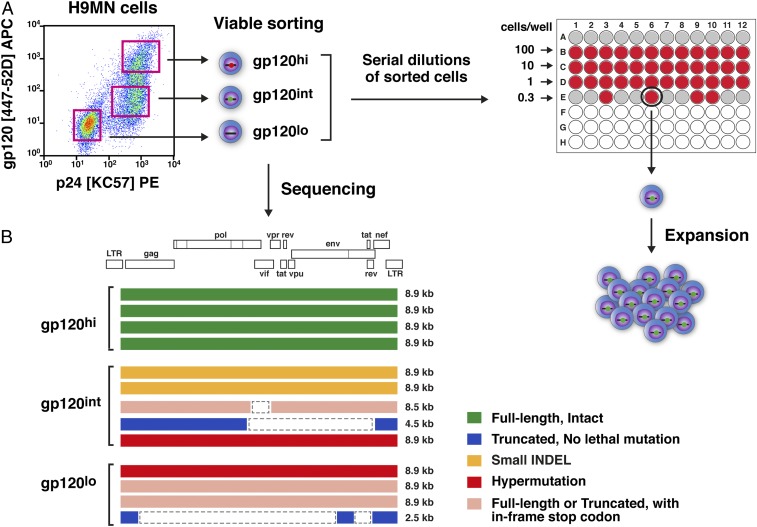
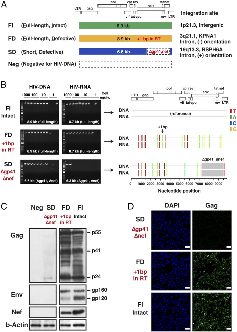
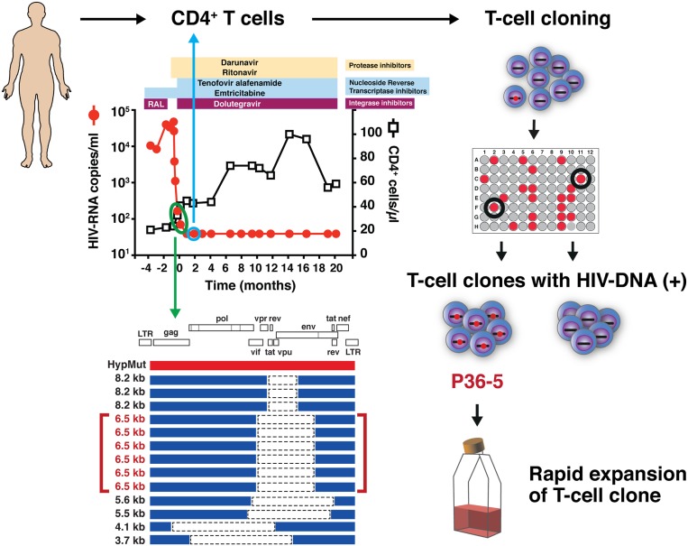
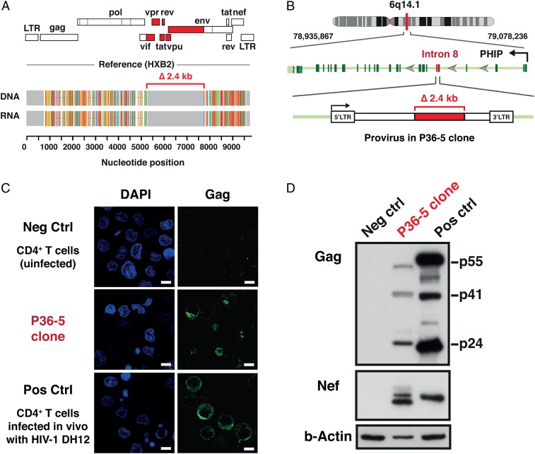
HIV-1 proviruses persist in the CD4 + T cells of HIV-infected individuals despite years of combination antiretroviral therapy (cART) with suppression of HIV-1 RNA levels <40 copies/mL. Greater than 95% of these proviruses detected in circulating peripheral blood mononuclear cells (PBMCs) are referred to as “defective” by virtue of having large internal deletions and lethal genetic mutations. As these defective proviruses are unable to encode intact and replication-competent viruses, they have long been thought of as biologically irrelevant “graveyard” of viruses with little significance to HIV-1 pathogenesis. Contrary to this notion, we have recently demonstrated that these defective proviruses are not silent, are capable of transcribing novel unspliced forms of HIV-RNA transcripts with competent open reading frames (ORFs), and can be found in the peripheral blood CD4 + T cells of patients at all stages of HIV-1 infection. In the present study, by an approach of combining serial dilutions of CD4 + T cells and T cell–cloning technologies, we are able to demonstrate that defective proviruses that persist in HIV-infected individuals during suppressive cART are translationally competent and produce the HIV-1 Gag and Nef proteins. The HIV-RNA transcripts expressed from these defective proviruses may trigger an element of innate immunity. Likewise, the viral proteins coded in the defective proviruses may form extracellular virus-like particles and may trigger immune responses. The persistent production of HIV-1 proteins in the absence of viral replication helps explain persistent immune activation despite HIV-1 levels below detection, and also presents new challenges to HIV-1 eradication.
Overview
The claims and statistics of this paper haven’t been checked yet.
Run the check — free, no account needed. It reads the open-access text and checks the sources it cites.
Integrity signals
Registry
Cites
Statistics
Figures
Verification
Claims the paper makes
The claims and statistics of this paper haven’t been checked yet.
Run the check — free, no account needed. It reads the open-access text and checks the sources it cites.
Claims other papers make about this one
Each is checked against this paper’s own text: does it say what the citing paper says it does?
The source backs part of the claim, not all of it — or backs it for a different population, condition or comparison.
In the citing paper
“Conversely, a multitude of studies showed that defective HIV-1 genomes inside cells may still produce individual viral transcripts or proteins, 33-35,55 which could be mistaken as indicators of a functional reservoir of infectious virus.”
From Revealing viral and cellular dynamics of HIV-1 at the single-cell level during early treatment periods (2023) · cites this paper as [35]
Experimental — accuracy not yet measured
Self-check
Percentages, sums and reported differences re-computed from the paper’s own numbers.
The self-check needs the paper’s full text.
Citations
Cites
Fiji: an open-source platform for biological-image analysis
Schindelin, J., Arganda-Carreras, I., Frise, E., Kaynig, V., Longair, M., Pietzsch, T., Preibisch, S., Rueden, C., Saalfeld, S., Schmid, B., et al. (2012) · Nature Methods · doi:10.1038/nmeth.2019 · Cited by 66,890
Gallo, R. C., Salahuddin, S. Z., Popovič, M., Shearer, G. M., Kaplan, M., Haynes, B. F., Palker, T. J., Redfield, R. R., Oleske, J. M., Safai, B., et al. (1984) · Science · doi:10.1126/science.6200936 · Cited by 3,639
. Inflammatory and coagulation biomarkers and mortality in patients with HIV infection.
Kuller, L. H., Tracy, R., Belloso, W., Wit, S. D., Drummond, F., Lane, H. C., Ledergerber, B., Lundgren, J., Neuhaus, J., Nixon, D., et al. (2008) · PLoS Medicine · doi:10.1371/journal.pmed.0050203 · Cited by 1,617
Self-citationReplication-competent noninduced proviruses in the latent reservoir increase barrier to HIV-1 cure
Ho, Y.-C., Shan, L., Hosmane, N. N., Wang, J., Laskey, S. B., Rosenbloom, D. I. S., Lai, J., Blankson, J. N., Siliciano, J. D., & Siliciano, R. F. (2013) · Cell · doi:10.1016/j.cell.2013.09.020 · Cited by 1,405
The accuracy of reverse transcriptase from HIV-1
Roberts, J. D., Bebenek, K., & Kunkel, T. A. (1988) · Science · doi:10.1126/science.2460925 · Cited by 997
Fidelity of HIV-1 reverse transcriptase
Preston, B. D., Poiesz, B. J., & Loeb, L. A. (1988) · Science · doi:10.1126/science.2460924 · Cited by 931
Defective proviruses rapidly accumulate during acute HIV-1 infection
Bruner, K. M., Murray, A. J., Pollack, R. A., Soliman, M. G., Laskey, S. B., Capoferri, A. A., Lai, J., Strain, M. C., Lada, S. M., Hoh, R., et al. (2016) · Nature Medicine · doi:10.1038/nm.4156 · Cited by 737
Shaw, G. M., Hahn, B. H., Arya, S. K., Groopman, J. E., Gallo, R. C., & Wong‐Staal, F. (1984) · Science · doi:10.1126/science.6095449 · Cited by 633
HIV-1 Reverse TranscriptionPreprint
Hu, W.-S., & Hughes, S. H. (2012) · Cold Spring Harbor Perspectives in Medicine · doi:10.1101/cshperspect.a006882 · Cited by 597
Absence of Detectable HIV-1 Viremia after Treatment Cessation in an Infant
Persaud, D., Gay, H., Ziemniak, C., Chen, Y. H., Piatak, M., Chun, T., Strain, M. C., Richman, D. D., & Luzuriaga, K. (2013) · New England Journal of Medicine · doi:10.1056/nejmoa1302976 · Cited by 580
Cited by
Sorted by citations
Research priorities for an HIV cure: International AIDS Society Global Scientific Strategy 2021
Deeks, S. G., Archin, N., Cannon, P., Collins, S., Jones, R. B., de Jong, M. A. W. P., Lambotte, O., Lamplough, R., Ndung’u, T., Sugarman, J., et al. (2021) · Nature Medicine · doi:10.1038/s41591-021-01590-5 · Cited by 377
The Biology of the HIV-1 Latent Reservoir and Implications for Cure Strategies
Cohn, L. B., Chomont, N., & Deeks, S. G. (2020) · Cell Host & Microbe · doi:10.1016/j.chom.2020.03.014 · Cited by 296
Prevention, treatment and cure of HIV infection
Landovitz, R. J., Scott, H. M., & Deeks, S. G. (2023) · Nature Reviews Microbiology · doi:10.1038/s41579-023-00914-1 · Cited by 153
Recommendations for measuring HIV reservoir size in cure-directed clinical trials
Abdel‐Mohsen, M., Richman, D. D., Siliciano, R. F., Nussenzweig, M. C., Howell, B. J., Martínez‐Picado, J., Chomont, N., Bar, K. J., Yu, X. G., Lichterfeld, M. D., et al. (2020) · Nature Medicine · doi:10.1038/s41591-020-1022-1 · Cited by 135
McMyn, N. F., Varriale, J., Fray, E. J., Zitzmann, C., Macleod, H. H. B., Lai, J., Singhal, A., Moskovljevic, M., Garcia, M. A., Lopez, B. M., et al. (2023) · Journal of Clinical Investigation · doi:10.1172/jci171554 · Cited by 143
Metadata
- Authors
- Imamichi, H., Smith, M., Adelsberger, J. W., Izumi, T., Scrimieri, F., Sherman, B. T., Rehm, C. A., Imamichi, T., Pau, A., Catalfamo, M., Fauci, A. S., & Lane, H. C.
- Affiliations
- National Institutes of Health (US)
- Frederick National Laboratory for Cancer Research (US)
- Department of Microbiology and Immunology, Georgetown University School of Medicine, Washington, DC 20057
- Corresponding author
- Fauci, Anthony S.
- Lane, H. Clifford
- ORCID
- Venue
- Proceedings of the National Academy of Sciences
- Publisher
- National Academy of Sciences
- Published
- 2020-02-06
- Volume
- 117
- Issue
- 7
- Pages
- 3704-3710
- ISSN
- 0027-8424 (ISSN-L)
- 1091-6490
- PMC
- PMC7035625
- OpenAlex
- W3005267901
- Cited by
- 251
- References
- 46
- Citations by year
- 2026: 38 · 2025: 41 · 2024: 40 · 2023: 41 · 2022: 33 · 2021: 38 · 2020: 19
- FWCI
- 17.40
- Type
- journal article
- Language
- en
- Open access
- hybrid
- doi.org
- Licence
- Locations
- Proceedings of the National Academy of Sciences (publishedVersion, cc-by-nc-nd)
- PubMed Central (publishedVersion, cc-by-nc-nd)
- PubMed (publishedVersion)
- Funders
- Division of Intramural Research, National Institute of Allergy and Infectious Diseases (N/A)
- Topics
- Life Sciences › Immunology and Microbiology › Virology › HIV Research and Treatment
- Life Sciences › Immunology and Microbiology › Immunology › T-cell and Retrovirus Studies
- Health Sciences › Medicine › Infectious Diseases › HIV/AIDS drug development and treatment
- Keywords
- HIV-1 Gag protein, HIV-1 Nef, CD4+ T cells, highly active antiretroviral therapy, viral persistence, open reading frames, virus-like particles, innate immunity, persistent immune activation, HIV-1 eradication
- MeSH terms
- Humans, Leukocytes, Mononuclear, Male, Middle Aged, Proviruses, Viral Proteins, CD4-Positive T-Lymphocytes, HIV-1, HIV Infections
About these checks
Registry, retraction, Cites and statistics results are computed, not judged, with one exception: a printed reference that no index lists is read once by a model before it counts as a citation error. Claims are extracted by a model and kept only when their quote appears verbatim in the text; each cited source is then checked, with a model’s help, for whether it says what the claim relies on. Those verdicts can be wrong — read the claim’s quote and the cited source before relying on one.
Statistics and arithmetic are recomputed, not judged.
Confidence bands are shown only for fields calibrated on our annotated test set. None is calibrated yet, so no band is shown.
What counts as a claim ›How we measure confidence ›How we check this ›
Are you an author, or do you see a problem with this page? Request a review
