

The DOI matches its Crossref record; 48 of the 48 references it cites were found in a registry or an index.
Cell entry mechanisms of SARS-CoV-2
Open accessAbstract
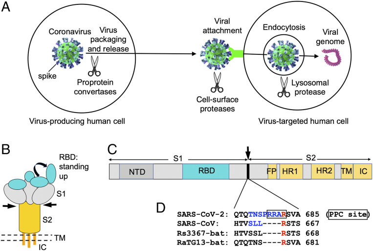
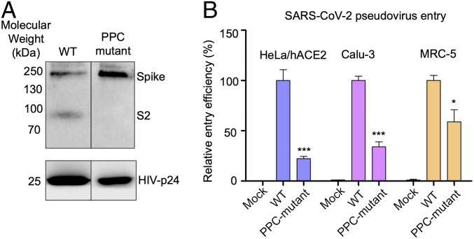
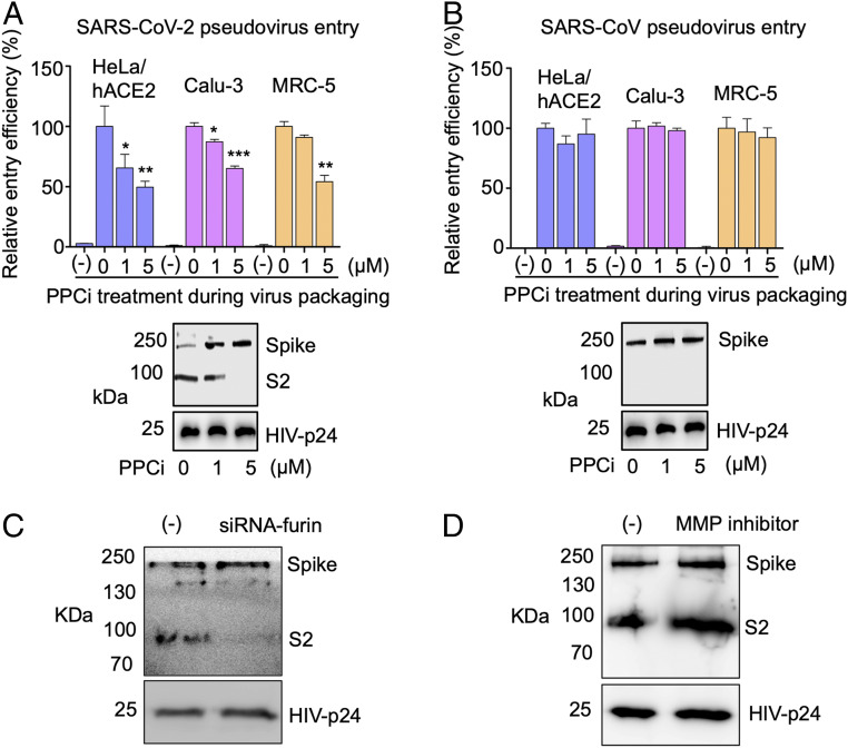
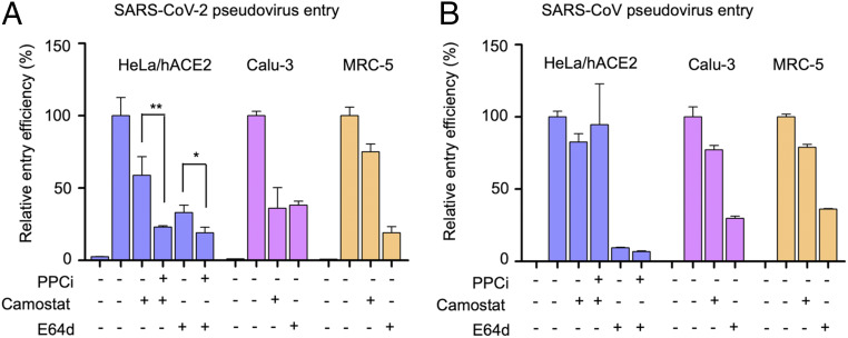
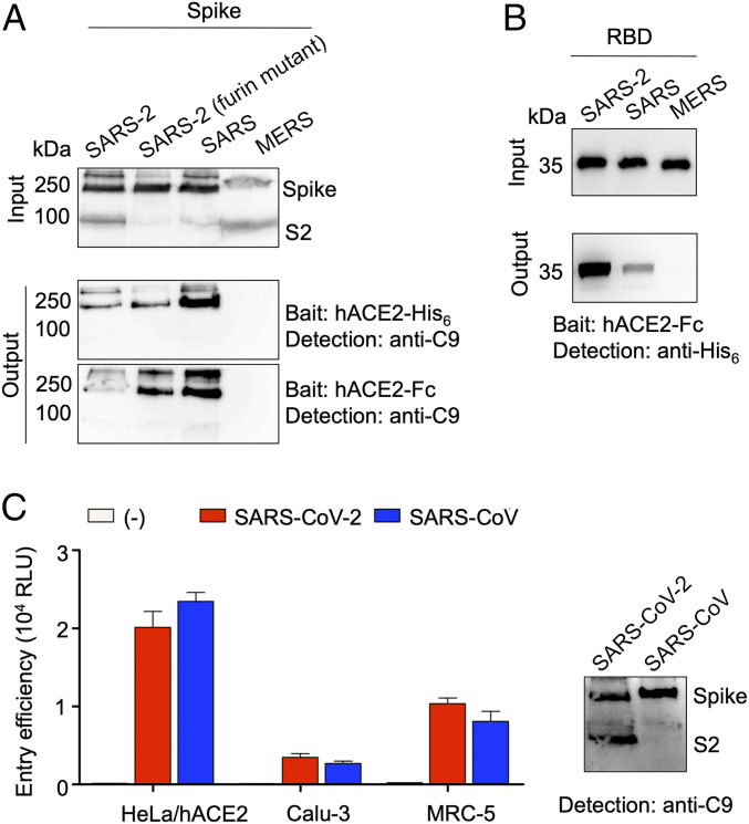
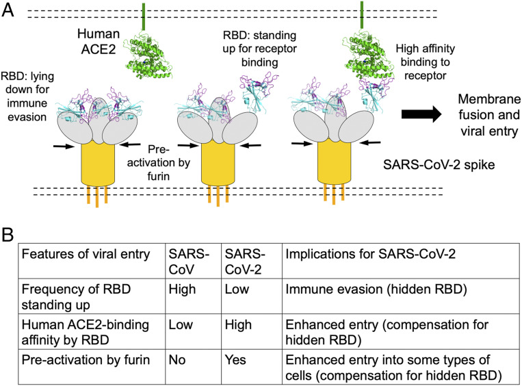
Significance: A key to curbing SARS-CoV-2 is to understand how it enters cells. SARS-CoV-2 and SARS-CoV both use human ACE2 as entry receptor and human proteases as entry activators. Using biochemical and pseudovirus entry assays and SARS-CoV as a comparison, we have identified key cell entry mechanisms of SARS-CoV-2 that potentially contribute to the immune evasion, cell infectivity, and wide spread of the virus. This study also clarifies conflicting reports from recent studies on cell entry of SARS-CoV-2. Finally, by highlighting the potency and the evasiveness of SARS-CoV-2, the study provides insight into intervention strategies that target its cell entry mechanisms.
Overview
The claims and statistics of this paper haven’t been checked yet.
Run the check — free, no account needed. It reads the open-access text and checks the sources it cites.
Integrity signals
Registry
Cites
Statistics
Figures
Verification
Claims the paper makes
The claims and statistics of this paper haven’t been checked yet.
Run the check — free, no account needed. It reads the open-access text and checks the sources it cites.
Claims other papers make about this one
Each is checked against this paper’s own text: does it say what the citing paper says it does?
In the citing paper
“The spike protein binds to its receptor on the host cell surface, angiotensin-converting enzyme 2 (ref.118).”
From mRNA vaccines for infectious diseases: principles, delivery and clinical translation (2021) · cites this paper as [118]
In the citing paper
“The attached spike protein is then cut open by the cell’s transmembrane serine protease 2, which induces a conformational change that exposes the spike protein’s fusion peptide and facilitates fusion with the cell or endosomal membrane118.”
From mRNA vaccines for infectious diseases: principles, delivery and clinical translation (2021) · cites this paper as [118]
Self-check
Percentages, sums and reported differences re-computed from the paper’s own numbers.
The self-check needs the paper’s full text.
Citations
Cites
Clinical features of patients infected with 2019 novel coronavirus in Wuhan, China
Huang, C., Wang, Y., Li, X., Ren, L., Zhao, J., Hu, Y., Zhang, L., Fan, G., Xu, J., Gu, X., et al. (2020) · The Lancet · doi:10.1016/s0140-6736(20)30183-5 · Cited by 48,884
Zhou, F., Yu, T., Du, R., Fan, G., Liu, Y., Liu, Z., Xiang, J., Wang, Y., Song, B., Gu, X., et al. (2020) · The Lancet · doi:10.1016/s0140-6736(20)30566-3 · Cited by 27,263
A pneumonia outbreak associated with a new coronavirus of probable bat origin
Zhou, P., Yang, X.-L., Wang, X.-G., Hu, B., Zhang, L., Zhang, W., Si, H.-R., Zhu, Y., Li, B., Huang, C.-L., et al. (2020) · Nature · doi:10.1038/s41586-020-2012-7 · Cited by 21,969
Hoffmann, M., Kleine-Weber, H., Schroeder, S., Krüger, N., Herrler, T., Erichsen, S., Schiergens, T. S., Herrler, G., Wu, N.-H., Nitsche, A., et al. (2020) · Cell · doi:10.1016/j.cell.2020.02.052 · Cited by 20,286
Early transmission dynamics in Wuhan, China, of novel coronavirus-infected pneumonia
Li, Q., Guan, X., Wu, P., Wang, X., Zhou, L., Tong, Y., Ren, R., Leung, K. S. M., Lau, E. H. Y., Wong, J. Y., et al. (2020) · New England Journal of Medicine · doi:10.1056/nejmoa2001316 · Cited by 16,575
Cryo-EM structure of the 2019-nCoV spike in the prefusion conformation
Wrapp, D., Wang, N., Corbett, K. S., Goldsmith, J. A., Hsieh, C.-L., Abiona, O., Graham, B. S., & McLellan, J. S. (2020) · Science · doi:10.1126/science.abb2507 · Cited by 8,897
Structure, function and antigenicity of the SARS-CoV-2 spike glycoprotein
Walls, A. C., Park, Y.-J., Tortorici, M. A., Wall, A., McGuire, A. T., & Veesler, D. (2020) · doi:10.1101/2020.02.19.956581 · Cited by 8,586
Virological assessment of hospitalized patients with COVID-2019
Wölfel, R., Corman, V. M., Guggemos, W., Seilmaier, M., Zange, S., Müller, M. A., Niemeyer, D., Jones, T. C., Vollmar, P., Rothe, C., et al. (2020) · Nature · doi:10.1038/s41586-020-2196-x · Cited by 6,661
Angiotensin-converting enzyme 2 is a functional receptor for the SARS coronavirus
Li, W., Moore, M. J., Vasilieva, N., Sui, J., Wong, S. K., Berne, M. A., Somasundaran, M., Sullivan, J. L., Luzuriaga, K., Greenough, T. C., et al. (2003) · Nature · doi:10.1038/nature02145 · Cited by 5,923
Wan, Y., Shang, J., Graham, R., Baric, R. S., & Li, F. (2020) · Journal of Virology · doi:10.1128/jvi.00127-20 · Cited by 4,341
Self-citation
Research works printed but not linked: 2
- R. Woelfel . Clinical presentation and virological assessment of hospitalized cases of coronavirus disease 2019 in a travel-associated transmission cluster. medRxiv:2020.2003.2005.20030502 (8 March 2020).
- 10.1016/j.cell.2020.02.058 · DOI · Paper page
Cited by
Sorted by citations
Characteristics of SARS-CoV-2 and COVID-19
Hu, B., Guo, H., Zhou, P., & Shi, Z.-L. (2021) · Nature Reviews Microbiology · doi:10.1038/s41579-020-00459-7 · Cited by 5,291
Coronavirus biology and replication: implications for SARS-CoV-2
V’kovski, P., Kratzel, A., Steiner, S., Stalder, H., & Thiel, V. (2020) · Nature Reviews Microbiology · doi:10.1038/s41579-020-00468-6 · Cited by 3,371
Mechanisms of SARS-CoV-2 entry into cells
Jackson, C. B., Farzan, M., Chen, B., & Choe, H. (2022) · Nature Reviews Molecular Cell Biology · doi:10.1038/s41580-021-00418-x · Cited by 3,022
Huang, Y., Yang, C., Xu, X.-F., Xü, W., & Liu, S. (2020) · Acta Pharmacologica Sinica · doi:10.1038/s41401-020-0485-4 · Cited by 2,288
mRNA vaccines for infectious diseases: principles, delivery and clinical translation
Chaudhary, N., Weissman, D., & Whitehead, K. A. (2021) · Nature Reviews Drug Discovery · doi:10.1038/s41573-021-00283-5 · Cited by 1,555
Metadata
- Authors
- Shang, J., Wan, Y., Luo, C., Ye, G., Geng, Q., Auerbach, A., & Li, F.
- Affiliations
- University of Minnesota (US)
- Corresponding author
- Li, Fang
- Venue
- Proceedings of the National Academy of Sciences
- Publisher
- National Academy of Sciences
- Published
- 2020-05-06
- Volume
- 117
- Issue
- 21
- Pages
- 11727-11734
- ISSN
- 0027-8424 (ISSN-L)
- 1091-6490
- PMC
- PMC7260975
- OpenAlex
- W3022435472
- Cited by
- 3,142
- References
- 48
- Citations by year
- 2026: 78 · 2025: 171 · 2024: 257 · 2023: 444 · 2022: 746 · 2021: 993 · 2020: 450
- FWCI
- 87.09
- Type
- journal article
- Language
- en
- Open access
- hybrid
- doi.org
- Licence
- Locations
- Proceedings of the National Academy of Sciences (publishedVersion, cc-by)
- PubMed Central (publishedVersion, cc-by)
- PubMed (publishedVersion)
- Funders
- Division of Intramural Research, National Institute of Allergy and Infectious Diseases (R01AI089728, R01AI110700)
- Topics
- Health Sciences › Medicine › Infectious Diseases › SARS-CoV-2 and COVID-19 Research
- Health Sciences › Medicine › Infectious Diseases › COVID-19 Clinical Research Studies
- Life Sciences › Agricultural and Biological Sciences › Animal Science and Zoology › Animal Virus Infections Studies
- Keywords
- SARS-CoV-2, COVID-19 pandemic, viral entry, spike protein, receptor binding domain, ACE2 receptor, furin protease, proteolytic activation, SARS-CoV, receptor binding affinity, immune evasion, pseudovirus entry
- MeSH terms
- Protein Domains, Receptors, Coronavirus, Angiotensin-Converting Enzyme 2, Cell Line, Humans, Peptidyl-Dipeptidase A, Receptors, Virus, Virus Activation, Severe acute respiratory syndrome-related coronavirus, Virus Internalization, Immune Evasion, Spike Glycoprotein, Coronavirus
About these checks
Registry, retraction, Cites and statistics results are computed, not judged, with one exception: a printed reference that no index lists is read once by a model before it counts as a citation error. Claims are extracted by a model and kept only when their quote appears verbatim in the text; each cited source is then checked, with a model’s help, for whether it says what the claim relies on. Those verdicts can be wrong — read the claim’s quote and the cited source before relying on one.
Statistics and arithmetic are recomputed, not judged.
Confidence bands are shown only for fields calibrated on our annotated test set. None is calibrated yet, so no band is shown.
What counts as a claim ›How we measure confidence ›How we check this ›
Are you an author, or do you see a problem with this page? Request a review
