

The DOI matches its Crossref record; 49 of the 49 references it cites were found in a registry or an index.
The Peroxisome Proliferator-activated Receptor δ Promotes Lipid Accumulation in Human Macrophages
Open accessAbstract
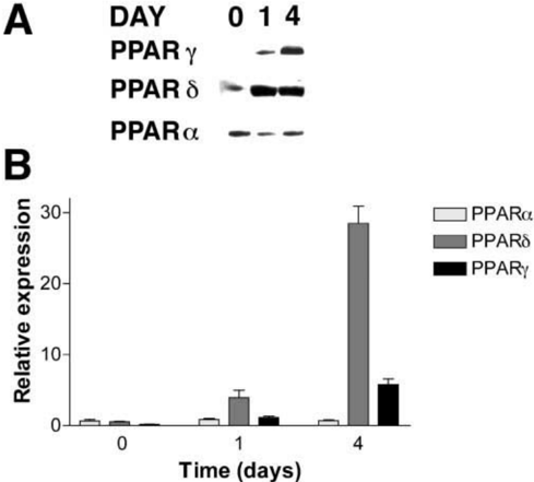
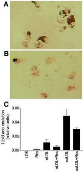
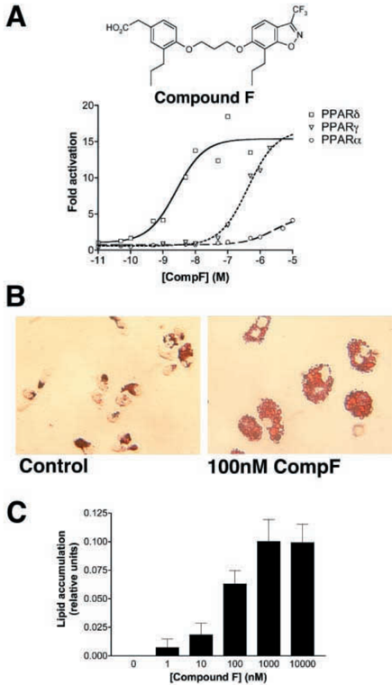
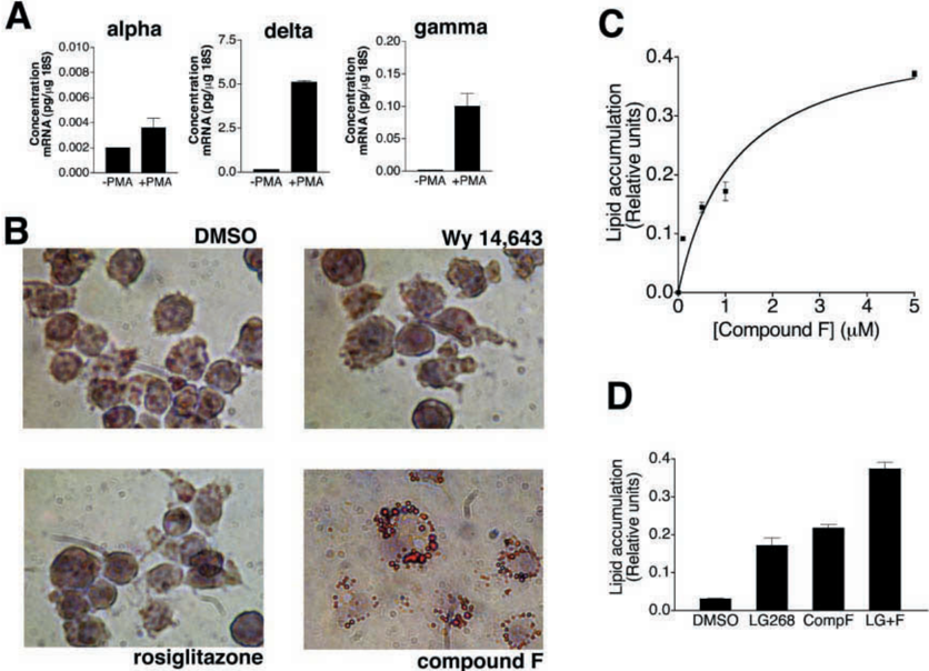
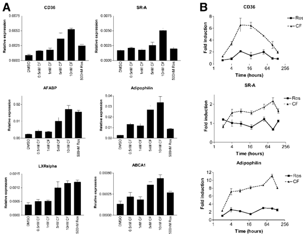
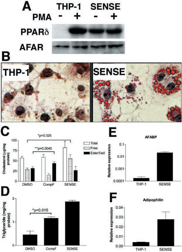
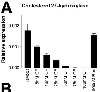
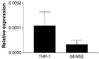
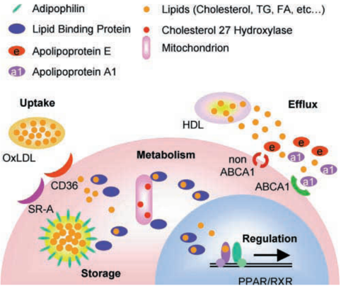
The peroxisome proliferator-activated receptors (PPARs) are a family of fatty acid-activated transcription factors which control lipid homeostasis and cellular differentiation. PPARalpha (NR1C1) controls lipid oxidation and clearance in hepatocytes and PPARgamma (NR1C3) promotes preadipocyte differentiation and lipogenesis. Drugs that activate PPARalpha are effective in lowering plasma levels of lipids and have been used in the management of hyperlipidemia. PPARgamma agonists increase insulin sensitivity and are used in the management of type 2 diabetes. In contrast, there are no marketed drugs that selectively target PPARdelta (NR1C2) and the physiological roles of PPARdelta are unclear. In this report we demonstrate that the expression of PPARdelta is increased during the differentiation of human macrophages in vitro. In addition, a highly selective agonist of PPARdelta (compound F) promotes lipid accumulation in primary human macrophages and in macrophages derived from the human monocytic cell line, THP-1. Compound F increases the expression of genes involved in lipid uptake and storage such as the class A and B scavenger receptors (SRA, CD36) and adipophilin. PPARdelta activation also represses key genes involved in lipid metabolism and efflux, i.e. cholesterol 27-hydroxylase and apolipoprotein E. We have generated THP-1 sublines that overexpress PPARdelta and have confirmed that PPARdelta is a powerful promoter of macrophage lipid accumulation. These data suggest that PPARdelta may play a role in the pathology of diseases associated with lipid-filled macrophages, such as atherosclerosis, arthritis, and neurodegeneration.
Overview
The claims and statistics of this paper haven’t been checked yet.
Run the check — free, no account needed. It reads the open-access text and checks the sources it cites.
Integrity signals
Registry
Cites
Statistics
Figures
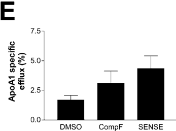
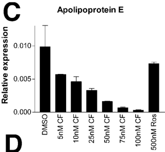
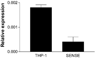
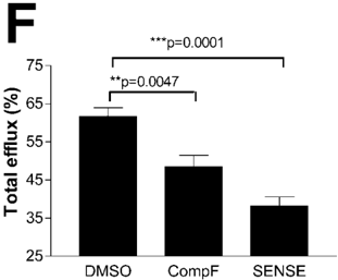
Verification
Claims the paper makes
The claims and statistics of this paper haven’t been checked yet.
Run the check — free, no account needed. It reads the open-access text and checks the sources it cites.
Claims other papers make about this one
Each is checked against this paper’s own text: does it say what the citing paper says it does?
The cited source says what the claim attributes to it.
In the citing paper
“This is likely reasoned by the abundant expression of PPARd, a hallmark of THP-1 cell differentiation (Vosper et al., 2001) (Supplemental Fig. 16).”
The cited source says what the claim attributes to it.
In the citing paper
“Consequently, the PPARd activation promotes lipid accumulation in THP-1 macrophages in the presence of serum (Vosper et al., 2001).”
The cited source says what the claim attributes to it.
In the citing paper
“As we did not use any lipoproteins as stimulus, the studies of Vosper et al. (2001) on PPARd and lipid accumulation in macrophages most accurately resemble our experimental setup.”
The cited source says what the claim attributes to it.
In the citing paper
“In line with our findings, the treatment with a PPARd agonist upregulated the expression of genes involved in lipid metabolism, including CD36 and PLIN2, in this study.”
Self-check
Percentages, sums and reported differences re-computed from the paper’s own numbers.
The self-check needs the paper’s full text.
Citations
Cites
Stimulation of adipogenesis in fibroblasts by PPARγ2, a lipid-activated transcription factor
Tontonoz, P., Hu, E., & Spiegelman, B. M. (1994) · Cell · doi:10.1016/0092-8674(94)90006-x · Cited by 3,564
The peroxisome proliferator-activated receptor-γ is a negative regulator of macrophage activation
Ricote, M., Li, A. C., Willson, T. M., Kelly, C. J., & Glass, C. K. (1998) · Nature · doi:10.1038/34178 · Cited by 3,525
PPAR-γ agonists inhibit production of monocyte inflammatory cytokines
Jiang, C., Ting, A. T., & Seed, B. (1998) · Nature · doi:10.1038/34184 · Cited by 2,899
15-Deoxy-Δ12,14-Prostaglandin J2 is a ligand for the adipocyte determination factor PPARγ
FORMAN, B. M., Tontonoz, P., Chen, J., Brun, R. P., Spiegelman, B. M., & Evans, R. M. (1995) · Cell · doi:10.1016/0092-8674(95)90193-0 · Cited by 2,857
Apolipoprotein E polymorphism and atherosclerosis.
Davignon, J., Gregg, R. E., & Sing, C. F. (1988) · Arteriosclerosis: An Official Journal of the American Heart Association, Inc. · doi:10.1161/01.atv.8.1.1 · Cited by 1,981
Roles of PPARs in health and disease
Kersten, S., Desvergne, B., & Wahli, W. (2000) · Nature · doi:10.1038/35013000 · Cited by 1,956
Oxidized LDL Regulates Macrophage Gene Expression through Ligand Activation of PPARγ
Nagy, L., Tontonoz, P., Alvarez, J. G. A., Chen, H.-W., & Evans, R. M. (1998) · Cell · doi:10.1016/s0092-8674(00)81574-3 · Cited by 1,861
PPARγ Promotes Monocyte/Macrophage Differentiation and Uptake of Oxidized LDL
Tontonoz, P., Nagy, L., Alvarez, J. G. A., Thomázy, V. A., & Evans, R. M. (1998) · Cell · doi:10.1016/s0092-8674(00)81575-5 · Cited by 1,815
Kliewer, S. A., Umesono, K., Noonan, D. J., Heyman, R. A., & Evans, R. M. (1992) · Nature · doi:10.1038/358771a0 · Cited by 1,746
Anti-inflammatory cyclopentenone prostaglandins are direct inhibitors of IκB kinase
Rossi, A., Kapahi, P., Natoli, G., Takahashi, T., Chen, Y., Karin, M., & Santoro, M. G. (2000) · Nature · doi:10.1038/47520 · Cited by 1,303
Research works printed but not linked: 3
- Cullen, 1987, Methods Enzymol., 152, 692
- Wu, 1994, Mol. Cell. Biol., 14, 2129
- Kang, 1997, Mutat. Res., 382, 57
Cited by
Sorted by citations
Items 101–200 of 270
Briand, F., Naik, S. U., Fuki, I. V., Millar, J. S., Macphee, C. H., Walker, M., Billheimer, J. T., Rothblat, G. H., & Rader, D. J. (2009) · Clinical and Translational Science · doi:10.1111/j.1752-8062.2009.00098.x · Cited by 66
Self-citationLim, H., Lee, S., Park, J., Lee, K., Choi, H., Chung, K., Lee, H.-H., & Park, H. (2008) · Atherosclerosis · doi:10.1016/j.atherosclerosis.2008.05.023 · Cited by 66
Payré, B., de Médina, P., Boubekeur, N., Mhamdi, L., Bertrand‐Michel, J., Tercé, F., Fourquaux, I., Goudounèche, D., Record, M., Poirot, M., et al. (2008) · Molecular Cancer Therapeutics · doi:10.1158/1535-7163.mct-08-0507 · Cited by 65
Wei, Z.-L., & Kozikowski, A. P. (2003) · The Journal of Organic Chemistry · doi:10.1021/jo035140g · Cited by 64
Biology and Therapeutic Applications of Peroxisome Proliferator- Activated Receptors
P. Menendez-Gutierrez, M., Roszer, T., & Ricote, M. (2012) · Current Topics in Medicinal Chemistry · doi:10.2174/156802612799436669 · Cited by 62
Liu, Y., Jia, X., Zhu, H., Zhang, Q., He, Y., Shen, Y., Xu, X., & Li, J. (2021) · Chemosphere · doi:10.1016/j.chemosphere.2021.131831 · Cited by 65
Genoula, M., Franco, J. L. M., Dupont, M., Kviatcovsky, D., Milillo, M. A., Schierloh, P., Moraña, E. J., Poggi, S., Palmero, D., Mata‐Espinosa, D., et al. (2018) · Frontiers in Immunology · doi:10.3389/fimmu.2018.00459 · Cited by 60
Xu, Y., Wang, Q., Cook, T. J., & Knipp, G. T. (2007) · Journal of Pharmaceutical Sciences · doi:10.1002/jps.20973 · Cited by 62
The peroxisome proliferator-activated receptor α (PPARα): role in hepatocarcinogenesis
Gonzalez, F. J. (2002) · Molecular and Cellular Endocrinology · doi:10.1016/s0303-7207(02)00098-9 · Cited by 59
Szántó, A., & Nagy, L. (2005) · Molecular Pharmacology · doi:10.1124/mol.104.006445 · Cited by 58
Peroxisome proliferator-activated receptors: Bridging metabolic syndrome with molecular nutrition
Guri, A. J., Hontecillas, R., & Bassaganya-Riera, J. (2006) · Clinical Nutrition · doi:10.1016/j.clnu.2006.08.006 · Cited by 57
Russell, T. D., Palmer, C. A., Orlicky, D. J., Bales, E. S., Chang, B. H., Chan, L., & McManaman, J. L. (2007) · Journal of Lipid Research · doi:10.1194/jlr.m700396-jlr200 · Cited by 57
Self-citationFürnsinn, C., Willson, T. M., & Brunmair, B. (2006) · Diabetologia · doi:10.1007/s00125-006-0492-0 · Cited by 56
Mechanisms involved in Jurkat cell death induced by oleic and linoleic acids
Cury‐Boaventura, M. F., Kanunfre, C. C., Gorjão, R., de Lima, T. M., & Curi, R. (2006) · Clinical Nutrition · doi:10.1016/j.clnu.2006.05.008 · Cited by 56
Yang, X., Li, D., Zhang, M., Feng, Y., Jin, X., Liu, D., Guo, Y., & Hu, Y. (2023) · Journal of Animal Science and Biotechnology/Journal of animal science and biotechnology · doi:10.1186/s40104-023-00900-w · Cited by 63
Chakraborty, A., Bodipati, N., Krstic–Demonacos, M., Peddinti, R. K., Ghosh, K., & Roy, P. (2012) · Molecular and Cellular Endocrinology · doi:10.1016/j.mce.2012.01.009 · Cited by 55
Regulatory functions of PPARβ in metabolism: implications for the treatment of metabolic syndrome
Grimaldi, P. A. (2007) · Biochimica et Biophysica Acta (BBA) - Molecular and Cell Biology of Lipids · doi:10.1016/j.bbalip.2007.02.006 · Cited by 56
Chen, S., Tsybouleva, N., Ballantyne, C. M., Gotto, A. M., & Marian, A. J. (2003) · Pharmacogenetics · doi:10.1097/00008571-200401000-00007 · Cited by 55
Macrophages, PPARs, and Cancer
Ginderachter, J. A. V., Movahedi, K., den Bossche, J. V., & BAETSELIER, P. D. (2008) · PPAR Research · doi:10.1155/2008/169414 · Cited by 54
Macrophages are Novel Sites of Expression and Regulation of Retinol Binding Protein-4 (RBP4)
Broch, M., Ramírez, R., Auguet, T., Alcaide, M. J., Aguilar, C., García‐España, A., & Richart, C. (2010) · Physiological Research · doi:10.33549/physiolres.931714 · Cited by 53
Bouhlel, M. A., Staels, B., & Chinetti‐Gbaguidi, G. (2008) · Journal of Internal Medicine · doi:10.1111/j.1365-2796.2007.01892.x · Cited by 51
Bojic, L. A., Sawyez, C. G., Telford, D. E., Edwards, J. Y., Hegele, R. A., & Huff, M. W. (2012) · Arteriosclerosis, Thrombosis, and Vascular Biology · doi:10.1161/atvbaha.112.255208 · Cited by 51
Hodgkinson, C. P., & Ye, S. (2003) · Biochemical and Biophysical Research Communications · doi:10.1016/s0006-291x(03)01416-5 · Cited by 51
Aspirin increases CD36, SR-BI, and ABCA1 expression in human THP-1 macrophages
Viñals, M. L., Bermúdez-Escobar, I. C., Llaverı́as, G., Alegret, M., SANCHEZ, R., Vázquez‐Carrera, M., & Laguna, J. C. (2005) · Cardiovascular Research · doi:10.1016/j.cardiores.2004.12.024 · Cited by 50
Jiménez, R., Sánchez, M., Zarzuelo-Romero, M. J., Romero, M., Quintela, A. M., López-Sepúlveda, R., Galindo, P., Gómez‐Guzmán, M., Haro, J. M., Zarzuelo, A., et al. (2009) · Journal of Pharmacology and Experimental Therapeutics · doi:10.1124/jpet.109.159806 · Cited by 50
Peroxisome Proliferator-Activated Receptors and the Cardiovascular System
Chen, Y. E., Fu, M., Zhang, J., Zhu, X., Lin, Y., Akinbami, M. A., & Song, Q. (2003) · Vitamins and hormones · doi:10.1016/s0083-6729(03)01005-7 · Cited by 50
Michalik, L., Desvergne, B., & Wahli, W. (2003) · Current Opinion in Lipidology · doi:10.1097/00041433-200304000-00003 · Cited by 49
Peroxisome proliferator-activated receptors: are they involved in atherosclerosis progression?
Puddu, P., Puddu, G. M., & Muscari, A. (2003) · International Journal of Cardiology · doi:10.1016/s0167-5273(02)00565-x · Cited by 47
New Therapeutic Target for Metabolic Syndrome: PPAR.DELTA.
Takahashi, S., Tanaka, T., & Sakai, J. (2007) · Endocrine Journal · doi:10.1507/endocrj.kr-99 · Cited by 46
Peroxisome proliferator-activated receptor-? as a target for wound healing drugs
Tan, N. S., Michalik, L., Desvergne, B., & Wahli, W. (2004) · Expert Opinion on Therapeutic Targets · doi:10.1517/14728222.8.1.39 · Cited by 46
Jakobsen, M. A., Petersen, R. K., Kristiansen, K., Lange, M., & Lillevang, S. T. (2006) · Scandinavian Journal of Immunology · doi:10.1111/j.1365-3083.2006.01745.x · Cited by 44
Cabrero, À., Cubero, M., Llaverı́as, G., Jové, M., Planavila, A., Alegret, M., Sánchez, R., Laguna, J. C., & Vázquez‐Carrera, M. (2003) · Metabolism · doi:10.1053/meta.2003.50100 · Cited by 44
Sarov‐Blat, L., Kiss, R. S., Haidar, B., Kavaslar, N., Jaye, M., Bertiaux, M., Steplewski, K. M., Hurle, M. R., Sprecher, D. L., McPherson, R., et al. (2007) · Arteriosclerosis Thrombosis and Vascular Biology · doi:10.1161/atvbaha.106.138990 · Cited by 44
Emerging roles of PPARδ in metabolism
Seedorf, U., & Aberle, J. (2007) · Biochimica et Biophysica Acta (BBA) - Molecular and Cell Biology of Lipids · doi:10.1016/j.bbalip.2007.04.017 · Cited by 41
Zhang, B., Wang, Z., Huang, C., Wang, D., Chang, D., Shi, X., Chen, Y., & Chen, H. (2022) · Frontiers in Veterinary Science · doi:10.3389/fvets.2022.1005643 · Cited by 40
Nikitin, A. G., Chistiakov, D. A., Minushkina, L. O., Zateyshchikov, D. A., & Nosikov, V. V. (2010) · Heart and Vessels · doi:10.1007/s00380-009-1159-9 · Cited by 40
PPAR‐δ in Vascular Pathophysiology
Wang, N. (2008) · PPAR Research · doi:10.1155/2008/164163 · Cited by 39
López‐Soriano, J., Chiellini, C., Maffei, M., Grimaldi, P. A., & Argilés, J. M. A. (2006) · Endocrine Reviews · doi:10.1210/er.2005-0012 · Cited by 40
Singh, A., & Chaudhary, R. (2025) · International Immunopharmacology · doi:10.1016/j.intimp.2025.114616 · Cited by 42
Chaves, J. M., Srivastava, K., Gupta, R., & Srivastava, O. P. (2008) · Biochemistry · doi:10.1021/bi8001902 · Cited by 39
Regulation of Milk Lipid Formation and Secretion in the Mouse Mammary Gland
McManaman, J. L., Palmer, C. A., Anderson, S. M., Schwertfeger, K. L., & Neville, M. C. (2004) · Advances in experimental medicine and biology · doi:10.1007/978-1-4757-4242-8_22 · Cited by 39
Self-citationPeroxisome proliferator-activated receptor β/δ as a therapeutic target for metabolic diseases
Bedu, E., Wahli, W., & Desvergne, B. (2005) · Expert Opinion on Therapeutic Targets · doi:10.1517/14728222.9.4.861 · Cited by 40
Okazaki, H., Osuga, J.-I., Tsukamoto, K., Iso-O, N., Kitamine, T., Tamura, Y., Tomita, S., Sekiya, M., Yahagi, N., Iizuka, Y., et al. (2002) · Journal of Biological Chemistry · doi:10.1074/jbc.m204016200 · Cited by 39
Fan, B., Ikuyama, S., Gu, J.-Q., Wei, P., Oyama, J.-I., Inoguchi, T., & Nishimura, J. (2009) · American Journal of Physiology-Endocrinology and Metabolism · doi:10.1152/ajpendo.00119.2009 · Cited by 38
Tanaka, T., Takeno, T., Watanabe, Y., Uchiyama, Y., Murakami, T., Yamashita, H., Suzuki, A., Aoi, R., Iwanari, H., Jiang, S.-Y., et al. (2002) · Journal of Atherosclerosis and Thrombosis · doi:10.5551/jat.9.233 · Cited by 38
Batt, K. V., Patel, L., Botham, K. M., & Suckling, K. E. (2004) · Journal of Molecular Medicine · doi:10.1007/s00109-004-0551-2 · Cited by 37
Self-citationPPARs in atherosclerosis: The spatial and temporal features from mechanism to druggable targets
Zheng, Y., Shao, M., Zheng, Y., Sun, W., Qin, S., Sun, Z., Zhu, L., Guan, Y., Wang, Q., Wang, Y., et al. (2024) · Journal of Advanced Research · doi:10.1016/j.jare.2024.03.020 · Cited by 41
PPARβ in macrophages and atherosclerosis
Chinetti, G., & Staels, B. (2016) · Biochimie · doi:10.1016/j.biochi.2016.12.008 · Cited by 37
Orphan nuclear receptors: therapeutic opportunities in skeletal muscle
Smith, A. G., & Muscat, G. E. O. (2006) · American Journal of Physiology-Cell Physiology · doi:10.1152/ajpcell.00476.2005 · Cited by 37
Peroxisome proliferator-activated receptor delta and cardiovascular disease
Ehrenborg, E., & Skogsberg, J. (2013) · Atherosclerosis · doi:10.1016/j.atherosclerosis.2013.08.027 · Cited by 36
BCL-6: a possible missing link for anti-inflammatory PPAR-δ signalling in pancreatic beta cells
Kharroubi, I., Lee, C., Hekerman, P., Darville, M. I., Evans, R. M., Eizirik, D. L., & Cnop, M. (2006) · Diabetologia · doi:10.1007/s00125-006-0366-5 · Cited by 36
Schubert, M., Becher, S., Wallert, M., Maeß, M. B., Abhari, M., Rennert, K., Mosig, A. S., Große, S., Heller, R., Grün, M., et al. (2020) · Molecular Pharmacology · doi:10.1124/mol.119.117887 · Cited by 35
Ravaux, L., Denoyelle, C., Monne, C., Limon, I., Raymondjean, M., & Hadri, K. E. (2007) · Molecular and Cellular Biology · doi:10.1128/mcb.00623-07 · Cited by 35
PPARβ Regulates Liver Regeneration by Modulating Akt and E2f Signaling
Liu, H., Fang, Y., Hu, Y. C., Gonzalez, F. J., Fang, J., & Wan, Y. Y. (2013) · PLoS ONE · doi:10.1371/journal.pone.0065644 · Cited by 34
Chen, W., Penumetcha, M., Santanam, N., Liu, Y.-G., Garelnabi, M., & Parthasarathy, S. (2005) · Biochimica et Biophysica Acta (BBA) - General Subjects · doi:10.1016/j.bbagen.2005.03.005 · Cited by 34
Coll, T., Barroso, E., Álvarez-Guardia, D., Serrano, L., Salvadó, L., Merlos, M., Palomer, X., & Vázquez‐Carrera, M. (2010) · PPAR Research · doi:10.1155/2010/368467 · Cited by 33
Cholesteryl ester hydroperoxides increase macrophage CD36 gene expression via PPARα
Jedidi, I., Couturier, M., Thérond, P., Gardès-Albert, M., Legrand, A., Barouki, R., Bonnefont‐Rousselot, D., & Aggerbeck, Μ. (2006) · Biochemical and Biophysical Research Communications · doi:10.1016/j.bbrc.2006.10.122 · Cited by 32
Chinetti, G., & Staels, B. (2009) · Biochimica et Biophysica Acta (BBA) - Molecular and Cell Biology of Lipids · doi:10.1016/j.bbalip.2009.01.009 · Cited by 32
Adipophilin protein expression in muscle – a possible protective role against insulin resistance
de Wilde, J., Smit, E., Snepvangers, F. J. M., de Wit, N. J. W., Mohren, R., Hulshof, M. F. M., & Mariman, E. C. M. (2009) · FEBS Journal · doi:10.1111/j.1742-4658.2009.07525.x · Cited by 31
Gouni-Berthold, I., Giannakidou, E., Faust, M., Berthold, H. K., & Krone, W. (2005) · Atherosclerosis · doi:10.1016/j.atherosclerosis.2005.03.016 · Cited by 31
PPAR-β/δ activation promotes phospholipid transfer protein expression
Chehaibi, K., Cedó, L., Metso, J., Palomer, X., Santos, D., Quesada, H., Slimane, M. N., Wahli, W., Julve, J., Vázquez‐Carrera, M., et al. (2015) · Biochemical Pharmacology · doi:10.1016/j.bcp.2015.01.016 · Cited by 30
Roles of Perilipins in Diseases and Cancers
Zhang, P., Meng, L., Song, L., Du, J., Du, S., Cui, W., Liu, C., & Li, F. (2017) · Current Genomics · doi:10.2174/1389202918666170915155948 · Cited by 31
Ahmed, N., Riley, C. B., & Quinn, M. A. (2008) · British Journal of Cancer · doi:10.1038/sj.bjc.6604306 · Cited by 30
Post-translationally modified human lens crystallin fragments show aggregation in vitro
Srivastava, O. P., Srivastava, K., Chaves, J. M., & Gill, A. (2017) · Biochemistry and Biophysics Reports · doi:10.1016/j.bbrep.2017.01.011 · Cited by 29
Jarvis, M. C., Gray, T. J. B., & Palmer, C. N. A. (2005) · Oncogene · doi:10.1038/sj.onc.1208983 · Cited by 29
Self-citationVaccination strategies in patients with renal cell carcinoma
Asemissen, A. M., & Brossart, P. (2009) · Cancer Immunology Immunotherapy · doi:10.1007/s00262-009-0706-7 · Cited by 28
Nuclear receptor mediated mechanisms of macrophage cholesterol metabolism
Nagy, Z. S., Czimmerer, Z., & Nagy, L. (2012) · Molecular and Cellular Endocrinology · doi:10.1016/j.mce.2012.04.003 · Cited by 27
Ito, F. (2020) · Current Molecular Pharmacology · doi:10.2174/1874467213666200320153410 · Cited by 30
Mammalian lipid droplets: structural, pathological, immunological and anti-toxicological roles
Hammoudeh, N., Soukkarieh, C., Murphy, D. J., & Hanano, A. (2023) · Progress in Lipid Research · doi:10.1016/j.plipres.2023.101233 · Cited by 26
Barrera, G., Toaldo, C., Pizzimenti, S., Cerbone, A., Pettazzoni, P., Dianzani, M. U., & Ferretti, C. (2008) · PPAR Research · doi:10.1155/2008/524671 · Cited by 27
PPAR-δ: A key nuclear receptor in vascular function and remodeling
Xiao, L., & Wang, N. (2022) · Journal of Molecular and Cellular Cardiology · doi:10.1016/j.yjmcc.2022.04.019 · Cited by 27
Yasugi, E., Horiuchi, A., Uemura, I., Okuma, E., Nakatsu, M., Saeki, K., Kamisaka, Y., Kagechika, H., Yasuda, K., & Yuo, A. (2006) · Development Growth & Differentiation · doi:10.1111/j.1440-169x.2006.00855.x · Cited by 26
Connolly, J. C., Ishihara, Y., Sawaya, E., Whitfield, V., Garrity, N., Sohata, R., Tsymbal, M., Lundberg, A., Merrill, M. A. L., DeWitt, J. C., et al. (2025) · Cardiovascular Toxicology · doi:10.1007/s12012-025-10048-w · Cited by 27
Liang, Y.-J., Liu, Y.-C., Chen, C., Lai, L., Shyu, K., Juang, S.-J., Wang, B.-W., & Leu, J.-G. (2009) · International Journal of Cardiology · doi:10.1016/j.ijcard.2009.03.100 · Cited by 25
Hiep, N. T., Choi, Y., Kim, N., Hong, S. S., Hong, S., Hwang, B. Y., Lee, H.-J., Lee, S., Jang, D. S., & Lee, D. (2012) · Journal of Natural Products · doi:10.1021/np200955z · Cited by 25
Burch, L. R., Donnelly, L. A., Doney, A. S. F., Brady, J. D., Tommasi, A. M., Whitley, A. L., Goddard, C. A., Morris, A. D., Hansen, M. K., & Palmer, C. N. A. (2010) · The Journal of Clinical Endocrinology & Metabolism · doi:10.1210/jc.2009-1201 · Cited by 24
Self-citationProfiling of membrane proteins from human macrophages: Comparison of two approaches
Slomianny, M., Dupont, A., Bouanou, F., Beseme, O., Guihot, A., Amouyel, P., Michalski, J., & Pinet, F. (2006) · PROTEOMICS · doi:10.1002/pmic.200500546 · Cited by 24
Water-Soluble Components of Sesame Oil Reduce Inflammation and Atherosclerosis
Narasimhulu, C. A., Selvarajan, K., Burge, K. Y., Litvinov, D. Y., Sengupta, B., & Parthasarathy, S. (2016) · Journal of Medicinal Food · doi:10.1089/jmf.2015.0154 · Cited by 23
Roles of omental and bone marrow adipocytes in tumor biology
Cha, Y. J., & Koo, J. S. (2019) · Adipocyte · doi:10.1080/21623945.2019.1643189 · Cited by 22
Nishikawa, K., Iwaya, K., Kinoshita, M., Fujiwara, Y., Akao, M., Sonoda, M., Thiruppathi, S., Suzuki, T., Hiroi, S., Seki, S., et al. (2015) · Molecular Nutrition & Food Research · doi:10.1002/mnfr.201400564 · Cited by 23
The Role of PPARδ Signaling in the Cardiovascular System
Ding, Y., Yang, K. D., & Yang, Q. (2014) · Progress in molecular biology and translational science · doi:10.1016/b978-0-12-800101-1.00014-4 · Cited by 23
Lundell, K., Thulin, P., Hamsten, A., & Ehrenborg, E. (2007) · BMC Molecular Biology · doi:10.1186/1471-2199-8-70 · Cited by 23
Cresser, J., Bonen, A., Chabowski, A., Stefanyk, L. E., Gulli, R. A., Ritchie, I. R. W., & Dyck, D. J. (2010) · American Journal of Physiology-Regulatory, Integrative and Comparative Physiology · doi:10.1152/ajpregu.00431.2009 · Cited by 23
Metformin inhibits monocyte adhesion to endothelial cells and foam cell formation
Mamputu, J., Wiernsperger, N. F., & Renier, G. (2003) · The British Journal of Diabetes · doi:10.1177/14746514030030041501 · Cited by 21
Perez, A., Thuillard, J.-L., Bentzen, C. L., & Niesor, E. J. (2003) · Cell Biology and Toxicology · doi:10.1023/a:1023307206125 · Cited by 21
Vosper, H., Khoudoli, G. A., & Palmer, C. N. (2003) · Nuclear Receptor · doi:10.1186/1478-1336-1-9 · Cited by 21
Self-citationLanatoside C Promotes Foam Cell Formation and Atherosclerosis
Shi, H., Mao, X., Zhong, Y., Liu, Y., Zhao, X., Yu, K., Zhu, R., Wei, Y., Zhu, J., Sun, H., et al. (2016) · Scientific Reports · doi:10.1038/srep20154 · Cited by 19
Wang, Q., Herrera‐Ruiz, D., Mathis, A. S., Cook, T. J., Bhardwaj, R. K., & Knipp, G. T. (2004) · Journal of Pharmaceutical Sciences · doi:10.1002/jps.20264 · Cited by 19
Wang, Z., Wang, M., Xu, X., Liu, Y., Chen, Q., Wu, B., & Zhang, Y. (2023) · Biochemical and Biophysical Research Communications · doi:10.1016/j.bbrc.2023.149128 · Cited by 17
Yang, Y., Chen, M., Loux, T., & Harmon, C. M. (2007) · Pediatric Surgery International · doi:10.1007/s00383-007-1942-6 · Cited by 17
Chu, C.-Y., Lee, H., Chu, C.-Y., Yin, Y., & Tseng, T. (2009) · Food and Chemical Toxicology · doi:10.1016/j.fct.2009.02.020 · Cited by 17
Guo, J., Lu, W., Liang, S., Choi, J., Kim, N., & Cui, X.-S. (2016) · Theriogenology · doi:10.1016/j.theriogenology.2016.11.018 · Cited by 17
Muralidhar, B., Carpenter, K. L. H., Müller, K. H., Skepper, J. N., & Arends, M. J. (2004) · Prostaglandins Leukotrienes and Essential Fatty Acids · doi:10.1016/j.plefa.2004.03.020 · Cited by 17
Luo, Y., Tanigawa, K., Kawashima, A., Ishido, Y., Ishii, N., & Suzuki, K. (2020) · PLoS neglected tropical diseases · doi:10.1371/journal.pntd.0008850 · Cited by 16
Røst, T. H., Moi, L. L. H., Berge, K., Staels, B., Mellgren, G., & Berge, R. K. (2009) · Biochimica et Biophysica Acta (BBA) - Molecular and Cell Biology of Lipids · doi:10.1016/j.bbalip.2009.06.005 · Cited by 16
Namgaladze, D., Morbitzer, D., von Knethen, A., & Brüne, B. (2009) · Arteriosclerosis Thrombosis and Vascular Biology · doi:10.1161/atvbaha.109.199232 · Cited by 16
Burch, L. R., Zhou, K., Donnelly, L. A., Doney, A. S. F., Brady, J. D., Goddard, C. A., Morris, A. D., Hansen, M. K., & Palmer, C. N. A. (2009) · The Journal of Clinical Endocrinology & Metabolism · doi:10.1210/jc.2009-0392 · Cited by 15
Self-citationTakahashi, K., Sasabe, N., Ohshima, K., Kitazato, K. T., Kato, R., Masuda, Y., Tsurumaki, M., Obama, T., Okudaira, S.-I., Aoki, J., et al. (2010) · Journal of Lipid Research · doi:10.1194/jlr.m004648 · Cited by 15
Yun, Y., Kim, H., & Song, Y. (2014) · Journal of Medicinal Food · doi:10.1089/jmf.2013.2943 · Cited by 15
Ligand Modulated Antagonism of PPARγ by Genomic and Non-Genomic Actions of PPARδ
Gustafsson, M. C. U., Knight, D. A., & Palmer, C. N. A. (2009) · PLoS ONE · doi:10.1371/journal.pone.0007046 · Cited by 14
Self-citation
Metadata
- Authors
- Vosper, H., Patel, L., Graham, T. L., Khoudoli, G. A., Hill, A., Macphee, C. H., Pinto, I., Smith, S. A., Suckling, K. E., Wolf, C. R., & Palmer, C. N. A.
- Affiliations
- Ninewells Hospital (GB)
- GlaxoSmithKline (United Kingdom) (GB)
- Corresponding author
- Palmer, Colin N.A.
- Venue
- Journal of Biological Chemistry
- Publisher
- Elsevier BV
- Published
- 2001-11
- Volume
- 276
- Issue
- 47
- Pages
- 44258-44265
- ISSN
- 0021-9258 (ISSN-L)
- OpenAlex
- W2103618523
- Cited by
- 268
- References
- 49
- Citations by year
- 2026: 2 · 2025: 4 · 2024: 3 · 2023: 3 · 2022: 3 · 2021: 3 · 2020: 4 · 2019: 3 · 2018: 7 · 2017: 7 · 2016: 6 · 2015: 7 · 2014: 2 · 2013: 10 · 2012: 7
- FWCI
- 8.32
- Type
- journal article
- Language
- en
- Open access
- hybrid
- doi.org
- Licence
- Locations
- Journal of Biological Chemistry (publishedVersion, cc-by)
- PubMed (publishedVersion)
- Discovery Research Portal (University of Dundee) (publishedVersion)
- Funders
- British Heart Foundation
- Topics
- Life Sciences › Biochemistry, Genetics and Molecular Biology › Molecular Biology › Peroxisome Proliferator-Activated Receptors
- Life Sciences › Biochemistry, Genetics and Molecular Biology › Biochemistry › Cholesterol and Lipid Metabolism
- Life Sciences › Biochemistry, Genetics and Molecular Biology › Clinical Biochemistry › Paraoxonase enzyme and polymorphisms
- Keywords
- PPARδ, peroxisome proliferator-activated receptors, macrophage lipid accumulation, macrophage differentiation, compound F, scavenger receptors, CD36, adipophilin, apolipoprotein E, lipid metabolism, atherosclerosis, THP-1 cells
- MeSH terms
- Animals, Cell Line, Cholesterol, Gene Expression Regulation, Humans, Macrophages, Transcription Factors, Receptors, Cytoplasmic and Nuclear, Lipid Metabolism
About these checks
Registry, retraction, Cites and statistics results are computed, not judged, with one exception: a printed reference that no index lists is read once by a model before it counts as a citation error. Claims are extracted by a model and kept only when their quote appears verbatim in the text; each cited source is then checked, with a model’s help, for whether it says what the claim relies on. Those verdicts can be wrong — read the claim’s quote and the cited source before relying on one.
Statistics and arithmetic are recomputed, not judged.
Confidence bands are shown only for fields calibrated on our annotated test set. None is calibrated yet, so no band is shown.
What counts as a claim ›How we measure confidence ›How we check this ›
Are you an author, or do you see a problem with this page? Request a review
