Cited by 122Mitochondrial Function and PathologyDNA Repair MechanismsDNA and Nucleic Acid Chemistry

The DOI matches its Crossref record; 72 of the 72 references it cites were found in a registry or an index.

Human Heart Mitochondrial DNA Is Organized in Complex Catenated Networks Containing Abundant Four-way Junctions and Replication Forks

Open access

Abstract

Analysis of human heart mitochondrial DNA (mtDNA) by electron microscopy and agarose gel electrophoresis revealed a complete absence of the -type replication intermediates seen abundantly in mtDNA from all other tissues. Instead only Y- and X-junctional forms were detected after restriction digestion. Uncut heart mtDNA was organized in tangled complexes of up to 20 or more genome equivalents, which could be resolved to genomic monomers, dimers, and linear fragments by treatment with the decatenating enzyme topoisomerase IV plus the cruciform-cutting T7 endonuclease I. Human and mouse brain also contained a population of such mtDNA forms, which were absent, however, from mouse, rabbit, or pig heart. Overexpression in transgenic mice of two proteins involved in mtDNA replication, namely human mitochondrial transcription factor A or the mouse Twinkle DNA helicase, generated abundant four-way junctions in mtDNA of heart, brain, and skeletal muscle. The organization of mtDNA of human heart as well as of mouse and human brain in complex junctional networks replicating via a presumed non- mechanism is unprecedented in mammals.

Overview

The claims and statistics of this paper haven’t been checked yet.

Run the check — free, no account needed. It reads the open-access text and checks the sources it cites.

A few free checks a day · papers already verified open instantly · results are public

Integrity signals

Registry

Crossref · 10.1074/jbc.m109.016600

Cites

72 of 72 references verified0 citation errors · 0 not checkable

Statistics

Figures

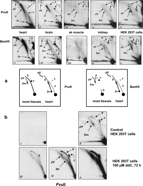
FIGURE 1. Two-dimensional AGE analysis of mtRIs from human tissues and cultured cells as indicated, treated with restriction enzymes cutting once in the mitochondrial genome. a, mtDNA was linearized with PvuII or BamHI as shown and hybridized with combined ND4 and OH region probes. The various arcs, interpreted in the panels below, represent dimeric linear molecules (2n), monomeric (c) and dimeric circles (2nc), Y intermediates (Y), double Y intermediates (dY), replication bubbles (b), i.e. standard initiation arc, X forms (X; i.e. dimeric fragments joined by four-way junctions), complex forms proposed to consist of multimeric fragments joined by multiple four-way junctions (dX), and θ molecules with broken arms attached to an intact circle (e; forming an eyebrow arc). For fuller explanation of the gels, see supplemental Fig. 1. Note that only bubble and double Y arcs can be considered as products of standard θ replication in such analyses, although RITOLS-type θ replication can also generate eyebrow arcs (Yasukawa et al. (20)). b, two-dimensional AGE of PvuII-digested mtDNA extracted from HEK 293T cells with or without 72 h of treatment with 100 μm ddC to induce widespread and prolonged replication stalling. Steady-state replication intermediates were greatly increased (panels i and iii are similar exposures as are panels ii and v), but the predominant forms were still θ bubbles and double Y forms with only a minority of molecules suffering fork regression or nicking to generate broken bubbles (bb). sk, skeletal.
FIGURE 1. Two-dimensional AGE analysis of mtRIs from human tissues and cultured cells as indicated, treated with restriction enzymes cutting once in the mitochondrial genome. a, mtDNA was linearized with PvuII or BamHI as shown and hybridized with combined ND4 and OH region probes. The various arcs, interpreted in the panels below, represent dimeric linear molecules (2n), monomeric (c) and dimeric circles (2nc), Y intermediates (Y), double Y intermediates (dY), replication bubbles (b), i.e. standard initiation arc, X forms (X; i.e. dimeric fragments joined by four-way junctions), complex forms proposed to consist of multimeric fragments joined by multiple four-way junctions (dX), and θ molecules with broken arms attached to an intact circle (e; forming an eyebrow arc). For fuller explanation of the gels, see supplemental Fig. 1. Note that only bubble and double Y arcs can be considered as products of standard θ replication in such analyses, although RITOLS-type θ replication can also generate eyebrow arcs (Yasukawa et al. (20)). b, two-dimensional AGE of PvuII-digested mtDNA extracted from HEK 293T cells with or without 72 h of treatment with 100 μm ddC to induce widespread and prolonged replication stalling. Steady-state replication intermediates were greatly increased (panels i and iii are similar exposures as are panels ii and v), but the predominant forms were still θ bubbles and double Y forms with only a minority of molecules suffering fork regression or nicking to generate broken bubbles (bb). sk, skeletal.CC BY-NC · PubMed Central
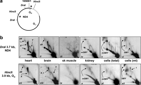
FIGURE 2. Two-dimensional AGE analysis of mtRIs in different human tissues and cultured HEK 293T cells, as indicated, using restriction enzymes cutting at multiple sites in the mitochondrial genome. a, schematic map of human mtDNA showing the locations of relevant restriction sites and probes (asterisks). The major NCR is shown as a dark gray bar, and rDNA is shown as a light gray bar. b, two-dimensional AGE blots, digests, and probes as shown. All DNA samples were total DNA except panels vi and xii, which used mtDNA extracted from sucrose gradient-purified mitochondria. The various arcs indicated (for clarity, and where obvious, not shown in every panel) represent X forms (X), Y intermediates (Y), putative double X forms (dX), “slow moving” Y arcs (SMY) created by non-digestion at restriction sites blocked by RNA or single strandedness, modified SMY arcs (mSMY) resulting from RNase H degradation, and termination intermediates (t) in which two oppositely moving forks are juxtaposed. For a fuller explanation of the gels, see supplemental Fig. 1. Bubble arcs, although not seen clearly at this exposure, were present in long exposures of panels iii–vi and ix–xii (e.g. see supplemental Fig. 3e). sk, skeletal.
FIGURE 2. Two-dimensional AGE analysis of mtRIs in different human tissues and cultured HEK 293T cells, as indicated, using restriction enzymes cutting at multiple sites in the mitochondrial genome. a, schematic map of human mtDNA showing the locations of relevant restriction sites and probes (asterisks). The major NCR is shown as a dark gray bar, and rDNA is shown as a light gray bar. b, two-dimensional AGE blots, digests, and probes as shown. All DNA samples were total DNA except panels vi and xii, which used mtDNA extracted from sucrose gradient-purified mitochondria. The various arcs indicated (for clarity, and where obvious, not shown in every panel) represent X forms (X), Y intermediates (Y), putative double X forms (dX), “slow moving” Y arcs (SMY) created by non-digestion at restriction sites blocked by RNA or single strandedness, modified SMY arcs (mSMY) resulting from RNase H degradation, and termination intermediates (t) in which two oppositely moving forks are juxtaposed. For a fuller explanation of the gels, see supplemental Fig. 1. Bubble arcs, although not seen clearly at this exposure, were present in long exposures of panels iii–vi and ix–xii (e.g. see supplemental Fig. 3e). sk, skeletal.CC BY-NC · PubMed Central
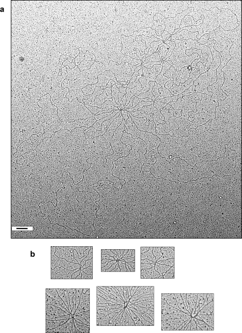
FIGURE 3. TEM of human heart mtDNA. Scale bars, 200 nm. a, tangled complex of monomeric and dimeric circles containing ∼20 genome equivalents. Some other tangled networks appeared even more complex. b, nexuses of five different tangles at higher magnification. c, panel i, three monomeric circles held together by junctions (arrows) (catenation, hemicatenation, and/or four-way recombination junctions). Panels ii–viii, examples of such junctions at higher magnification. Panels ii and iii are from the same complex as shown in panel i. Panels iv–vi are from other paired circular molecules. Panels vii and viii are similar junctions seen in the multimeric tangles. d, Southern blot of purified mtDNA (1 μg of nucleic acid) alongside total DNA (20 μg) undigested (U) or digested with PvuII (P) or EcoRI (E; E2 is an independent mtDNA sample from a second individual) and either stained with ethidium bromide (EtBr) or blot-hybridized with probes for 18 S rDNA (18S) or cytochrome b (cyt b; Ref. 19).
FIGURE 3. TEM of human heart mtDNA. Scale bars, 200 nm. a, tangled complex of monomeric and dimeric circles containing ∼20 genome equivalents. Some other tangled networks appeared even more complex. b, nexuses of five different tangles at higher magnification. c, panel i, three monomeric circles held together by junctions (arrows) (catenation, hemicatenation, and/or four-way recombination junctions). Panels ii–viii, examples of such junctions at higher magnification. Panels ii and iii are from the same complex as shown in panel i. Panels iv–vi are from other paired circular molecules. Panels vii and viii are similar junctions seen in the multimeric tangles. d, Southern blot of purified mtDNA (1 μg of nucleic acid) alongside total DNA (20 μg) undigested (U) or digested with PvuII (P) or EcoRI (E; E2 is an independent mtDNA sample from a second individual) and either stained with ethidium bromide (EtBr) or blot-hybridized with probes for 18 S rDNA (18S) or cytochrome b (cyt b; Ref. 19).CC BY-NC · PubMed Central
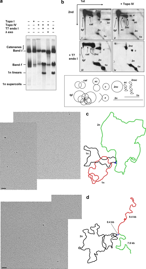
FIGURE 4. Electrophoretic and TEM analysis of human heart mtDNA. a, one-dimensional AGE of undigested cardiac muscle mtDNA treated with the enzymes as indicated (topoisomerase (Topo) I, topoisomerase IV, T7 endonuclease (endo) I, and λ-exonuclease (exo)). The identity of the various species is inferred by the effects of enzymatic treatments on their relative amounts and by their migration properties on two-dimensional AGE (b). Bands t and f are each composites of several species. λ-Exonuclease digests molecules with exposed 5′ phosphorylated ends. Because it removes or modifies the residual material running just behind open circles after topoisomerase IV treatment, i.e. 2n linear molecules plus species x (b, panel ii), we infer that species x has exposed ends. b, two-dimensional AGE of cardiac muscle mtDNA untreated (panel i), treated with topoisomerase IV (panel ii), treated with T7 endonuclease I (panel iii), and treated with topoisomerase IV + T7 endonuclease I (panel iv). The topology of the various forms, annotated on the gel images and inferred from the treatments and electrophoretic mobilities, is shown below the gel panels. tgl, tangled complexes (see Fig. 3); cat, catenanes (can also include >2 monomeric or dimeric circles); 1n and 2n, monomeric and dimeric linear molecules, respectively; c and 2nc, monomeric and dimeric open circles, respectively; sc and 2nsc, supercoiled monomeric and dimeric circles, respectively. xc, circular molecules joined by four-way junctions; x, suggested to be circles joined to linear molecules by four-way junctions. Arrows indicate directions of first and second dimension electrophoresis. c and d, examples of forms seen by TEM following topoisomerase IV treatment of heart mtDNA alongside interpretations. Distinct circles and linear segments are indicated in different colors with inferred contour lengths in kb or (for circles) genome lengths. Scale bars, 200 nm.
FIGURE 4. Electrophoretic and TEM analysis of human heart mtDNA. a, one-dimensional AGE of undigested cardiac muscle mtDNA treated with the enzymes as indicated (topoisomerase (Topo) I, topoisomerase IV, T7 endonuclease (endo) I, and λ-exonuclease (exo)). The identity of the various species is inferred by the effects of enzymatic treatments on their relative amounts and by their migration properties on two-dimensional AGE (b). Bands t and f are each composites of several species. λ-Exonuclease digests molecules with exposed 5′ phosphorylated ends. Because it removes or modifies the residual material running just behind open circles after topoisomerase IV treatment, i.e. 2n linear molecules plus species x (b, panel ii), we infer that species x has exposed ends. b, two-dimensional AGE of cardiac muscle mtDNA untreated (panel i), treated with topoisomerase IV (panel ii), treated with T7 endonuclease I (panel iii), and treated with topoisomerase IV + T7 endonuclease I (panel iv). The topology of the various forms, annotated on the gel images and inferred from the treatments and electrophoretic mobilities, is shown below the gel panels. tgl, tangled complexes (see Fig. 3); cat, catenanes (can also include >2 monomeric or dimeric circles); 1n and 2n, monomeric and dimeric linear molecules, respectively; c and 2nc, monomeric and dimeric open circles, respectively; sc and 2nsc, supercoiled monomeric and dimeric circles, respectively. xc, circular molecules joined by four-way junctions; x, suggested to be circles joined to linear molecules by four-way junctions. Arrows indicate directions of first and second dimension electrophoresis. c and d, examples of forms seen by TEM following topoisomerase IV treatment of heart mtDNA alongside interpretations. Distinct circles and linear segments are indicated in different colors with inferred contour lengths in kb or (for circles) genome lengths. Scale bars, 200 nm.CC BY-NC · PubMed Central
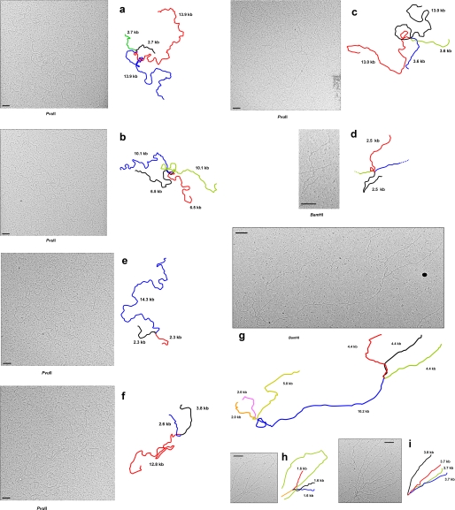
FIGURE 5. TEM of PvuII- and BamHI-digested human cardiac muscle mtDNA. a–i, images of various junctional molecules alongside interpretations are shown. In each case except f at least two branches (plus two other branches if entirely within the field) are of equal length. Note that many junctions appear to be composite, i.e. to consist of two adjacent four-way (and/or three-way) junctions. Distinct linear segments emanating from junctions are indicated in different colors with inferred contour lengths in kb. Scale bars, 200 nm.
FIGURE 5. TEM of PvuII- and BamHI-digested human cardiac muscle mtDNA. a–i, images of various junctional molecules alongside interpretations are shown. In each case except f at least two branches (plus two other branches if entirely within the field) are of equal length. Note that many junctions appear to be composite, i.e. to consist of two adjacent four-way (and/or three-way) junctions. Distinct linear segments emanating from junctions are indicated in different colors with inferred contour lengths in kb. Scale bars, 200 nm.CC BY-NC · PubMed Central
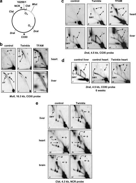
FIGURE 6. Two-dimensional AGE analysis of mtRIs in Twinkle and TFAM transgenic mice and non-transgenic controls. a, summary map of mouse mtDNA (nomenclature as in Fig. 2) showing relevant probes and restriction fragments. b, MluI-linearized mtDNA from 10-month-old Twinkle transgenic mice, non-transgenic littermate controls, and 8-week-old TFAM transgenic mice with tissues and probe as indicated. The various arcs represent monomeric circles (c), X forms (X), replication bubbles (b), double Y (dY) intermediates, and forms proposed to comprise three or more fragments joined by four-way junctions (dX). For clarity, and where obvious, these are not indicated in each individual gel panel. In the TFAM mouse samples an additional arc is seen running below the trajectory of the standard initiation arc, which probably arose by degradation of lagging strand RNA during shipment of the samples. c, DraI-digested mtDNA probed for the 4.5-kb COXI-containing fragment; mice, tissues, and arcs are indicated as in b. Y, standard Y arcs; SMY, slow moving Y-like arcs arising from restriction site blockage due to lagging strand RNA (Yang et al. (16)). d, similar DraI digests from 6-week-old mice. The mtDNA alterations in hearts of transgenic Twinkle mice are seen already in young animals. e, ClaI-digested mtDNA probed for the 4.3-kb NCR-containing fragment; mice, tissues, and arcs are indicated as in a–e. Note that a double Y arc is seen in non-transgenic heart DNA where it probably corresponds with termination intermediates. A much more prominent X arc is evident in Twinkle transgenic mice in both heart and brain (and also control brain), but this arc may not extend to the double-stranded DNA linear arc except in the case of Twinkle-transgenic mouse brain, suggesting that junction formation in this fragment occurs preferentially in the center of the fragment near the OH-distal boundary of the NCR.
FIGURE 6. Two-dimensional AGE analysis of mtRIs in Twinkle and TFAM transgenic mice and non-transgenic controls. a, summary map of mouse mtDNA (nomenclature as in Fig. 2) showing relevant probes and restriction fragments. b, MluI-linearized mtDNA from 10-month-old Twinkle transgenic mice, non-transgenic littermate controls, and 8-week-old TFAM transgenic mice with tissues and probe as indicated. The various arcs represent monomeric circles (c), X forms (X), replication bubbles (b), double Y (dY) intermediates, and forms proposed to comprise three or more fragments joined by four-way junctions (dX). For clarity, and where obvious, these are not indicated in each individual gel panel. In the TFAM mouse samples an additional arc is seen running below the trajectory of the standard initiation arc, which probably arose by degradation of lagging strand RNA during shipment of the samples. c, DraI-digested mtDNA probed for the 4.5-kb COXI-containing fragment; mice, tissues, and arcs are indicated as in b. Y, standard Y arcs; SMY, slow moving Y-like arcs arising from restriction site blockage due to lagging strand RNA (Yang et al. (16)). d, similar DraI digests from 6-week-old mice. The mtDNA alterations in hearts of transgenic Twinkle mice are seen already in young animals. e, ClaI-digested mtDNA probed for the 4.3-kb NCR-containing fragment; mice, tissues, and arcs are indicated as in a–e. Note that a double Y arc is seen in non-transgenic heart DNA where it probably corresponds with termination intermediates. A much more prominent X arc is evident in Twinkle transgenic mice in both heart and brain (and also control brain), but this arc may not extend to the double-stranded DNA linear arc except in the case of Twinkle-transgenic mouse brain, suggesting that junction formation in this fragment occurs preferentially in the center of the fragment near the OH-distal boundary of the NCR.CC BY-NC · PubMed Central
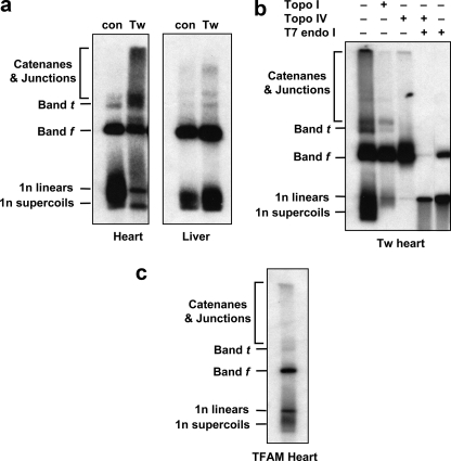
FIGURE 7. One-dimensional AGE analysis of mtDNA in Twinkle and TFAM transgenic mice and non-transgenic controls. a, one-dimensional AGE of undigested mtDNA from 10-month-old animals as indicated (Tw, Twinkle transgenic; con, non-transgenic littermate control mice). The identity of the various molecular forms is based on the enzymatic digestions of b plus the analysis of similar forms seen in human tissue mtDNA (Fig. 4). Bands f and t are composites of circular and junctional forms. b, enzymatic analysis of heart mtDNA from Twinkle transgenic mice using topoisomerases (Topo) I and IV and T7 endonuclease (endo) I as indicated. c, one-dimensional AGE of undigested mtDNA from 8-week-old TFAM transgenic mouse hearts. The same high molecular weight forms are seen as in Twinkle transgenic mouse hearts.
FIGURE 7. One-dimensional AGE analysis of mtDNA in Twinkle and TFAM transgenic mice and non-transgenic controls. a, one-dimensional AGE of undigested mtDNA from 10-month-old animals as indicated (Tw, Twinkle transgenic; con, non-transgenic littermate control mice). The identity of the various molecular forms is based on the enzymatic digestions of b plus the analysis of similar forms seen in human tissue mtDNA (Fig. 4). Bands f and t are composites of circular and junctional forms. b, enzymatic analysis of heart mtDNA from Twinkle transgenic mice using topoisomerases (Topo) I and IV and T7 endonuclease (endo) I as indicated. c, one-dimensional AGE of undigested mtDNA from 8-week-old TFAM transgenic mouse hearts. The same high molecular weight forms are seen as in Twinkle transgenic mouse hearts.CC BY-NC · PubMed Central

Verification

Claims the paper makes

The claims and statistics of this paper haven’t been checked yet.

Run the check — free, no account needed. It reads the open-access text and checks the sources it cites.

A few free checks a day · papers already verified open instantly · results are public

Claims other papers make about this one

Each is checked against this paper’s own text: does it say what the citing paper says it does?

Self-check

Percentages, sums and reported differences re-computed from the paper’s own numbers.

The self-check needs the paper’s full text.

Citations

Cites

Show all 70 references

Research works printed but not linked: 1

  1. Han, 1994, Chromosoma, 103, 162

Cited by

Sorted by citations

See all 122

Metadata

Authors
Pohjoismäki, J. L. O., Goffart, S., Tyynismaa, H., Willcox, S., Ide, T., Kang, D., Suomalainen, A., Karhunen, P. J., Griffith, J. D., Holt, I. J., & Jacobs, H. T.
Affiliations
  • Tampere University Hospital (FI)
  • University of Helsinki (FI)
  • University of North Carolina at Chapel Hill (US)
  • Kyushu University (JP)
  • University of Tampere (FI)
  • MRC Mitochondrial Biology Unit (GB)
Corresponding author
Jacobs, Howard T.
Venue
Journal of Biological Chemistry
Publisher
Elsevier BV
Published
2009-08
Volume
284
Issue
32
Pages
21446-21457
ISSN
0021-9258 (ISSN-L)
OpenAlex
W2014114000
Cited by
122
References
70
Citations by year
2026: 1 · 2025: 1 · 2024: 9 · 2023: 4 · 2022: 8 · 2021: 6 · 2020: 8 · 2019: 10 · 2018: 10 · 2017: 4 · 2016: 8 · 2015: 10 · 2014: 5 · 2013: 9 · 2012: 10
FWCI
3.89
Type
journal article
Language
en
Open access
Locations
  • Journal of Biological Chemistry (publishedVersion, cc-by)
  • PubMed Central (publishedVersion, cc-by-nc)
  • PubMed (publishedVersion)
  • Carolina Digital Repository (University of North Carolina at Chapel Hill) (submittedVersion)
  • UCL Discovery (University College London) (submittedVersion)
  • UNC Libraries
Funders
  • Foundation for Cardiovascular Research
  • European Commission
  • Academy of Finland
  • Suomen Kulttuurirahasto
  • Sydäntutkimussäätiö
  • Tays
  • National Institutes of Health
  • Medical Research Council
  • National Institute of General Medical Sciences (GM-31819, R01GM031819)
Topics
  • Life Sciences › Biochemistry, Genetics and Molecular Biology › Molecular Biology › Mitochondrial Function and Pathology
  • Life Sciences › Biochemistry, Genetics and Molecular Biology › Molecular Biology › DNA Repair Mechanisms
  • Life Sciences › Biochemistry, Genetics and Molecular Biology › Molecular Biology › DNA and Nucleic Acid Chemistry
Keywords
mitochondrial DNA, heart, four-way junction, replication fork, topoisomerase IV, T7 endonuclease I, TFAM, Twinkle helicase, mitochondrial DNA replication, brain, skeletal muscle, species differences
MeSH terms
Adult, Aged, Animals, Brain, Child, Preschool, DNA, Mitochondrial, Female, Heart, Humans, Male, Mice, Transgenic, Middle Aged, Nucleic Acid Conformation, Rabbits, Species Specificity, Swine, Tissue Distribution, Mice

About these checks

Registry, retraction, Cites and statistics results are computed, not judged, with one exception: a printed reference that no index lists is read once by a model before it counts as a citation error. Claims are extracted by a model and kept only when their quote appears verbatim in the text; each cited source is then checked, with a model’s help, for whether it says what the claim relies on. Those verdicts can be wrong — read the claim’s quote and the cited source before relying on one.

Statistics and arithmetic are recomputed, not judged.

Confidence bands are shown only for fields calibrated on our annotated test set. None is calibrated yet, so no band is shown.

What counts as a claim ›How we measure confidence ›How we check this ›

Are you an author, or do you see a problem with this page? Request a review