

The DOI matches its Crossref record; 44 of the 44 references it cites were found in a registry or an index.
Glycomics Profiling of Chinese Hamster Ovary Cell Glycosylation Mutants Reveals N-Glycans of a Novel Size and Complexity
Open accessAbstract
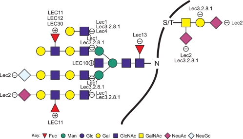
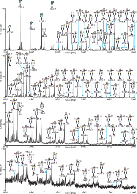
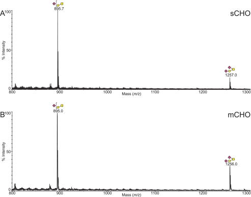
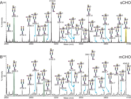
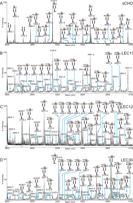
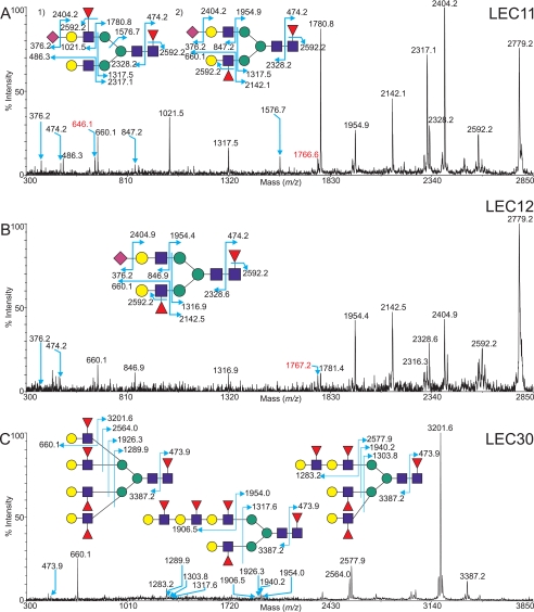
Identifying biological roles for mammalian glycans and the pathways by which they are synthesized has been greatly facilitated by investigations of glycosylation mutants of cultured cell lines and model organisms. Chinese hamster ovary (CHO) glycosylation mutants isolated on the basis of their lectin resistance have been particularly useful for glycosylation engineering of recombinant glycoproteins. To further enhance the application of these mutants, and to obtain insights into the effects of altering one specific glycosyltransferase or glycosylation activity on the overall expression of cellular glycans, an analysis of the N-glycans and major O-glycans of a panel of CHO mutants was performed using glycomic analyses anchored by matrix-assisted laser desorption ionization-time of flight/time of flight mass spectrometry. We report here the complement of the major N-glycans and O-glycans present in nine distinct CHO glycosylation mutants. Parent CHO cells grown in monolayer versus suspension culture had similar profiles of N- and O-GalNAc glycans, although the profiles of glycosylation mutants Lec1, Lec2, Lec3.2.8.1, Lec4, LEC10, LEC11, LEC12, Lec13, and LEC30 were consistent with available genetic and biochemical data. However, the complexity of the range of N-glycans observed was unexpected. Several of the complex N-glycan profiles contained structures of m/z approximately 13,000 representing complex N-glycans with a total of 26 N-acetyllactosamine (Gal beta1-4GlcNAc)(n) units. Importantly, the LEC11, LEC12, and LEC30 CHO mutants exhibited unique complements of fucosylated complex N-glycans terminating in Lewis(x) and sialyl-Lewis(x) determinants. This analysis reveals the larger-than-expected complexity of N-glycans in CHO cell mutants that may be used in a broad variety of functional glycomics studies and for making recombinant glycoproteins.
Overview
The claims and statistics of this paper haven’t been checked yet.
Run the check — free, no account needed. It reads the open-access text and checks the sources it cites.
Integrity signals
Registry
Cites
Statistics
Figures
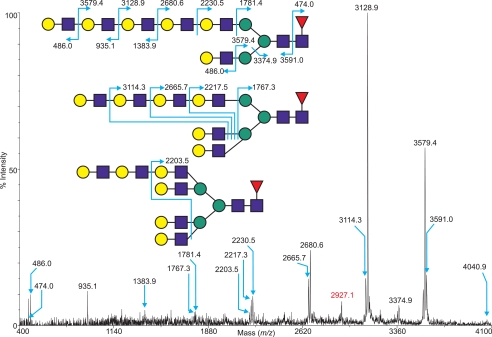
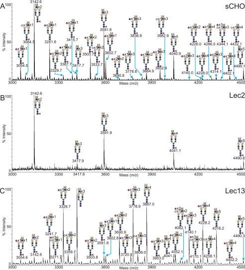
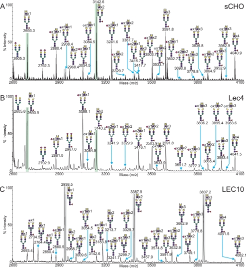
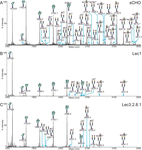
Verification
Claims the paper makes
The claims and statistics of this paper haven’t been checked yet.
Run the check — free, no account needed. It reads the open-access text and checks the sources it cites.
Claims other papers make about this one
Each is checked against this paper’s own text: does it say what the citing paper says it does?
The source backs part of the claim, not all of it — or backs it for a different population, condition or comparison.
In the citing paper
“Advances in various omics technologies have significantly improved our understanding of CHO cells at different levels and enabled the rational targeting of glycosylation effectors for improved biopharmaceutical production [27,71,72,150–152].”
From Glycoengineering Chinese hamster ovary cells: a short history (2021) · cites this paper as [27]
Experimental — accuracy not yet measuredThe source backs part of the claim, not all of it — or backs it for a different population, condition or comparison.
In the citing paper
“when NeuAc is α2,3-linked (main sialylated species in CHO-derived glycoproteins) the conformational stability of the CH2 domain of IgG can be reduced, most likely a result of a steric effect between the protein backbone and the α2,3-sialylated 6-arm [27,28].”
From Glycoengineering Chinese hamster ovary cells: a short history (2021) · cites this paper as [27]
Experimental — accuracy not yet measuredThe check wasn’t confident either way, so it gives no verdict — never disputed.
In the citing paper
“CHO cells do not express alpha-1,3-galactosyltransferases, which produce the immunogenic alpha-galactosyl epitope (although they have the gene), and the expression of CMP-N-acetylneuraminic acid hydroxylase, which produces CMP-NeuGc, is virtually absent [27,71,72].”
From Glycoengineering Chinese hamster ovary cells: a short history (2021) · cites this paper as [27]
Experimental — accuracy not yet measured
Self-check
Percentages, sums and reported differences re-computed from the paper’s own numbers.
The self-check needs the paper’s full text.
Citations
Cites
A method for the analysis of sugars in plant cell-wall polysaccharides by gas-liquid chromatography
Albersheim, P., Nevins, D. J., English, P. D., & Karr, A. L. (1967) · Carbohydrate Research · doi:10.1016/s0008-6215(00)80510-8 · Cited by 1,648
Monoclonal antibody defining a stage-specific mouse embryonic antigen (SSEA-1).
Solter, D., & Knowles, B. B. (1978) · Proceedings of the National Academy of Sciences · doi:10.1073/pnas.75.11.5565 · Cited by 1,295
GlycoWorkbench: A Tool for the Computer-Assisted Annotation of Mass Spectra of Glycans
Ceroni, A., Maaß, K., Geyer, H., Geyer, R., Dell, A., & Haslam, S. M. (2008) · Journal of Proteome Research · doi:10.1021/pr7008252 · Cited by 1,055
Self-citationSialic acids in human health and disease
Varki, A. P. (2008) · Trends in Molecular Medicine · doi:10.1016/j.molmed.2008.06.002 · Cited by 1,032
Fucose: biosynthesis and biological function in mammals
Becker, D. J., & Lowe, J. B. (2003) · Glycobiology · doi:10.1093/glycob/cwg054 · Cited by 931
Glycomics: an integrated systems approach to structure-function relationships of glycans
Raman, R., Raguram, S., Venkataraman, G., Paulson, J. C., & Sasisekharan, R. (2005) · Nature Methods · doi:10.1038/nmeth807 · Cited by 443
Wada, Y., Azadi, P., Costello, C. E., Dell, A., Dwek, R. A., Geyer, H., Geyer, R., Kakehi, K., Karlsson, N. G., Kato, K., et al. (2007) · Glycobiology · doi:10.1093/glycob/cwl086 · Cited by 400
Self-citationBardor, M., Nguyen, D., Diaz, S. L., & Varki, A. P. (2004) · Journal of Biological Chemistry · doi:10.1074/jbc.m412040200 · Cited by 390
Moloney, D. J., Shair, L. H., Lu, F. M., Xia, J. L., Locke, R. D., Matta, K. L., & Haltiwanger, R. S. (2000) · Journal of Biological Chemistry · doi:10.1074/jbc.275.13.9604 · Cited by 348
Structure of a Heterophilic Adhesion Complex between the Human CD2 and CD58 (LFA-3) Counterreceptors
Wang, J., Smolyar, A., Tan, K., Liu, J., Kim, M., Sun, Z., Wagner, G., & Reinherz, E. L. (1999) · Cell · doi:10.1016/s0092-8674(00)80790-4 · Cited by 245
Research works printed but not linked: 1
- Stanley, 1989, Mol. Cell. Biol., 9, 377
Cited by
Sorted by citations
Unravelling the Role of O-glycans in Influenza A Virus Infection
Mayr, J., Lau, K., Lai, J. C. C., Gagarinov, I. A., Shi, Y. Y., McAtamney, S., Chan, R. W. Y., Nicholls, J. M., von Itzstein, M., & Haselhorst, T. (2018) · Scientific Reports · doi:10.1038/s41598-018-34175-3 · Cited by 1,068
Precision Mapping of an In Vivo N-Glycoproteome Reveals Rigid Topological and Sequence Constraints
Zielinska, D. F., Gnad, F., Wiśniewski, J. R., & Mann, M. (2010) · Cell · doi:10.1016/j.cell.2010.04.012 · Cited by 905
Cell surface protein glycosylation in cancer
Christiansen, M. N., Chik, J., Lee, L., Anugraham, M., Abrahams, J. L., & Packer, N. H. (2014) · PROTEOMICS · doi:10.1002/pmic.201300387 · Cited by 518
Golgi GlycosylationPreprint
Stanley, P. (2011) · Cold Spring Harbor Perspectives in Biology · doi:10.1101/cshperspect.a005199 · Cited by 462
Self-citationGalectin-3 Coordinates a Cellular System for Lysosomal Repair and Removal
Jia, J., Claude-Taupin, A., Gu, Y., Choi, S. W., Peters, R., Bissa, B., Mudd, M. H., Allers, L., Pallikkuth, S., Lidke, K. A., et al. (2020) · Developmental Cell · doi:10.1016/j.devcel.2019.10.025 · Cited by 434
Enzymes in the Synthesis of Glycoconjugates
Schmaltz, R. M., Hanson, S. R., & Wong, C. (2011) · Chemical Reviews · doi:10.1021/cr200113w · Cited by 291
Terminal N-Linked Galactose Is the Primary Receptor for Adeno-associated Virus 9
Shen, S., Bryant, K. D., Brown, S. M., Randell, S. H., & Asokan, A. (2011) · Journal of Biological Chemistry · doi:10.1074/jbc.m110.210922 · Cited by 297
Malaker, S. A., Pedram, K., Ferracane, M. J., Bensing, B. A., Krishnan, V., Pett, C., Yu, J., Woods, E. C., Kramer, J. R., Westerlind, U., et al. (2019) · Proceedings of the National Academy of Sciences · doi:10.1073/pnas.1813020116 · Cited by 251
Engineered CHO cells for production of diverse, homogeneous glycoproteins
Yang, Z., Wang, S., Halim, A., Schulz, M. A., Frodin, M., Rahman, S. H., Vester-Christensen, M. B., Behrens, C., Kristensen, C., Vakhrushev, S. Y., et al. (2015) · Nature Biotechnology · doi:10.1038/nbt.3280 · Cited by 254
Glycotherapy: New Advances Inspire a Reemergence of Glycans in Medicine
Hudak, J. E., & Bertozzi, C. R. (2013) · Chemistry & Biology · doi:10.1016/j.chembiol.2013.09.010 · Cited by 232
Mbua, N. E., Guo, J., Wolfert, M. A., Steet, R., & Boons, G. (2011) · ChemBioChem · doi:10.1002/cbic.201100117 · Cited by 190
The emerging CHO systems biology era: harnessing the ‘omics revolution for biotechnology
Kildegaard, H. F., Baycin-Hizal, D., Lewis, N. E., & Betenbaugh, M. J. (2013) · Current Opinion in Biotechnology · doi:10.1016/j.copbio.2013.02.007 · Cited by 165
Ho, S. C. L., Bardor, M., Feng, H., Mariati, Tong, Y. W., Song, Z., Yap, M. G. S., & Yang, Y. (2011) · Journal of Biotechnology · doi:10.1016/j.jbiotec.2011.09.023 · Cited by 153
Emerging Technologies for Making Glycan-Defined Glycoproteins
Wang, L., & Lomino, J. V. (2011) · ACS Chemical Biology · doi:10.1021/cb200429n · Cited by 138
An 'omics approach towards CHO cell engineering
Datta, P., Linhardt, R. J., & Sharfstein, S. T. (2013) · Biotechnology and Bioengineering · doi:10.1002/bit.24841 · Cited by 136
Houel, S., Hilliard, M., Yu, Y. Q., McLoughlin, N., Millán‐Martín, S., Rudd, P. M., Williams, J. P., & Chen, W. (2013) · Analytical Chemistry · doi:10.1021/ac402726h · Cited by 127
Londrigan, S. L., Turville, S. G., Tate, M. D., Deng, Y., Brööks, A. G., & Reading, P. C. (2010) · Journal of Virology · doi:10.1128/jvi.01705-10 · Cited by 125
How does mild hypothermia affect monoclonal antibody glycosylation?
Sou, S. N., Sellick, C., Lee, K., Mason, A., Kyriakopoulos, S., Polizzi, K. M., & Kontoravdi, C. (2015) · Biotechnology and Bioengineering · doi:10.1002/bit.25524 · Cited by 107
García-Vallejo, J. J., Ilarregui, J. M., Kalay, H., Chamorro, S., Koning, N., Unger, W. W. J., Ambrosini, M., Montserrat, V., Fernandes, R. J., Bruijns, S. C. M., et al. (2014) · The Journal of Experimental Medicine · doi:10.1084/jem.20122192 · Cited by 110
Exploring the N-glycosylation Pathway in Chlamydomonas reinhardtii Unravels Novel Complex Structures
Mathieu‐Rivet, E., Scholz, M., Arias, C., Dardelle, F., Schulze, S., Mauff, F. L., Teo, G., Hochmal, A. K., Blanco-Rivero, A., Loutelier‐Bourhis, C., et al. (2013) · Molecular & Cellular Proteomics · doi:10.1074/mcp.m113.028191 · Cited by 109
Song, Y., Aglipay, J. A., Bernstein, J. D., Goswami, S., & Stanley, P. (2010) · Cancer Research · doi:10.1158/0008-5472.can-09-2719 · Cited by 104
Self-citationHarvey, D. J. (2010) · Mass Spectrometry Reviews · doi:10.1002/mas.20265 · Cited by 100
Monitoring Dynamic Glycosylation in Vivo Using Supersensitive Click Chemistry
Jiang, H., Zheng, T., Aguilar, A. L., Feng, L., Kopp, F., Marlow, F. L., & Wu, P. (2014) · Bioconjugate Chemistry · doi:10.1021/bc400502d · Cited by 102
Glycosylation control technologies for recombinant therapeutic proteins
Gupta, S. K., & Shukla, P. (2018) · Applied Microbiology and Biotechnology · doi:10.1007/s00253-018-9430-6 · Cited by 99
Bush, D. R., Zang, L., Belov, A. M., Ivanov, A. R., & Karger, B. L. (2016) · Analytical Chemistry · doi:10.1021/acs.analchem.5b03218 · Cited by 92
The VP8* Domain of Neonatal Rotavirus Strain G10P[11] Binds to Type II Precursor Glycans
Ramani, S., Cortes-Penfield, N. W., Hu, L., Crawford, S. E., Czakó, R., Smith, D. F., Kang, G., Ramig, R. F., LePendu, J., Prasad, B. V. V., et al. (2013) · Journal of Virology · doi:10.1128/jvi.03518-12 · Cited by 89
The bisecting GlcNAc in cell growth control and tumor progression
Miwa, H. E., Song, Y., Alvarez, R., Cummings, R. D., & Stanley, P. (2012) · Glycoconjugate Journal · doi:10.1007/s10719-012-9373-6 · Cited by 86
Self-citationTracking N‐Acetyllactosamine on Cell‐Surface Glycans In Vivo
Zheng, T., Jiang, H., Gros, M., del Amo, D. S., Sundaram, S., Lauvau, G. S., Marlow, F. L., Liu, Y., Stanley, P., & Wu, P. (2011) · Angewandte Chemie International Edition · doi:10.1002/anie.201100265 · Cited by 85
Self-citationCHO microRNA engineering is growing up: Recent successes and future challenges
Jadhav, V., Hackl, M., Druz, A., Shridhar, S., Chung, C., Heffner, K. M., Kreil, D. P., Betenbaugh, M., Shiloach, J., Barron, N., et al. (2013) · Biotechnology Advances · doi:10.1016/j.biotechadv.2013.07.007 · Cited by 94
Direct quality control of glycoengineered erythropoietin variants
Čaval, T., Tian, W., Yang, Z., Clausen, H., & Heck, A. J. R. (2018) · Nature Communications · doi:10.1038/s41467-018-05536-3 · Cited by 82
Harvey, D. J. (2014) · Mass Spectrometry Reviews · doi:10.1002/mas.21411 · Cited by 84
Using glyco-engineering to produce therapeutic proteins
Dicker, M., & Strasser, R. (2015) · Expert Opinion on Biological Therapy · doi:10.1517/14712598.2015.1069271 · Cited by 73
Buffone, A., Mondal, N., Gupta, R., McHugh, K. P., Lau, J. T. Y., & Neelamegham, S. (2012) · Journal of Biological Chemistry · doi:10.1074/jbc.m112.400929 · Cited by 81
Applications and continued evolution of glycan imaging mass spectrometry
McDowell, C. T., Lu, X., Mehta, A. S., Angel, P. M., & Drake, R. R. (2021) · Mass Spectrometry Reviews · doi:10.1002/mas.21725 · Cited by 77
Cancer glycan epitopes: biosynthesis, structure and function
Pearce, O. M. T. (2018) · Glycobiology · doi:10.1093/glycob/cwy023 · Cited by 78
Integrated analysis of glycan and RNA in single cells
Minoshima, F., Ozaki, H., Odaka, H., & Tateno, H. (2021) · iScience · doi:10.1016/j.isci.2021.102882 · Cited by 70
Influenza A virus entry into cells lacking sialylated N-glycans
de Vries, E., de Vries, R. P., Wienholts, M. J., Floris, C. E., Jacobs, M.-S., van den Heuvel, A., Rottier, P. J. M., & de Haan, C. A. M. (2012) · Proceedings of the National Academy of Sciences · doi:10.1073/pnas.1200987109 · Cited by 74
Goey, C. H., Alhuthali, S., & Kontoravdi, C. (2018) · Biotechnology Advances · doi:10.1016/j.biotechadv.2018.03.021 · Cited by 72
Glycoengineering in CHO cells: advances in systems biology
Tejwani, V., Andersen, M. R., Nam, J. H., & Sharfstein, S. T. (2018) · Biotechnology Journal · doi:10.1002/biot.201700234 · Cited by 70
CD44 and HCELL: Preventing hematogenous metastasis at step 1
Jacobs, P. P., & Sackstein, R. (2011) · FEBS Letters · doi:10.1016/j.febslet.2011.07.039 · Cited by 71
Selective N-glycan editing on living cell surfaces to probe glycoconjugate function
Tang, F., Zhou, M., Qin, K., Shi, W., Yashinov, A., Yang, Y., LiYun, Y., Guan, D., Zhao, L., Tang, Y., et al. (2020) · Nature Chemical Biology · doi:10.1038/s41589-020-0551-8 · Cited by 68
Chan, K. F., Shahreel, W., Wan, C., Teo, G., Hayati, N., Tay, S. J., Tong, W. H., Yang, Y., Rudd, P. M., Zhang, P., et al. (2015) · Biotechnology Journal · doi:10.1002/biot.201500331 · Cited by 70
The use of ‘Omics technology to rationally improve industrial mammalian cell line performance
Lewis, A. M., Abu‐Absi, N. R., Borys, M., & Li, Z. J. (2015) · Biotechnology and Bioengineering · doi:10.1002/bit.25673 · Cited by 67
Analysis of site-specific N-glycan remodeling in the endoplasmic reticulum and the Golgi
Hang, I., Lin, C.-W., Grant, O. C., Fleurkens, S., Villiger, T. K., Soos, M., Morbidelli, M., Woods, R. J., Gauss, R., & Aebi, M. (2015) · Glycobiology · doi:10.1093/glycob/cwv058 · Cited by 67
Tashima, Y., & Stanley, P. (2014) · Journal of Biological Chemistry · doi:10.1074/jbc.m113.492512 · Cited by 66
Self-citationMass Spectrometric Analysis of Mutant Mice
North, S. J., Jang‐Lee, J., Harrison, R., Canis, K., Ismail, M. N., Trollope, A., Antonopoulos, A., Pang, P., Grassi, P., Chalabi, S., et al. (2010) · Methods in enzymology on CD-ROM/Methods in enzymology · doi:10.1016/s0076-6879(10)78002-2 · Cited by 62
Self-citationPoly-LacNAc as an Age-Specific Ligand for Rotavirus P[11] in Neonates and Infants
Liu, Y., Huang, P., Jiang, B., Tan, M., Morrow, A. L., & Jiang, X. (2013) · PLoS ONE · doi:10.1371/journal.pone.0078113 · Cited by 67
Ng, W. C., Londrigan, S. L., Nasr, N., Cunningham, A. L., Turville, S. G., Brööks, A. G., & Reading, P. C. (2015) · Journal of Virology · doi:10.1128/jvi.01447-15 · Cited by 62
Bacterial glycosyltransferase-mediated cell-surface chemoenzymatic glycan modification
Hong, S., Shi, Y., Wu, N. C., Grande, G., Douthit, L., Wang, H., Zhou, W., Sharpless, K. B., Wilson, I. A., Xie, J., et al. (2019) · Nature Communications · doi:10.1038/s41467-019-09608-w · Cited by 61
Precision genome editing: A small revolution for glycobiology
Steentoft, C., Bennett, E., Schjoldager, K. T.-B. G., Vakhrushev, S. Y., Wandall, H. H., & Clausen, H. (2014) · Glycobiology · doi:10.1093/glycob/cwu046 · Cited by 62
Rahman, S. A., Bergström, E., Watson, C. J., Wilson, K. S., Ashford, D. A., Thomas, J. R., Ungár, D., & THOMAS‐OATES, J. E. (2014) · Journal of Proteome Research · doi:10.1021/pr401043r · Cited by 58
Hirakawa, J., Tsuboi, K., Sato, K., Kobayashi, M., Watanabe, S., Takakura, A., Imai, Y., Ito, Y., Fukuda, M., & Kawashima, H. (2010) · Journal of Biological Chemistry · doi:10.1074/jbc.m110.167296 · Cited by 57
Lakshmanan, M., Kok, Y. J., Lee, A., Kyriakopoulos, S., Lim, H. L., Teo, G., Poh, S. L., Tang, W. Q., Hong, J. K., Tan, A. H., et al. (2019) · Biotechnology and Bioengineering · doi:10.1002/bit.27014 · Cited by 57
Zhang, X., Wang, Y., Qian, Y., Wu, X., Zhang, Z., Liu, X., Zhao, R., Zhou, L., Ruan, Y., Xu, J., et al. (2014) · PLoS ONE · doi:10.1371/journal.pone.0087978 · Cited by 55
Allam, H., Aoki, K., Benigno, B. B., McDonald, J. F., Mackintosh, S. G., Tiemeyer, M., & Abbott, K. L. (2014) · Journal of Proteome Research · doi:10.1021/pr501174p · Cited by 54
Bloem, K., Vuist, I. M., van der Plas, A.-J., Knippels, L. M. J., Garssen, J., García-Vallejo, J. J., van Vliet, S. J., & van Kooyk, Y. (2013) · PLoS ONE · doi:10.1371/journal.pone.0066266 · Cited by 52
Vanier, G., Lucas, P., Loutelier‐Bourhis, C., Vanier, J., Plasson, C., Walet-Balieu, M.-L., Chan, P., Rémy‐Jouet, I., Richard, V. J., Bernard, S., et al. (2017) · Scientific Reports · doi:10.1038/s41598-017-10698-z · Cited by 51
Model-based analysis of N-glycosylation in Chinese hamster ovary cells
Krambeck, F. J., Bennun, S. V., Andersen, M. R., & Betenbaugh, M. J. (2017) · PLoS ONE · doi:10.1371/journal.pone.0175376 · Cited by 49
Bryan, L. L. K., CLYNES, M. M., & Meleady, P. (2021) · Biotechnology Advances · doi:10.1016/j.biotechadv.2021.107757 · Cited by 49
Bennun, S. V., Hızal, D. B., Heffner, K. M., Can, Ö., Zhang, H., & Betenbaugh, M. J. (2016) · Journal of Molecular Biology · doi:10.1016/j.jmb.2016.07.005 · Cited by 47
A testis-specific regulator of complex and hybrid N-glycan synthesis
Huang, H.-H., & Stanley, P. (2010) · The Journal of Cell Biology · doi:10.1083/jcb.201004102 · Cited by 47
Self-citationTharmalingam, T., Adamczyk, B., Doherty, M., Royle, L., & Rudd, P. M. (2012) · Glycoconjugate Journal · doi:10.1007/s10719-012-9443-9 · Cited by 45
Noël, M., Gilormini, P., Cogez, V., Yamakawa, N., Vicogne, D., Lion, C. J., Biot, C., Guérardel, Y., & Harduin‐Lepers, A. (2017) · ChemBioChem · doi:10.1002/cbic.201700024 · Cited by 45
Comprehensive Glycoproteomic Analysis of Chinese Hamster Ovary Cells
Yang, G., Hu, Y., Sun, S., OuYang, C., Yang, W., Wang, Q., Betenbaugh, M. J., & Zhang, H. (2018) · Analytical Chemistry · doi:10.1021/acs.analchem.8b03520 · Cited by 45
Londrigan, S. L., Tate, M. D., Brööks, A. G., & Reading, P. C. (2011) · Journal of Leukocyte Biology · doi:10.1189/jlb.1011492 · Cited by 45
Huang, C., Liu, Y., Wu, H., Sun, D., & Li, Y. (2017) · Analytical and Bioanalytical Chemistry · doi:10.1007/s00216-017-0302-1 · Cited by 44
Sun, Y., Senger, K., Baginski, T. K., Mazloom, A., Chinn, Y., Pantua, H., Hamidzadeh, K., Ramani, S. R., Luis, E., Tom, I., et al. (2012) · Journal of Biological Chemistry · doi:10.1074/jbc.m111.286633 · Cited by 44
A Versatile Technology for Cellular Glycomics Using Lectin Microarray
Tateno, H., Kuno, A., Itakura, Y., & Hirabayashi, J. (2010) · Methods in enzymology on CD-ROM/Methods in enzymology · doi:10.1016/s0076-6879(10)78008-3 · Cited by 43
Galactosyltransferase 4 is a major control point for glycan branching in N-linked glycosylation
McDonald, A. G., Hayes, J. M., Bezak, T., Gluchowska, S. A., Cosgrave, E. F. J., Struwe, W. B., Stroop, C. J. M., Kok, H. J., van de Laar, T., Rudd, P. M., et al. (2014) · Journal of Cell Science · doi:10.1242/jcs.151878 · Cited by 44
Intracellular galectins sense cytosolically exposed glycans as danger and mediate cellular responses
Hong, M.-H., Weng, I., Li, F.-Y., Lin, W., & Liu, F. (2021) · Journal of Biomedical Science · doi:10.1186/s12929-021-00713-x · Cited by 42
Metabolism of Vertebrate Amino Sugars with N-Glycolyl Groups
Bergfeld, A. K., Pearce, O. M. T., Diaz, S. L., Lawrence, R., Vocadlo, D. J., Choudhury, B., Esko, J. D., & Varki, A. P. (2012) · Journal of Biological Chemistry · doi:10.1074/jbc.m112.363499 · Cited by 43
Influence of protein/glycan interaction on site‐specific glycan heterogeneity
Losfeld, M., Scibona, E., Lin, C., Villiger, T. K., Gauss, R., Morbidelli, M., & Aebi, M. (2017) · The FASEB Journal · doi:10.1096/fj.201700403r · Cited by 43
del Val, I. J., Polizzi, K. M., & Kontoravdi, C. (2016) · Scientific Reports · doi:10.1038/srep28547 · Cited by 41
Lectin inhibition assays for the analysis of bioactive milk sialoglycoconjugates
Koliwer‐Brandl, H., Siegert, N., Umnus, K., Kelm, A., Tolkach, A., Kulozik, U., Kuballa, J., Cartellieri, S., & Kelm, S. (2011) · International Dairy Journal · doi:10.1016/j.idairyj.2011.01.005 · Cited by 40
Biological Functions and Large-Scale Profiling of Protein Glycosylation in Human Semen
Lan, R., Xin, M., Hao, Z., You, S., Xu, Y., Wu, J., Dang, L., Zhang, X., & Sun, S. (2020) · Journal of Proteome Research · doi:10.1021/acs.jproteome.9b00795 · Cited by 40
Glycoengineering design options for IgG1 in CHO cells using precise gene editing
Schulz, M. A., Tian, W., Mao, Y., Coillie, J. V., Sun, L., Larsen, J. S., Chen, Y., Kristensen, C., Vakhrushev, S. Y., Clausen, H., et al. (2018) · Glycobiology · doi:10.1093/glycob/cwy022 · Cited by 41
N-glycome of the Lysosomal Glycocalyx is Altered in Niemann-Pick Type C Disease (NPC) Model Cells
Košiček, M., Gudelj, I., Horvatić, A., Jovic, T., Vučković, F., Lauc, G., & Hecimovic, S. K. (2018) · Molecular & Cellular Proteomics · doi:10.1074/mcp.ra117.000129 · Cited by 38
Glycoengineering Chinese hamster ovary cells: a short history
Donini, R., Haslam, S. M., & Kontoravdi, C. (2021) · Biochemical Society Transactions · doi:10.1042/bst20200840 · Cited by 37
Self-citationA Computational Framework for the Automated Construction of Glycosylation Reaction Networks
Liu, G., & Neelamegham, S. (2014) · PLoS ONE · doi:10.1371/journal.pone.0100939 · Cited by 37
Hou, X., Tashima, Y., & Stanley, P. (2011) · Journal of Biological Chemistry · doi:10.1074/jbc.m111.317578 · Cited by 37
Self-citationBisected, complex N-glycans and galectins in mouse mammary tumor progression and human breast cancer
Miwa, H. E., Koba, W. R., Fine, E. J., Giricz, O., Kenny, P. A., & Stanley, P. (2013) · Glycobiology · doi:10.1093/glycob/cwt075 · Cited by 36
Self-citationLucas, P., Mathieu‐Rivet, E., Song, P. C. T., Oltmanns, A., Loutelier‐Bourhis, C., Plasson, C., Afonso, C., Hippler, M., Lerouge, P., Mati‐Baouche, N., et al. (2019) · The Plant Journal · doi:10.1111/tpj.14620 · Cited by 35
Recent Advances in Cellular Glycomic Analyses
Furukawa, J., Fujitani, N., & Shinohara, Y. (2013) · Biomolecules · doi:10.3390/biom3010198 · Cited by 32
Intramolecular Glycan–Protein Interactions in Glycoproteins
Barb, A. W., Borgert, A. J., Liu, M., Bárány, G., & Live, D. H. (2010) · Methods in enzymology on CD-ROM/Methods in enzymology · doi:10.1016/s0076-6879(10)78018-6 · Cited by 32
Xie, Y., Mota, L. M., Bergin, A., O’Flaherty, R., Jones, A. W., Morgan, B., & Butler, M. (2021) · Analytical Biochemistry · doi:10.1016/j.ab.2021.114205 · Cited by 31
Origin of cytoplasmic GDP-fucose determines its contribution to glycosylation reactions
Sosicka, P., Ng, B. G., Pepi, L. E., Shajahan, A., Wong, M., Scott, D. A., Matsumoto, K., Xia, Z., Lebrilla, C. B., Haltiwanger, R. S., et al. (2022) · The Journal of Cell Biology · doi:10.1083/jcb.202205038 · Cited by 29
Viral glycoproteomes: technologies for characterization and outlook for vaccine design
Bagdonaite, I., Vakhrushev, S. Y., Joshi, H. J., & Wandall, H. H. (2018) · FEBS Letters · doi:10.1002/1873-3468.13177 · Cited by 30
Sun, S., Huang, C., Wang, Y., Liu, Y., Zhang, J., Zhou, J., Gao, F., Yang, F., Chen, R., Mulloy, B., et al. (2018) · Analytical Chemistry · doi:10.1021/acs.analchem.8b03967 · Cited by 30
Zhang, P., Chan, K. F., Haryadi, R., Bardor, M., & Song, Z. (2012) · Advances in biochemical engineering, biotechnology · doi:10.1007/10_2012_163 · Cited by 31
Blundell, P. A., Lu, D., Dell, A., Haslam, S., & Pleass, R. J. (2020) · The Journal of Immunology · doi:10.4049/jimmunol.1901145 · Cited by 29
“For example, only N-glycans attached to Asn-297 are available for sampling in either the N563A/C575A or C309L/N563A/C575A, mutants that, therefore, also allow the contribution of disulfide bonding to glycosylation at Asn-297 to be elucidated.”
BackgroundInfluentialSelf-citationMendoza, M., Lu, D., Ballesteros, Á., Blois, S. M., Abernathy, K., Feng, C., Dimitroff, C. J., Zmuda, J. F., Panico, M., Dell, A., et al. (2020) · Glycobiology · doi:10.1093/glycob/cwaa034 · Cited by 30
Self-citationJung, J., Schmidt, E. N., Chang, H.-C., Jame-Chenarboo, Z., Enterina, J. R., McCord, K. A., Gray, T. E., Kageler, L., Laurent, C. D. S., Wang, C., et al. (2025) · ACS Chemical Biology · doi:10.1021/acschembio.4c00677 · Cited by 28
CHOGlycoNET: Comprehensive glycosylation reaction network for CHO cells
Kotidis, P., Donini, R., Arnsdorf, J., Hansen, A. H., Voldborg, B. G. R., Chiang, A. W. T., Haslam, S. M., Betenbaugh, M. J., del Val, I. J., Lewis, N. E., et al. (2023) · Metabolic Engineering · doi:10.1016/j.ymben.2022.12.009 · Cited by 27
Self-citationCell surface lectin array: parameters affecting cell glycan signature
Landemarre, L., Cancellieri, P., & Duverger, E. (2012) · Glycoconjugate Journal · doi:10.1007/s10719-012-9433-y · Cited by 30
Metabolic flux control in glycosylation
McDonald, A. G., Hayes, J. M., & Davey, G. P. (2016) · Current Opinion in Structural Biology · doi:10.1016/j.sbi.2016.08.007 · Cited by 25
Trabjerg, E., Kartberg, F., Christensen, S., & Rand, K. D. (2017) · Journal of Biological Chemistry · doi:10.1074/jbc.m117.803320 · Cited by 26
Matsumura, R., Hirakawa, J., Sato, K., Ikeda, T., Nagai, M., Fukuda, M., Imai, Y., & Kawashima, H. (2015) · Journal of Biological Chemistry · doi:10.1074/jbc.m115.650051 · Cited by 25
Bubka, M., Link‐Lenczowski, P., Janik, M., Pocheć, E., & Lityń́ska, A. (2014) · Biochimie · doi:10.1016/j.biochi.2014.04.003 · Cited by 25
Technologies for glycomic characterization of biopharmaceutical erythropoietins
Hua, S. S., Oh, M. J., Özcan, S., Seo, Y. S., Grimm, R., & An, H. J. (2015) · TrAC Trends in Analytical Chemistry · doi:10.1016/j.trac.2015.02.004 · Cited by 25
Hong, S., Sahai‐Hernandez, P., Chapla, D. G., Moremen, K. W., Traver, D. J., & Wu, P. (2019) · Angewandte Chemie International Edition · doi:10.1002/anie.201907410 · Cited by 25
Metadata
- Authors
- North, S. J., Huang, H.-H., Sundaram, S., Jang-Lee, J., Etienne, A. T., Trollope, A., Chalabi, S., Dell, A., Stanley, P., & Haslam, S. M.
- Affiliations
- Imperial College London (GB)
- Albert Einstein College of Medicine (US)
- Corresponding author
- Stanley, Pamela
- Haslam, Stuart M.
- Venue
- Journal of Biological Chemistry
- Publisher
- Elsevier BV
- Published
- 2010-02
- Volume
- 285
- Issue
- 8
- Pages
- 5759-5775
- ISSN
- 0021-9258 (ISSN-L)
- PMC
- PMC2820803
- OpenAlex
- W2033604747
- Cited by
- 207
- References
- 44
- Citations by year
- 2026: 5 · 2025: 7 · 2024: 8 · 2023: 6 · 2022: 10 · 2021: 12 · 2020: 11 · 2019: 15 · 2018: 17 · 2017: 16 · 2016: 8 · 2015: 11 · 2014: 21 · 2013: 17 · 2012: 13
- FWCI
- 5.78
- Type
- journal article
- Language
- en
- Open access
- hybrid
- doi.org
- Licence
- Locations
- Journal of Biological Chemistry (publishedVersion, cc-by)
- PubMed Central (publishedVersion, cc-by-nc)
- PubMed (publishedVersion)
- Funders
- Wellcome Trust
- National Institutes of Health
- Directorate for Biological Sciences
- U.S. Public Health Service (NCI RO1-36434)
- Biotechnology and Biological Sciences Research Council (BB/F008309/1, B19088)
- National Cancer Institute
- National Institute of General Medical Sciences (GM 62116, U54GM062116)
- Topics
- Life Sciences › Biochemistry, Genetics and Molecular Biology › Molecular Biology › Glycosylation and Glycoproteins Research
- Life Sciences › Biochemistry, Genetics and Molecular Biology › Molecular Biology › Viral Infectious Diseases and Gene Expression in Insects
- Physical Sciences › Chemistry › Organic Chemistry › Carbohydrate Chemistry and Synthesis
- Keywords
- glycosylation mutants, CHO cells, N-glycans, O-glycans, glycomics, lectin resistance, glycosyltransferases, N-acetyllactosamine, fucosylated N-glycans, CD15, sialyl Lewis X
- MeSH terms
- Animals, Carbohydrate Sequence, Cricetulus, Glycosylation, Cricetinae, Mutation, Polysaccharides, CHO Cells
About these checks
Registry, retraction, Cites and statistics results are computed, not judged, with one exception: a printed reference that no index lists is read once by a model before it counts as a citation error. Claims are extracted by a model and kept only when their quote appears verbatim in the text; each cited source is then checked, with a model’s help, for whether it says what the claim relies on. Those verdicts can be wrong — read the claim’s quote and the cited source before relying on one.
Statistics and arithmetic are recomputed, not judged.
Confidence bands are shown only for fields calibrated on our annotated test set. None is calibrated yet, so no band is shown.
What counts as a claim ›How we measure confidence ›How we check this ›
Are you an author, or do you see a problem with this page? Request a review
