

The DOI matches its Crossref record; 27 of the 27 references it cites were found in a registry or an index.
Detection of Proteome Diversity Resulted from Alternative Splicing is Limited by Trypsin Cleavage Specificity
Open accessAbstract
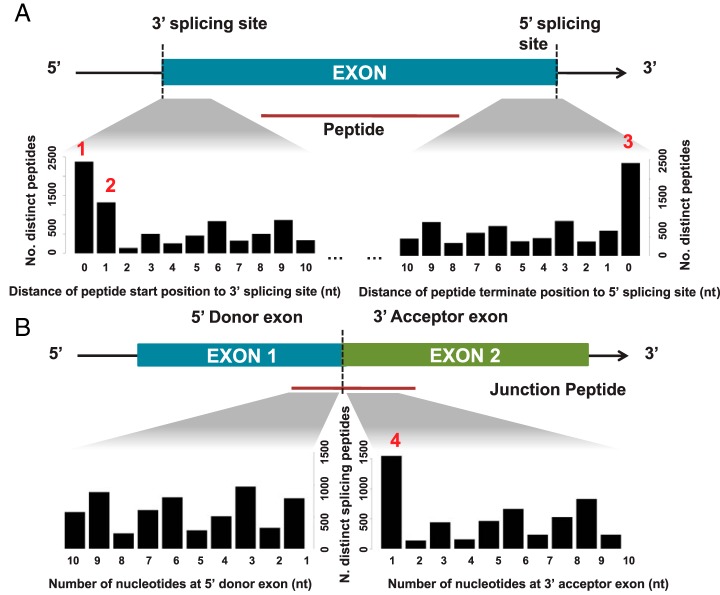
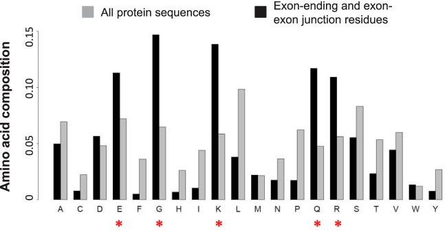
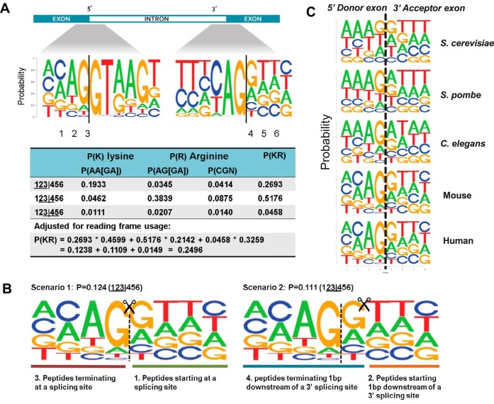
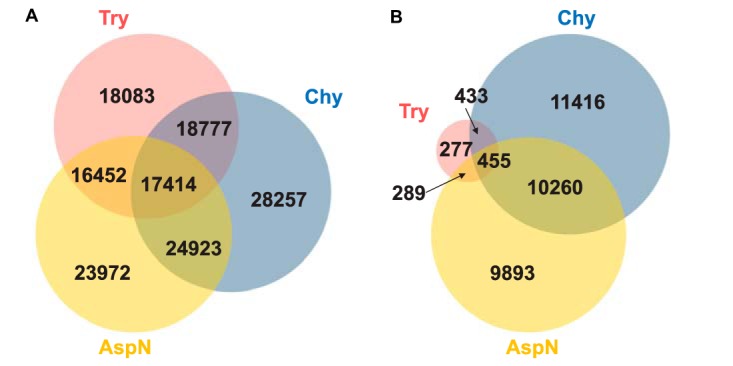
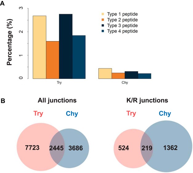
Alternative splicing dramatically increases transcriptome complexity but its contribution to proteome diversity remains controversial. Exon-exon junction spanning peptides provide direct evidence for the translation of specific splice isoforms and are critical for delineating protein isoform complexity. Here we found that junction-spanning peptides are underrepresented in publicly available mass spectrometry-based shotgun proteomics data sets. Further analysis showed that evolutionarily conserved preferential nucleotide usage at exon boundaries increases the occurrence of lysine- and arginine-coding triplets at the end of exons. Because both lysine and arginine residues are cleavage sites of trypsin, the nearly exclusive use of trypsin as the protein digestion enzyme in shotgun proteomic analyses hinders the detection of junction-spanning peptides. To study the impact of enzyme selection on splice junction detectability, we performed in-silico digestion of the human proteome using six proteases. The six enzymes created a total of 161,125 detectable junctions, and only 1,029 were common across all enzyme digestions. Chymotrypsin digestion provided the largest number of detectable junctions. Our experimental results further showed that combination of a chymotrypsin-based human proteome analysis with a trypsin-based analysis increased detection of junction-spanning peptides by 37% over the trypsin-only analysis and identified over a thousand junctions that were undetectable in fully tryptic digests. Our study demonstrates that detection of proteome diversity resulted from alternative splicing is limited by trypsin cleavage specificity, and that complementary digestion schemes will be essential to comprehensively analyze the translation of alternative splicing isoforms.
Overview
The claims and statistics of this paper haven’t been checked yet.
Run the check — free, no account needed. It reads the open-access text and checks the sources it cites.
Integrity signals
Registry
Cites
Statistics
Figures
Verification
Claims the paper makes
The claims and statistics of this paper haven’t been checked yet.
Run the check — free, no account needed. It reads the open-access text and checks the sources it cites.
Claims other papers make about this one
Each is checked against this paper’s own text: does it say what the citing paper says it does?
In the citing paper
“despite the fact that the number of peptides containing exon-exon splice junctions are reduced with trypsin digestion due to the overabundance of basic amino acid residues at splice junctions [51].”
From Deep proteomic network analysis of Alzheimer’s disease brain reveals alterations in RNA binding proteins and RNA splicing associated with disease (2018) · cites this paper as [51]
Self-check
Percentages, sums and reported differences re-computed from the paper’s own numbers.
The self-check needs the paper’s full text.
Citations
Cites
Alternative isoform regulation in human tissue transcriptomes
Wang, E. T., Sandberg, R., Luo, S., Khrebtukova, I., Zhang, L., Mayr, C., Kingsmore, S. F., Schroth, G. P., & Burge, C. B. (2008) · Nature · doi:10.1038/nature07509 · Cited by 5,181
A cross-platform toolkit for mass spectrometry and proteomics
Chambers, M., MacLean, B., Burke, R., Amodei, D., Ruderman, D., Neumann, S., Gatto, L., Fischer, B., Pratt, B. S., Egertson, J. D., et al. (2012) · Nature Biotechnology · doi:10.1038/nbt.2377 · Cited by 4,189
Self-citationMass-spectrometry-based draft of the human proteome
Wilhelm, M., Schlegl, J., Hahne, H., Gholami, A. M., Lieberenz, M., Savitski, M. M., Ziegler, E., Butzmann, L., Gessulat, S., Marx, H., et al. (2014) · Nature · doi:10.1038/nature13319 · Cited by 1,820
Proteogenomic characterization of human colon and rectal cancer
CPTAC, T. N., Zhang, B., Wang, J., Wang, X., Zhu, J., Liu, Q., Shi, Z., Chambers, M. C., Zimmerman, L. J., Shaddox, K. F., et al. (2014) · Nature · doi:10.1038/nature13438 · Cited by 1,462
Self-citationTrypsin Cleaves Exclusively C-terminal to Arginine and Lysine Residues
Olsen, J. V., Ong, S., & Mann, M. (2004) · Molecular & Cellular Proteomics · doi:10.1074/mcp.t400003-mcp200 · Cited by 1,309
MS-GF+ makes progress towards a universal database search tool for proteomics
Kim, S., & Pevzner, P. A. (2014) · Nature Communications · doi:10.1038/ncomms6277 · Cited by 1,098
Tabb, D. L., Fernando, C., & Chambers, M. (2007) · Journal of Proteome Research · doi:10.1021/pr0604054 · Cited by 550
Self-citationHow did alternative splicing evolve?
Ast, G. (2004) · Nature Reviews Genetics · doi:10.1038/nrg1451 · Cited by 544
Glatter, T., Ludwig, C., Ahrné, E., Aebersold, R. H., Heck, A. J. R., & Schmidt, A. (2012) · Journal of Proteome Research · doi:10.1021/pr300273g · Cited by 364
Alternative Splicing May Not Be the Key to Proteome Complexity
Tress, M. L., Abascal, F., & Valencia, A. (2016) · Trends in Biochemical Sciences · doi:10.1016/j.tibs.2016.08.008 · Cited by 341
Cited by
Sorted by citations
Comprehensive Analysis of Alternative Splicing Across Tumors from 8,705 Patients
Kahles, A., Lehmann, K.-V., Toussaint, N. C., Hüser, M., Stark, S. G., Sachsenberg, T. T., Stegle, O., Kohlbacher, O., Sander, C., Johnson, R., et al. (2018) · Cancer Cell · doi:10.1016/j.ccell.2018.07.001 · Cited by 914
“Furthermore, a recent study showed that junction-spanning peptides resulting from alternative splicing are underrepresented in protein MS datasets due to the cleavage specificity of trypsin (Wang et al., 2017).”
BackgroundAlternative Splicing Regulatory Networks: Functions, Mechanisms, and Evolution
Ule, J., & Blencowe, B. J. (2019) · Molecular Cell · doi:10.1016/j.molcel.2019.09.017 · Cited by 780
“, based on an in silico analysis) to miss detection of the majority of splice junction-spanning peptides (Wang et al., 2018), and mass spectrometry (MS) analysis methods, such as those employed by Tress et al.”
MethodAlternative mRNA splicing in cancer immunotherapy
Frankiw, L., Baltimore, D., & Li, G. (2019) · Nature reviews. Immunology · doi:10.1038/s41577-019-0195-7 · Cited by 253
Pervasive translation of circular RNAs driven by short IRES-like elements
Fan, X., Yang, Y., Chen, C., & Wang, Z. (2022) · Nature Communications · doi:10.1038/s41467-022-31327-y · Cited by 241
Johnson, E. C. B., Dammer, E. B., Duong, D. M., Yin, L., Thambisetty, M., Troncoso, J. C., Lah, J. J., Levey, A. I., & Seyfried, N. T. (2018) · Molecular Neurodegeneration · doi:10.1186/s13024-018-0282-4 · Cited by 225
Alternative Splicing and Protein Diversity: Plants Versus Animals
Chaudhary, S., Khokhar, W., Jabre, I., Reddy, A. S. N., Byrne, L. J., Wilson, C. M., & Syed, N. H. (2019) · Frontiers in Plant Science · doi:10.3389/fpls.2019.00708 · Cited by 209
Global detection of human variants and isoforms by deep proteome sequencing
Sinitcyn, P. G., Richards, A., Weatheritt, R. J., Brademan, D. R., Marx, H., Shishkova, E., Meyer, J. G., Hebert, A. S., Westphall, M. S., Blencowe, B. J., et al. (2023) · Nature Biotechnology · doi:10.1038/s41587-023-01714-x · Cited by 193
“7b) 65 .”
BackgroundInfluentialProteomics in aging research: A roadmap to clinical, translational research
Moaddel, R., Ubaida‐Mohien, C., Tanaka, T., Lyashkov, A., Basisty, N., Schilling, B., Semba, R. D., Franceschi, C., Gorospe, M., & Ferrucci, L. (2021) · Aging Cell · doi:10.1111/acel.13325 · Cited by 139
Perspective on Alternative Splicing and Proteome Complexity in Plants
Chaudhary, S., Jabre, I., Reddy, A. S. N., Staiger, D., & Syed, N. H. (2019) · Trends in Plant Science · doi:10.1016/j.tplants.2019.02.006 · Cited by 135
“Among these five enzymes, the highest number of detectable junctions including exon-ending and exon-exon junctions were observed in chymotrypsin digestion, making it a protease of choice in LC-MS/MS studies, especially to predict RNA splicing derived peptides [101].”
PDV: an integrative proteomics data viewer
Li, K., Vaudel, M., Zhang, B., Ren, Y., & Wen, B. (2018) · Bioinformatics · doi:10.1093/bioinformatics/bty770 · Cited by 126
Self-citationActionable Cytopathogenic Host Responses of Human Alveolar Type 2 Cells to SARS-CoV-2
Hekman, R. M., Hume, A. J., Goel, R. K., Abo, K. M., Huang, J., Blum, B. C., Werder, R. B., Suder, E. L., Paul, I., Phanse, S., et al. (2020) · Molecular Cell · doi:10.1016/j.molcel.2020.11.028 · Cited by 115
Proteomics Using Protease Alternatives to Trypsin Benefits from Sequential Digestion with Trypsin
Dau, T., Bartolomucci, G., & Rappsilber, J. (2020) · Analytical Chemistry · doi:10.1021/acs.analchem.0c00478 · Cited by 105
Saltzman, A. B., Leng, M., Bhatt, B., Singh, P., Chan, D. W., Dobrolecki, L. E., Chandrasekaran, H., Choi, J. M., Jain, A., Jung, S. Y., et al. (2018) · Molecular & Cellular Proteomics · doi:10.1074/mcp.tir118.000850 · Cited by 104
“Peptides that differentiate protein isoforms are greatly un-derrepresented in proteomic profiling data due to their low frequency and an inherent tryptic bias against exon junctions (24).”
BackgroundEnhanced protein isoform characterization through long-read proteogenomics
Miller, R., Jordan, B. T., Mehlferber, M., Jeffery, E. D., Chatzipantsiou, C., Kaur, S., Millikin, R. J., Dai, Y., Tiberi, S., Castaldi, P. J., et al. (2022) · Genome biology · doi:10.1186/s13059-022-02624-y · Cited by 95
“Alternative isoforms are particularly susceptible, because their isoform-specific regions comprise a small fraction of the proteome and suffer from a negative detection bias in traditional MS-based proteomics workflows using tryptic digestion [51].”
BackgroundSplice-Junction-Based Mapping of Alternative Isoforms in the Human Proteome
Lau, E., Han, Y., Williams, D. R., Thomas, C. T., Shrestha, R., Wu, J. C., & Lam, M. P. Y. (2019) · Cell Reports · doi:10.1016/j.celrep.2019.11.026 · Cited by 67
“However, a lower score distribution for these sequences could also be due to the lower abundance of alternative isoforms (Blencowe, 2017) and the known enrichment of lysine at splice junctions producing miscleavages (Wang et al., 2018b) whose scoring is penalized by Percolator (The et al., 2016).”
BackgroundŠalovská, B., Zhu, H., Gandhi, T., Frank, M., Li, W., Rosenberger, G., Wu, C., Germain, P., Zhou, H., Hodný, Z., et al. (2020) · Molecular Systems Biology · doi:10.15252/msb.20199170 · Cited by 66
“Our first goal was to assess the role of protein degradation in protein abundance buffering (Mueller et al, 2015; Liu et al, 2016; Wang et al, 2018a).”
BackgroundMethodUncovering the impacts of alternative splicing on the proteome with current omics techniques
Reixachs‐Solé, M., & Eyras, E. (2022) · Wiley Interdisciplinary Reviews - RNA · doi:10.1002/wrna.1707 · Cited by 65
“Regarding splicing products, different digestion enzymes used in proteomics are biased toward identifying certain splicing junctions with small overlap among them (Wang et al., 2018).”
BackgroundContributions of alternative splicing to muscle type development and function
Nikonova, E. V., Kao, S.-Y., & Spletter, M. L. (2020) · Seminars in Cell and Developmental Biology · doi:10.1016/j.semcdb.2020.02.003 · Cited by 64
Cancer-Specific Splicing Changes and the Potential for Splicing-Derived Neoantigens
Escobar‐Hoyos, L. F., & Abdel-Wahab, O. I. (2018) · Cancer Cell · doi:10.1016/j.ccell.2018.07.008 · Cited by 58
“Moreover, it has also been noted that the enzymes most commonly used to generate peptides from full-length proteins for mass spectrometry have a tendency to cleave proteins at EEJs, thereby obscuring detection of neopeptides translated from neojunctions (Wang et al., 2018).”
Reixachs‐Solé, M., Ruiz‐Orera, J., Albà, M. M., & Eyras, E. (2020) · Nature Communications · doi:10.1038/s41467-020-15634-w · Cited by 57
Assessing the functional relevance of splice isoforms
del Pozo, F. C., Gómez, L. M., Walsh, T., Rodrı́guez, J. M., Domenico, T. D., Abascal, F., Vázquez, J., & Tress, M. L. (2021) · NAR Genomics and Bioinformatics · doi:10.1093/nargab/lqab044 · Cited by 46
“There are fewer alternative isoforms detected in proteomics experiments than would be expected from transcription levels (16), even when technical issues are taken into account (28).”
BackgroundThe evolution of alternative splicing in glioblastoma under therapy
Wang, L., Shamardani, K., Babikir, H. A., Catalan, F. L., Nejo, T., Chang, S., Phillips, J. J., Okada, H., & Diaz, A. A. (2021) · Genome biology · doi:10.1186/s13059-021-02259-5 · Cited by 51
Steering research on mRNA splicing in cancer towards clinical translation
Anczuków, O., Allain, F. H. ‐., Angarola, B. L., Black, D. L., Brooks, A. N., Cheng, C., Conesa, A., Crosse, E. I., Eyras, E., Guccione, E., et al. (2024) · Nature reviews. Cancer · doi:10.1038/s41568-024-00750-2 · Cited by 50
“However, measurements of protein diversity by mass spectrometry 156 are affected by the biases of the digestion enzymes used in the process, which can limit variant detection 170 .”
Blank‐Landeshammer, B., Teichert, I., Märker, R., Nowrousian, M., Kück, U., & Sickmann, A. (2019) · mBio · doi:10.1128/mbio.02367-19 · Cited by 40
“In addition to a described occurrence of peculiar amino acid sequences found at splice sites that were unfavorable for LC-MS-based analysis (44), we did not expect to find equivalent evidence for the translation of AS mRNA variants into protein compared to their transcript abundances.”
BackgroundAlternative Splicing in Apicomplexan Parasites
Yeoh, L. M., Lee, V. V., McFadden, G. I., & Ralph, S. A. (2019) · mBio · doi:10.1128/mbio.02866-18 · Cited by 35
“in peptide cleavage techniques in protein preparation (56) and sensitivity limits of some”
MethodHumphrey, J., Brophy, E., Kosoy, R., Zeng, B., Coccia, E., Mattei, D., Ravi, A., Naito, T., Efthymiou, A. G., Navarro, E., et al. (2025) · Nature Genetics · doi:10.1038/s41588-025-02099-0 · Cited by 37
“Although we observed little peptide support for our novel isoforms, as previously observed 65 , this is likely due in part to the technical disfavoring of peptides that span exon junctions 66 .”
Re-evaluating the impact of alternative RNA splicing on proteomic diversity
Manuel, J. M., Guilloy, N., Khatir, I., Roucou, X., & Laurent, B. (2023) · Frontiers in Genetics · doi:10.3389/fgene.2023.1089053 · Cited by 32
“The standard protease used in shotgun proteomics is the trypsin that digests at K or R residues, hereby producing short peptides (around six amino acids) and limiting the proteome coverage and detection of isoform proteins (Wang et al., 2018).”
BackgroundLi, M., Zheng, H., Lin, M., Zhu, W., & Zhang, J. (2020) · Food Chemistry · doi:10.1016/j.foodchem.2020.126712 · Cited by 32
Proteogenomics reveals sex-biased aging genes and coordinated splicing in cardiac aging
Han, Y., Wennersten, S. A., Wright, J. M., Ludwig, R. W., Lau, E., & Lam, M. P. Y. (2022) · American Journal of Physiology-Heart and Circulatory Physiology · doi:10.1152/ajpheart.00244.2022 · Cited by 31
Cifani, P., Dhabaria, A., Chen, Z., Yoshimi, A., Kawaler, E. A., Abdel-Wahab, O. I., Poirier, J. T., & Kentsis, A. (2018) · Journal of Proteome Research · doi:10.1021/acs.jproteome.8b00295 · Cited by 34
Protein isoform-centric therapeutics: expanding targets and increasing specificity
Kjer-Hansen, P., Phan, T. G., & Weatheritt, R. J. (2024) · Nature Reviews Drug Discovery · doi:10.1038/s41573-024-01025-z · Cited by 27
“Techniques that modulate exon usage (thereby causing isoform switching) without altering gene product amounts therefore offer the clearest insight into individual isoform function, with antisense oligonucleotides being the current gold standard 240,241 .”
Agosto, L. M., Gazzara, M. R., Radens, C. M., Sidoli, S., Baeza, J., Garcia, B. A., & Lynch, K. W. (2019) · Genome Research · doi:10.1101/gr.248435.119 · Cited by 30
APPRIS principal isoforms and MANE Select transcripts define reference splice variants
Pozo, F., Rodrı́guez, J. M., Gómez, L. M., Vázquez, J., & Tress, M. L. (2022) · Bioinformatics · doi:10.1093/bioinformatics/btac473 · Cited by 27
Transcription Factor Dynamics in Cross-Regulation of Plant Hormone Signaling PathwaysPreprint
Yin, L., Zander, M., Huang, S. C., Xie, M., Song, L., Guzmán, J. P. S., Hann, E. C., Shanbhag, B. K., Ng, S., Jain, S., et al. (2023) · bioRxiv (Cold Spring Harbor Laboratory) · doi:10.1101/2023.03.07.531630 · Cited by 25
Ran, X., Hu, F., Mao, N., Ruan, Y., Yi, F., Niu, X., Huang, S., Li, S., You, L., Zhang, F., et al. (2021) · Porcine Health Management · doi:10.1186/s40813-021-00226-x · Cited by 22
“AS is the main reason leading to change the different transcripts together with proteome varieties [47].”
BackgroundLin, X., Zhu, Z., Lin, D., Bao, Q., Gao, Y., Liu, Q., Liu, A., Lin, L., & Lin, X. (2021) · Talanta · doi:10.1016/j.talanta.2021.122647 · Cited by 21
Harnessing the power of proteomics in precision diabetes medicine
Kurgan, N., Larsen, J. K., & Deshmukh, A. S. (2024) · Diabetologia · doi:10.1007/s00125-024-06097-5 · Cited by 22
“In addition, transcriptomics is currently more fit for discovering genetic variants and alternative splicing events that are difficult to detect by proteomics due to the limited sequence coverage [31].”
Expression and splicing mediate distinct biological signals
Dam, S. H., Olsen, L. R., & Vitting‐Seerup, K. (2023) · BMC Biology · doi:10.1186/s12915-023-01724-w · Cited by 20
“8 Furthermore, peptides generated by standard bottom-up proteomics are heavily depleted for peptides 1 that cross exon-exon junctions, making it harder to detect splicing differences [30].”
BackgroundMulti-omics approach to identifying isoform variants as therapeutic targets in cancer patients
Shaw, T. I., Zhao, B., Li, Y., Wang, H., Wang, L., Manley, B. J., Stewart, P. A., & Karolak, A. M. (2022) · Frontiers in Oncology · doi:10.3389/fonc.2022.1051487 · Cited by 16
“Combining chymotrypsin with trypsin has partially alleviated this issue and resulted in increased junction peptide detection by >30% (69).”
BackgroundSøreide, K., Roalsø, M. T. T., & Aunan, J. R. (2020) · Journal of Pancreatic Cancer · doi:10.1089/pancan.2019.0014 · Cited by 17
“teomic analyses hinders the detection of transcriptomic alterations, such as seen in junction-spanning peptides.(60) In proteomic sciences, when cellular protein fractions corresponding to cytosol, membrane, nucleus, and cytoskeleton are further prepared for mass-spectrometry,”
BackgroundAn extended catalogue of tandem alternative splice sites in human tissue transcriptomes
Mironov, A. A., Denisov, S., Greß, A., Kalinina, O. V., & Pervouchine, D. D. (2021) · PLoS Computational Biology · doi:10.1371/journal.pcbi.1008329 · Cited by 17
“Direct measurement of this proportion by, for example, shotgun proteomics is not instructive for many reasons, including limited coverage and low sensitivity of such experiments [77], as well as the fact that the cleavage site consensus of a widely used trypsin protease overlaps with the amino acid sequence induced by…”
BackgroundAnnalora, A. J., Jozic, M., Marcus, C. B., & Iversen, P. L. (2018) · Toxicology and Applied Pharmacology · doi:10.1016/j.taap.2018.12.009 · Cited by 15
The Zea mays PeptideAtlas: A New Maize Community Resource
van Wijk, K. J., Leppert, T., Sun, Z., Guzchenko, I., Debley, E. L., Sauermann, G., Routray, P., Mendoza, L., Sun, Q., & Deutsch, E. W. (2024) · Journal of Proteome Research · doi:10.1021/acs.jproteome.4c00320 · Cited by 14
Hornburg, D., Kruse, T., Anderl, F., Daschkin, C., Semper, R. P., Klar, K., Guenther, A., Mejías‐Luque, R., Schneiderhan‐Marra, N., Mann, M., et al. (2019) · Scientific Reports · doi:10.1038/s41598-019-53493-8 · Cited by 14
Proteomic Analysis of Oil-Roasted Cashews Using a Customized Allergen-Focused Protein Database
Chen, S., & Downs, M. L. (2022) · Journal of Proteome Research · doi:10.1021/acs.jproteome.2c00095 · Cited by 13
Ait-Hamlat, A., Zea, D. J., Labeeuw, A., Polit, L., Richard, H., & Laine, É. (2020) · Journal of Molecular Biology · doi:10.1016/j.jmb.2020.01.032 · Cited by 14
The peptide woods are lovely, dark and deep: Hunting for novel cancer antigens
Oreper, D., Klaeger, S., Jhunjhunwala, S., & Delamarre, L. (2023) · Seminars in Immunology · doi:10.1016/j.smim.2023.101758 · Cited by 13
Origins and Evolution of Human Tandem Duplicated Exon Substitution Events
Gómez, L. M., Cerdán-Vélez, D., Abascal, F., & Tress, M. L. (2022) · Genome Biology and Evolution · doi:10.1093/gbe/evac162 · Cited by 11
Integrative Proteogenomics for Differential Expression and Splicing Variation in a DM1 Mouse Model
Solovyeva, E. M., Utzinger, S., Vissières, A., Mitchelmore, J., Ahrné, E., Hermes, E., Poetsch, T., Ronco, M., Bidinosti, M., Merkl, C., et al. (2023) · Molecular & Cellular Proteomics · doi:10.1016/j.mcpro.2023.100683 · Cited by 13
Mehlferber, M., Jeffery, E. D., Saquing, J., Jordan, B. T., Sheynkman, L., Murali, M., Genet, G., Acharya, B. R., Hirschi, K. K., & Sheynkman, G. (2022) · RNA Biology · doi:10.1080/15476286.2022.2141938 · Cited by 11
“Therefore, sampling peptides from the small space of unique amino acids that can directly confirm the presence of a protein isoform is limited by the space of “informa-tive” (i.e. unique-to-an-isoform) peptides[55].”
BackgroundJeong, S.-K., Kim, C.-Y., & Paik, Y. (2018) · Journal of Proteome Research · doi:10.1021/acs.jproteome.8b00548 · Cited by 11
Assessing conservation of alternative splicing with evolutionary splicing graphsPreprint
Zea, D. J., Laskina, S., Baudin, A., Richard, H., & Laine, É. (2021) · Genome Research · doi:10.1101/gr.274696.120 · Cited by 15
Cagnin, C., Garcia, B. D. F., Rocha, T. D. S., & Prudêncio, S. H. (2024) · Biology · doi:10.3390/biology13100772 · Cited by 11
“According to Wang et al. [46], the proteases most used in in vitro studies for the simulation of gastrointestinal digestion are pepsin, pancreatin (which is a mixture of several digestive enzymes, including trypsin and chymotrypsin), and chymotrypsin.”
BackgroundMethodCrowl, S., Coleman, M. B., Chaphiv, A., Jordan, B. T., & Naegle, K. M. (2025) · Cell Systems · doi:10.1016/j.cels.2025.101318 · Cited by 10
Li, Y., Zhang, C., Luo, F., & Peng, B. (2018) · Applied Microbiology and Biotechnology · doi:10.1007/s00253-018-9419-1 · Cited by 10
Tools and tactics for studying alternative splicing
Sousa‐Luís, R., & Carmo‐Fonseca, M. (2026) · Nature Reviews Genetics · doi:10.1038/s41576-026-00952-4 · Cited by 8
Kim, C.-Y., Na, K., Park, S., Jeong, S.-K., Cho, J.-Y., Shin, H., Lee, M. J., Han, G., & Paik, Y. (2019) · Molecular & Cellular Proteomics · doi:10.1074/mcp.ra119.001456 · Cited by 10
“Studies have shown that the percentages of Lys and Arg that constitute the exon boundary are relatively high and that chymotrypsin and Asp-N greatly improves the peptide coverage of the exon-exon splice junction containing K/R residue (66).”
BackgroundMethodBeyond the gene: decoding alternative isoforms
Mattioli, K., & Bulyk, M. L. (2025) · Trends in Genetics · doi:10.1016/j.tig.2025.11.003 · Cited by 9
Impact of alternative splicing on Arabidopsis proteome
Reyes, A. V., Zhang, C., Karunadasa, S. S., Shrestha, R., Grismer, T. S., Byun, D., & Xu, S. (2026) · Nucleic Acids Research · doi:10.1093/nar/gkag400 · Cited by 7
“As noted previously, IR-derived peptides are often challenging to detect because exon–intron junctions are frequently enriched in K/R residues, which can obscure the identification of AS-specific peptides [ 42 , 44 ].”
Li, N., Jia, W., Wang, J., Shao, Q., Feng, X., Li, Z., Sun, W., Kang, M., Hu, D., Xing, L., et al. (2024) · The EPMA Journal · doi:10.1007/s13167-024-00366-4 · Cited by 8
A ratiometric catalog of protein isoform shifts in the cardiac fetal gene program
Han, Y., Binti, S., Wennersten, S. A., Pandi, B., Ng, D. C. M., Lau, E., & Lam, M. P. Y. (2025) · JCI Insight · doi:10.1172/jci.insight.184309 · Cited by 5
“At present, the identification of splicing-derived isoform proteins (spliceoforms) remains hindered by experimental and data analytical challenges (42, 59, 60).”
APOER2 splicing repertoire in Alzheimer’s disease: Insights from long-read RNA sequencing
Gallo, C. M., Kistler, S. A., Natrakul, A., Labadorf, A. T., Beffert, U., & Ho, A. (2024) · PLoS Genetics · doi:10.1371/journal.pgen.1011348 · Cited by 6
Hümmer, S., Borao, S., Guerra-Moreno, Á., Cozzuto, L., Hidalgo, E., & Ayté, J. (2021) · Cell Reports · doi:10.1016/j.celrep.2021.109893 · Cited by 5
Validating Amino Acid Variants in Proteogenomics Using Sequence Coverage by Multiple Reads
Levitsky, L. I., Kuznetsova, K. G., Kliuchnikova, A. A., Ilina, I. Y., Goncharov, A. O., Lobas, A. A., Ivanov, M. V., Lazarev, V. N., Ziganshin, R., Gorshkov, M. V., et al. (2022) · Journal of Proteome Research · doi:10.1021/acs.jproteome.2c00033 · Cited by 5
Humphrey, J., Brophy, E., Kosoy, R., Zeng, B., Coccia, E., Mattei, D., Ravi, A., Efthymiou, A. G., Navarro, E., Muller, B. Z., et al. (2023) · medRxiv · doi:10.1101/2023.12.01.23299073 · Cited by 5
“Secondly, we observed very little peptide support for our novel isoforms, likely due to trypsin digestion in shotgun proteomics disfavouring the creation of peptides that span exon junctions (Wang et al. 2018).”
Alternative splicing in plants: emerging order from chaos
Jabre, I., Khokhar, W., Reddy, A. S. N., Wilson, C. M., & Syed, N. H. (2026) · Trends in Plant Science · doi:10.1016/j.tplants.2026.04.001 · Cited by 5
Puliasis, S. S., Lewandowska, D., Hemsley, P. A., & Zhang, R. (2023) · Journal of Proteome Research · doi:10.1021/acs.jproteome.3c00135 · Cited by 4
Jin, L., Wang, F., Wang, X., Harvey, B. P., Bi, Y., Hu, C., Cui, B., Darcy, A. T., Maull, J. W., Phillips, B. R., et al. (2023) · Proteomes · doi:10.3390/proteomes11040032 · Cited by 7
Self-citationIS-PRM-Based Peptide Targeting Informed by Long-Read Sequencing for Alternative Proteome Detection
Korchak, J. A., Jeffery, E. D., Bandyopadhyay, S., Jordan, B. T., Lehe, M. D., Watts, E. F., Fenix, A. M., Wilhelm, M., & Sheynkman, G. (2024) · Journal of the American Society for Mass Spectrometry · doi:10.1021/jasms.4c00119 · Cited by 5
Pandi, B., Brenman, S., Black, A., Ng, D. C. M., Lau, E., & Lam, M. P. Y. (2024) · Journal of Proteome Research · doi:10.1021/acs.jproteome.3c00789 · Cited by 3
Horokhovskyi, Y., Roetschke, H. P., Cormican, J. A., Pašen, M., Garazhian, S., Mishto, M., & Liepe, J. (2025) · Molecular & Cellular Proteomics · doi:10.1016/j.mcpro.2025.101039 · Cited by 2
“…with proteins that are enzymatically digested into peptides—for example, by trypsin, which mainly cleaves proteins after arginine (R) and lysin (K) (41–43), separated by chromatography, and measured with tandem MS. Peptide sequences are matched to a reference protein sequence database for…”
Detecting Differential Alternative Splicing in Mass Spectrometry-based Proteomics DataPreprint
Ammar, C., Csaba, G., Hadziahmetovic, A., Vasilopoulou, C. G., Gruber, M., Mann, M., & Zimmer, R. (2023) · bioRxiv (Cold Spring Harbor Laboratory) · doi:10.1101/2023.09.19.558203 · Cited by 3
Decoding the Cryptic Proteome Between Antigens and Novel Functional Proteins
Bawden, E., Amigorena, S., & Arribas, Y. A. (2025) · European Journal of Immunology · doi:10.1002/eji.70102 · Cited by 3
“In addition, trypsin digestion poses limitations on the identification of cryptic peptides derived from splicing junctions because exon boundaries exhibit high usage of arginine and lysine [14].”
Wang, Z.-Y., Zhang, W., Zhang, N., Wu, P.-M., Su, Q., Ma, C., Wang, W., & Chen, M. (2026) · Journal of Experimental Botany · doi:10.1093/jxb/erag052 · Cited by 2
Lehe, M. D., Almofeez, R., Jeffery, E. D., & Sheynkman, G. (2026) · Journal of Mass Spectrometry · doi:10.1002/jms.70024 · Cited by 3
Lobas, A. A., Solovyeva, E. M., Levitsky, L. I., Goncharov, A. O., Lyssuk, E. Y., Larin, S. S., Moshkovskii, S. A., & Gorshkov, M. V. (2023) · International Journal of Molecular Sciences · doi:10.3390/ijms24032466 · Cited by 2
“There are a number of reasons for such dichotomy in mass spectrometry-based bottom-up proteomics, including (i) low protein sequence coverage translating into a low probability to detect splicing-specific peptides, (ii) a statistically increased probability of arginine or lysine being encoded at exon boundaries [37],…”
BackgroundNarykov, O., Johnson, N. T., & Korkin, D. (2018) · bioRxiv (Cold Spring Harbor Laboratory) · doi:10.1101/256834 · Cited by 2
“and carrying out other basic functions (2-4).”
MethodZhang, F., Tang, L., Ran, X., Mao, N., Ruan, Y., Yi, F., Liu, C., Niu, X., Huang, S., Li, S., et al. (2019) · bioRxiv (Cold Spring Harbor Laboratory) · doi:10.1101/547810 · Cited by 2
Expression and Splicing Mediate Distinct Biological SignalsPreprint
Dam, S. H., Olsen, L. R., & Vitting‐Seerup, K. (2022) · bioRxiv (Cold Spring Harbor Laboratory) · doi:10.1101/2022.08.29.505720 · Cited by 2
Zhao, F., Yan, J., Liu, Z., Pan, D., Wang, Y., Li, L., Suo, S., Dang, K., Li, D., Dang, Y., et al. (2025) · Food Research International · doi:10.1016/j.foodres.2025.118157 · Cited by 1
Assessing Conservation of Alternative Splicing with Evolutionary Splicing GraphsPreprint
Zea, D. J., Laskina, S., Baudin, A., Richard, H., & Laine, É. (2020) · bioRxiv (Cold Spring Harbor Laboratory) · doi:10.1101/2020.11.14.382820 · Cited by 1
Zhong, J. J., Morin, G. B., & Chen, D. D. Y. (2022) · Wiley eBooks · doi:10.1002/9783527833092.ch3 · Cited by 1
Gallo, C. M., Kistler, S. A., Natrakul, A., Labadorf, A. T., Beffert, U., & Ho, A. (2023) · bioRxiv (Cold Spring Harbor Laboratory) · doi:10.1101/2023.01.12.523804 · Cited by 1
“Although detection of lowly expressed isoforms at the protein level can be technically challenging (Blakeley et al., 2010; X. Wang et al., 2018), novel methods including the integration of RNA-seq and mass spectrometry data (Agosto et al., 2019; Han et al., 2020) are rapidly increasing the…”
MethodAit-hamlat, A., Zea, D. J., Labeeuw, A., Polit, L., Richard, H., & Laine, É. (2017) · bioRxiv (Cold Spring Harbor Laboratory) · doi:10.1101/119891
Proteomics and Metabolomics in Pain
Schmidt, M. (2020) · Elsevier eBooks · doi:10.1016/b978-0-12-809324-5.24256-9
Data regarding the sensibility to proteolysis of a natural apolipoprotein A-I mutant.
Gaddi, G. M., Gisonno, R. A., Rosu, S. A., Cortez, M. F., Finarelli, G. S., Ramella, N. A., & Tricerri, M. A. (2020) · Data in Brief · doi:10.1016/j.dib.2020.105960
“Together with trypsin, this enzyme is a potent tool for structural studies involving mass spectrometry and cross-linking [2] , as the cleavage pattern is well-defined (it selectively hydrolyzes peptide bonds on the C-terminal side of tyrosine, phenylalanine, tryptophan, and leucine) [3] .”
BackgroundSegelcke, D., van der Burgt, M., Kappert, C., Garcia, D. S., Sondermann, J. R., Bigalke, S., Pradier, B., Gómez‐Varela, D., Zahn, P. K., Schmidt, M., et al. (2022) · bioRxiv (Cold Spring Harbor Laboratory) · doi:10.1101/2022.03.07.482980
Additional file 1 of The evolution of alternative splicing in glioblastoma under therapy
Wang, L., Shamardani, K., Babikir, H. A., Catalan, F. L., Nejo, T., Chang, S., Phillips, J. J., Okada, H., & Diaz, A. A. (2021) · Figshare · doi:10.6084/m9.figshare.13647119
Ran, X., Hu, F., Mao, N., Ruan, Y., Yi, F., Niu, X., Huang, S., Li, S., You, L., Zhang, F., et al. (2021) · Figshare · doi:10.6084/m9.figshare.16557251
Ran, X., Hu, F., Mao, N., Ruan, Y., Yi, F., Niu, X., Huang, S., Li, S., You, L., Zhang, F., et al. (2021) · Figshare · doi:10.6084/m9.figshare.16557254
Additional file 7 of The evolution of alternative splicing in glioblastoma under therapy
Wang, L., Shamardani, K., Babikir, H. A., Catalan, F. L., Nejo, T., Chang, S., Phillips, J. J., Okada, H., & Diaz, A. A. (2021) · Figshare · doi:10.6084/m9.figshare.13647140
Additional file 6 of The evolution of alternative splicing in glioblastoma under therapy
Wang, L., Shamardani, K., Babikir, H. A., Catalan, F. L., Nejo, T., Chang, S., Phillips, J. J., Okada, H., & Diaz, A. A. (2021) · Figshare · doi:10.6084/m9.figshare.13647137
Ran, X., Hu, F., Mao, N., Ruan, Y., Yi, F., Niu, X., Huang, S., Li, S., You, L., Zhang, F., et al. (2021) · Figshare · doi:10.6084/m9.figshare.16557245
Additional file 8 of The evolution of alternative splicing in glioblastoma under therapy
Wang, L., Shamardani, K., Babikir, H. A., Catalan, F. L., Nejo, T., Chang, S., Phillips, J. J., Okada, H., & Diaz, A. A. (2021) · Figshare · doi:10.6084/m9.figshare.13647143
Ran, X., Hu, F., Mao, N., Ruan, Y., Yi, F., Niu, X., Huang, S., Li, S., You, L., Zhang, F., et al. (2021) · Figshare · doi:10.6084/m9.figshare.16557263
Ran, X., Hu, F., Mao, N., Ruan, Y., Yi, F., Niu, X., Huang, S., Li, S., You, L., Zhang, F., et al. (2021) · Figshare · doi:10.6084/m9.figshare.16557248
Additional file 3 of The evolution of alternative splicing in glioblastoma under therapy
Wang, L., Shamardani, K., Babikir, H. A., Catalan, F. L., Nejo, T., Chang, S., Phillips, J. J., Okada, H., & Diaz, A. A. (2021) · Figshare · doi:10.6084/m9.figshare.13647128
Ran, X., Hu, F., Mao, N., Ruan, Y., Yi, F., Niu, X., Huang, S., Li, S., You, L., Zhang, F., et al. (2021) · Figshare · doi:10.6084/m9.figshare.16557260
Additional file 5 of The evolution of alternative splicing in glioblastoma under therapy
Wang, L., Shamardani, K., Babikir, H. A., Catalan, F. L., Nejo, T., Chang, S., Phillips, J. J., Okada, H., & Diaz, A. A. (2021) · Figshare · doi:10.6084/m9.figshare.13647134
Additional file 9 of The evolution of alternative splicing in glioblastoma under therapy
Wang, L., Shamardani, K., Babikir, H. A., Catalan, F. L., Nejo, T., Chang, S., Phillips, J. J., Okada, H., & Diaz, A. A. (2021) · Figshare · doi:10.6084/m9.figshare.13647146
Metadata
- Authors
- Wang, X., Codreanu, S. G., Wen, B., Li, K., Chambers, M. C., Liebler, D. C., & Zhang, B.
- Affiliations
- Baylor College of Medicine (US)
- Vanderbilt University (US)
- BGI Group (China) (CN)
- Corresponding author
- Zhang, Bing
- ORCID
- Venue
- Molecular & Cellular Proteomics
- Publisher
- Elsevier BV
- Published
- 2018-03
- Volume
- 17
- Issue
- 3
- Pages
- 422-430
- ISSN
- 1535-9476 (ISSN-L)
- PMC
- PMC5836368
- OpenAlex
- W2773925504
- Cited by
- 97
- References
- 27
- Citations by year
- 2026: 10 · 2025: 6 · 2024: 11 · 2023: 13 · 2022: 13 · 2021: 11 · 2020: 11 · 2019: 11 · 2018: 10
- FWCI
- 4.45
- Type
- journal article
- Language
- en
- Open access
- hybrid
- doi.org
- Licence
- Locations
- Molecular & Cellular Proteomics (publishedVersion, cc-by)
- PubMed Central (publishedVersion, cc-by)
- PubMed (publishedVersion)
- Funders
- HHS NIH National Cancer Institute (NCI) (U24CA159988, U24CA210954)
- Cancer Prevention and Research Institute of Texas (CPRIT) (RR160027)
- Topics
- Physical Sciences › Chemistry › Spectroscopy › Advanced Proteomics Techniques and Applications
- Physical Sciences › Chemistry › Spectroscopy › Mass Spectrometry Techniques and Applications
- Life Sciences › Biochemistry, Genetics and Molecular Biology › Molecular Biology › RNA Research and Splicing
- Keywords
- alternative splicing, proteome diversity, protease selection, chymotrypsin digestion, protein isoforms, in silico digestion, human proteome
- MeSH terms
- Exons, Humans, Neoplasm Proteins, Neoplasms, Peptide Hydrolases, Peptides, Alternative Splicing, Proteome, Cell Line, Tumor
About these checks
Registry, retraction, Cites and statistics results are computed, not judged, with one exception: a printed reference that no index lists is read once by a model before it counts as a citation error. Claims are extracted by a model and kept only when their quote appears verbatim in the text; each cited source is then checked, with a model’s help, for whether it says what the claim relies on. Those verdicts can be wrong — read the claim’s quote and the cited source before relying on one.
Statistics and arithmetic are recomputed, not judged.
Confidence bands are shown only for fields calibrated on our annotated test set. None is calibrated yet, so no band is shown.
What counts as a claim ›How we measure confidence ›How we check this ›
Are you an author, or do you see a problem with this page? Request a review
