

The DOI matches its Crossref record; 36 of the 36 references it cites were found in a registry or an index.
Brain delivery of quercetin-loaded exosomes improved cognitive function in AD mice by inhibiting phosphorylated tau-mediated neurofibrillary tangles
Open accessAbstract
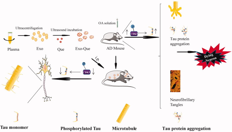
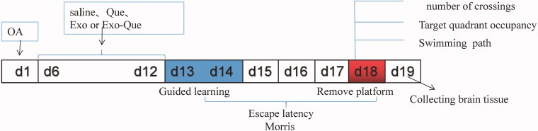
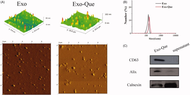
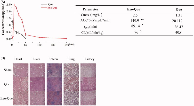
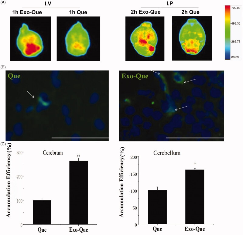
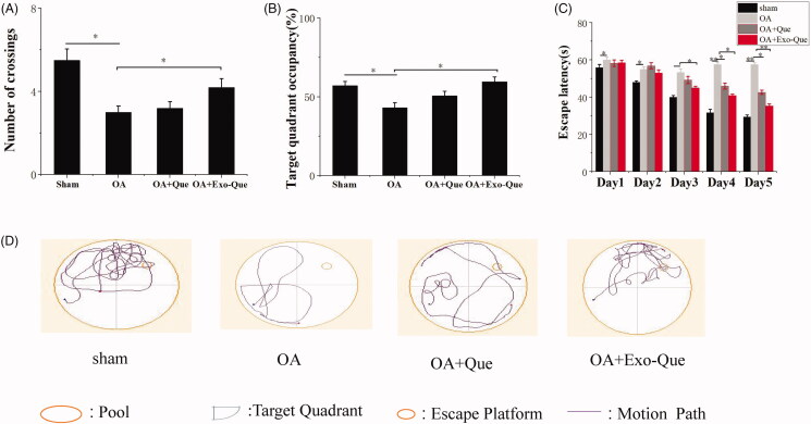
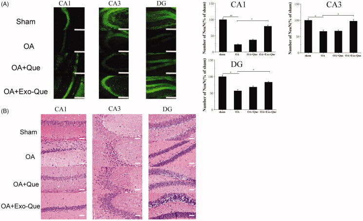
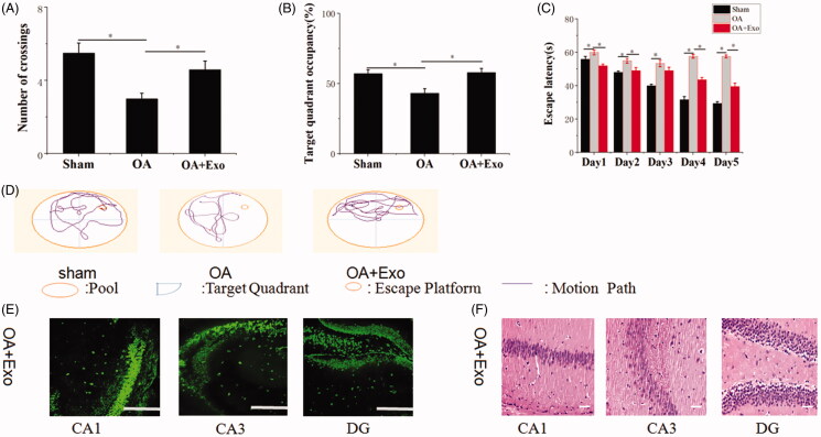
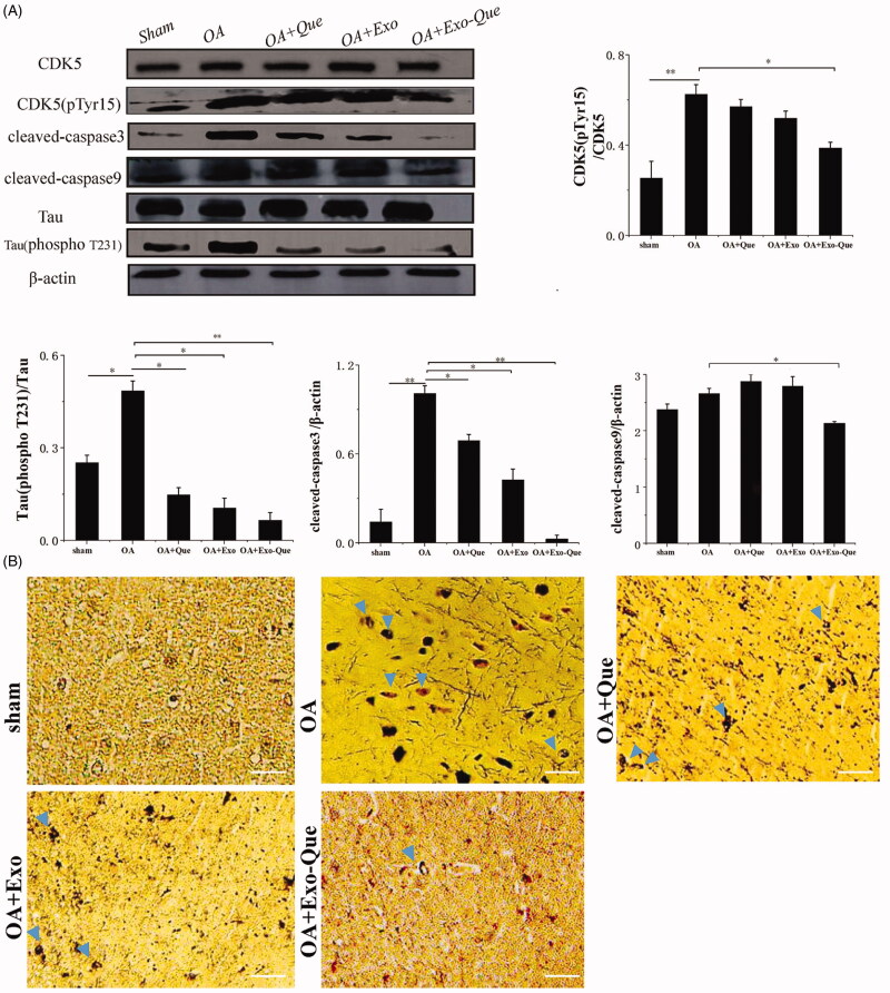
It is reported that quercetin (Que) can prevent tau pathology and induce neuroprotection by improving cognitive and functional symptoms in the treatment of Alzheimer's disease (AD). However, its clinical application has been limited due to its poor brain targeting and bioavailability. Exosomes are considered as cargo carriers for intercellular communication and especially serve as a natural and important drug brain delivery platform for achieving better treatment of central neurological diseases. Here, we developed plasma exosomes (Exo) loaded with Que (Exo-Que) to improve the drug bioavailability, enhance the brain targeting of Que and potently ameliorate cognitive dysfunction in okadaic acid (OA)-induced AD mice. Our results showed that Exo-Que improved brain targeting of Que as well as significantly enhanced bioavailability of Que. Furthermore, compared with free Que, Exo-Que better relieved the symptoms of AD by inhibiting cyclin-dependent kinase 5 (CDK5)-mediated phosphorylation of Tau and reducing formation of insoluble neurofibrillary tangles (NFTs), suggesting its therapeutic potential for better treatment of AD.
Overview
The claims and statistics of this paper haven’t been checked yet.
Run the check — free, no account needed. It reads the open-access text and checks the sources it cites.
Integrity signals
Registry
Cites
Statistics
Figures
Verification
Claims the paper makes
The claims and statistics of this paper haven’t been checked yet.
Run the check — free, no account needed. It reads the open-access text and checks the sources it cites.
Claims other papers make about this one
Each is checked against this paper’s own text: does it say what the citing paper says it does?
The source backs part of the claim, not all of it — or backs it for a different population, condition or comparison.
In the citing paper
“Exo-Que significantly enhanced Qc’s pharmacokinetic profile, increased its accumulation in the brain, and improved the memory and spatial learning of SD rats.”
From Advances in Strategies to Transport Nanoparticles Across the Blood–Brain Barrier for Drug Delivery into the Brain for the Treatment of Alzheimer’s Disease (2026) · cites this paper as [115]
Experimental — accuracy not yet measuredThe cited source says what the claim attributes to it.
In the citing paper
“Their administration was safe without systematic toxicity in vivo.”
From Advances in Strategies to Transport Nanoparticles Across the Blood–Brain Barrier for Drug Delivery into the Brain for the Treatment of Alzheimer’s Disease (2026) · cites this paper as [115]
The cited source says what the claim attributes to it.
In the citing paper
“Ultimately, exosomes without Qc exerted a synergistic neuroprotection in rescuing memory deficits.”
From Advances in Strategies to Transport Nanoparticles Across the Blood–Brain Barrier for Drug Delivery into the Brain for the Treatment of Alzheimer’s Disease (2026) · cites this paper as [115]
The cited source says what the claim attributes to it.
In the citing paper
“Moreover, it showed neuroprotective properties, suppressed the apoptosis of neurons in the hippocampus, and inhibited the formation of NFTs by limiting CDK5-mediated phosphorylation of tau protein, all these consequences derived from OA-injection.”
From Advances in Strategies to Transport Nanoparticles Across the Blood–Brain Barrier for Drug Delivery into the Brain for the Treatment of Alzheimer’s Disease (2026) · cites this paper as [115]
Self-check
Percentages, sums and reported differences re-computed from the paper’s own numbers.
The self-check needs the paper’s full text.
Citations
Cites
Lane, C. A., Hardy, J., & Schott, J. M. (2018) · European Journal of Neurology · doi:10.1111/ene.13439 · Cited by 2,616
Applying extracellular vesicles based therapeutics in clinical trials – an ISEV position paper
Lener, T., Gimona, M., Aigner, L., Börger, V., Buzas, E., Camussi, G., Chaput, N., Chatterjee, D., Court, F. A., del Portillo, H. A., et al. (2015) · Journal of Extracellular Vesicles · doi:10.3402/jev.v4.30087 · Cited by 1,491
Surface functionalized exosomes as targeted drug delivery vehicles for cerebral ischemia therapy
Tian, T., Zhang, H.-X., He, C.-P., Fan, S., Zhu, Y.-L., Qi, C., Huang, N.-P., Xiao, Z.-D., Lu, Z.-H., Tannous, B. A., et al. (2018) · Biomaterials · doi:10.1016/j.biomaterials.2017.10.012 · Cited by 1,154
The blood-brain barrier in Alzheimer’s disease
Zenaro, E., Piacentino, G., & Constantin, G. (2017) · Neurobiology of Disease · doi:10.1016/j.nbd.2016.07.007 · Cited by 767
Dopamine-loaded blood exosomes targeted to brain for better treatment of Parkinson’s disease
Qu, M., Lin, Q., Huang, L., Fu, Y., Wang, L., He, S., Fu, Y., Yang, S., Zhang, Z., Zhang, L., et al. (2018) · Journal of Controlled Release · doi:10.1016/j.jconrel.2018.08.035 · Cited by 543
Evidence-Based Clinical Use of Nanoscale Extracellular Vesicles in Nanomedicine
Fais, S., O’Driscoll, L., Borras, F. E., Buzas, E., Camussi, G., Cappello, F., Carvalho, J., Cordeiro da Silva, A., Del Portillo, H., El Andaloussi, S., et al. (2016) · ACS Nano · doi:10.1021/acsnano.5b08015 · Cited by 481
Blood-Brain Barrier Dysfunction and the Pathogenesis of Alzheimer’s Disease
Yamazaki, Y., & Kanekiyo, T. (2017) · International Journal of Molecular Sciences · doi:10.3390/ijms18091965 · Cited by 451
β-catenin mutation and overexpression in hepatocellular carcinoma
Wong, C., Fan, S. T., & Ng, I. O. (2001) · Cancer · doi:10.1002/1097-0142(20010701)92:1 · Cited by 353
Wang, H., Sui, H., Zheng, Y., Jiang, Y., Shi, Y., Liang, J., & Zhao, L. (2019) · Nanoscale · doi:10.1039/c9nr01255a · Cited by 324
Self-citationLuo, Z.-W., Li, F.-X.-Z., Liu, Y.-W., Rao, S.-S., Yin, H., Huang, J., Chen, C.-Y., Hu, Y., Zhang, Y., Tan, Y.-J., et al. (2019) · Nanoscale · doi:10.1039/c9nr02791b · Cited by 281
Cited by
Sorted by citations
Items 101–200 of 239
Phukan, B. C., Roy, R., Gahatraj, I., Bhattacharya, P., & Borah, A. (2023) · Phytotherapy Research · doi:10.1002/ptr.8012 · Cited by 22
Cellular organelles as drug carriers for disease treatment
Hu, J., Liu, Y., Du, Y., Peng, X., & Liu, Z. (2023) · Journal of Controlled Release · doi:10.1016/j.jconrel.2023.09.038 · Cited by 22
Zhang, S., Yang, Y., Lv, X., Zhou, X., Zhao, W., Meng, L., Zhu, S., Zhang, Z., & Wang, Y. (2024) · Brain Sciences · doi:10.3390/brainsci14111049 · Cited by 22
Zhang, L., Shi, C., Lan, Y., Zhang, X., Ji, X., Li, L., He, X., Tan, Y., Zhao, N., & Lu, C. C. (2025) · International Journal of Nanomedicine · doi:10.2147/ijn.s542641 · Cited by 28
Shala, A., Arduino, I., Salihu, M. B., & Denora, N. (2023) · Pharmaceutics · doi:10.3390/pharmaceutics15030963 · Cited by 21
Astrocyte-Derived Extracellular Vesicles for Ischemic Stroke: Therapeutic Potential and Prospective
Wang, X., Li, A., Fan, H., Li, Y., Yang, N., & Tang, Y. (2023) · Aging and Disease · doi:10.14336/ad.2023.0823-1 · Cited by 21
A review on the use of extracellular vesicles for the delivery of drugs and biological therapeutics
Amina, S. J., Azam, T., Dagher, F., & Guo, B. (2024) · Expert Opinion on Drug Delivery · doi:10.1080/17425247.2024.2305115 · Cited by 21
Stress-Related Roles of Exosomes and Exosomal miRNAs in Common Neuropsychiatric Disorders
Chamakioti, M., Chrousos, G. P., Kassi, E. N., Vlachakis, D., & Yapijakis, C. (2024) · International Journal of Molecular Sciences · doi:10.3390/ijms25158256 · Cited by 19
Dey, A., Ghosh, S., Rajendran, R. L., Bhuniya, T., Das, P., Bhattacharjee, B., Das, S., Mahajan, A. A., Samant, A., Anand, K., et al. (2024) · International Journal of Molecular Sciences · doi:10.3390/ijms25179690 · Cited by 20
Exosomes as Neurological Nanosized Machines
Pishavar, E., Trentini, M., Zanotti, F., Camponogara, F., Tiengo, E., Zanolla, I., Bonora, M., & Zavan, B. (2022) · ACS Nanoscience Au · doi:10.1021/acsnanoscienceau.1c00062 · Cited by 19
Basurto‐Islas, G., Diaz, M. C., Zavala-Ocampo, L. M., Martínez‐Herrera, M., & López-Camacho, P. Y. (2025) · Archiv der Pharmazie · doi:10.1002/ardp.202400721 · Cited by 20
Iglesias‐Aguirre, C. E., Ávila‐Gálvez, M. Á., Hazas, M. L. D. L., Dávalos, A., & Espı́n, J. C. (2022) · Nutrients · doi:10.3390/nu14173632 · Cited by 18
Liu, X., Geng, X., Shi, Y., Liang, J., & Zhao, L. (2023) · Biomaterials Advances · doi:10.1016/j.bioadv.2023.213379 · Cited by 18
Self-citationMishra, K., Rana, R., Tripathi, S., Siddiqui, S., Yadav, P. K., Yadav, P. N., & Chourasia, M. K. (2023) · Neurochemical Research · doi:10.1007/s11064-023-03955-3 · Cited by 18
Yang, C., Xu, T., Lu, Y., Liu, J., Chen, C., Wang, H., & Chen, X. (2024) · Neuroscience · doi:10.1016/j.neuroscience.2024.05.028 · Cited by 19
Non-pharmacological treatment of Alzheimer’s disease: an update
Wang, S., Xu, H., Liu, G., & Chen, L.-M. (2025) · Frontiers in Aging Neuroscience · doi:10.3389/fnagi.2025.1527242 · Cited by 19
Diagnostic and Therapeutic Roles of Extracellular Vesicles in Aging‐Related Diseases
Sun, Z., Hou, X., Zhang, J., Li, J., Wu, P., Yan, L., & Qian, H. (2022) · Oxidative Medicine and Cellular Longevity · doi:10.1155/2022/6742792 · Cited by 16
Tang, X., He, Y., Liu, J., Xu, J., & Peng, Q. (2024) · Colloids and Surfaces B Biointerfaces · doi:10.1016/j.colsurfb.2024.113938 · Cited by 17
The role of exosomes in diagnosis, pathophysiology, and management of Alzheimer's Disease
Daksh, R., Mathew, M. S., Bosco, A. M., Sojan, C., Tom, A. A., Bojja, S. L., & Nampoothiri, M. (2025) · Biochemical and Biophysical Research Communications · doi:10.1016/j.bbrc.2025.151526 · Cited by 16
Exosomes in Age-Related Cognitive Decline: Mechanistic Insights and Improving Outcomes
Duggan, M. R., Lu, A., Foster, T. C., Wimmer, M. E., & Parikh, V. (2022) · Frontiers in Aging Neuroscience · doi:10.3389/fnagi.2022.834775 · Cited by 15
Wei, C., Li, S., Zhu, Y., Chen, W., Li, C., & Xu, R. (2022) · Frontiers in Aging Neuroscience · doi:10.3389/fnagi.2022.902092 · Cited by 15
Raghav, A., Giri, R., Agarwal, S., Kala, S., & Jeong, G. (2022) · Frontiers in Immunology · doi:10.3389/fimmu.2022.1040027 · Cited by 16
Rondanelli, M., Riva, A., Petrangolini, G., Gasparri, C., & Perna, S. (2023) · Biomedicine & Pharmacotherapy · doi:10.1016/j.biopha.2023.115453 · Cited by 16
Recent Progress in Nano-TCM Active Ingredient Co-Delivery Systems for Inflammation-Mediated Diseases
Sun, C., Chen, J., Bai, S., Liao, W., Chen, J., Guo, W., Zeng, L., & Yu, L. (2025) · International Journal of Nanomedicine · doi:10.2147/ijn.s526731 · Cited by 16
Bibliometric and visualized analysis of the applications of exosomes based drug delivery
Wei, B., Huang, H., Cao, Q., Song, X., & Zhang, Z. (2024) · Biomedicine & Pharmacotherapy · doi:10.1016/j.biopha.2024.116803 · Cited by 14
Alhodieb, F. S., Rahman, M. A., Barkat, M. A., Alanezi, A. A., Barkat, H. A., Hadi, H. A., Harwansh, R. K., & Mittal, V. (2023) · Nanomedicine · doi:10.2217/nnm-2022-0108 · Cited by 13
Liu, X., Hao, Y., Huang, Z., Shi, Y., Su, C., & Zhao, L. (2023) · Drug Delivery and Translational Research · doi:10.1007/s13346-023-01408-6 · Cited by 13
Self-citationGao, C., Liu, Y., Zhang, T., Luo, Y., Gao, J., Chu, J., Gong, B., Chen, X., Yin, T., Zhang, J., et al. (2023) · International Journal of Nanomedicine · doi:10.2147/ijn.s436774 · Cited by 16
The landscape of extracellular vesicles combined with intranasal delivery towards brain diseases
Li, J., Zhang, H., Jiang, Y., Li, N., Zhu, A., Zhang, Y., Feng, K., Zeng, W., Di, L., & Wang, R. (2024) · Nano Today · doi:10.1016/j.nantod.2024.102169 · Cited by 15
Self-citationDeng, Z., Chen, H., Chen, J., Du, Z., Zhou, W., & Yuan, Z. (2025) · Biochimica et Biophysica Acta (BBA) - Molecular Basis of Disease · doi:10.1016/j.bbadis.2025.167960 · Cited by 14
Wang, J., Ma, X., Si, X., & Wang, H. (2023) · Journal of Biomaterials Applications · doi:10.1177/08853282231194317 · Cited by 12
Jiyah, A., Muhammad, S. A., Ibrahim, A., Bulama, I., Ibrahim, A., Abbas, A. Y., Imam, M. U., Saidu, Y., & Bilbis, L. S. (2024) · Journal of Drug Delivery Science and Technology · doi:10.1016/j.jddst.2024.105618 · Cited by 13
Surface engineering of nanoparticles for imparting multifunctionality
Choudhury, H., Gorain, B., Pandey, M., Nirmal, J., & Kesharwani, P. (2021) · Elsevier eBooks · doi:10.1016/b978-0-12-820757-4.00001-6 · Cited by 12
Huang, C., Wang, C., Lin, C., Li, H., Yen, A., & Peng, C. (2020) · BMC Complementary Medicine and Therapies · doi:10.1186/s12906-020-03163-4 · Cited by 12
Shahlaei, M., Afkhami, H., Ahmadieh-Yazdi, A., Mirmazloumi, S. H., Sahraei, S. S., Akbari, M., Yang, P., Manoochehri, H., Tanzadehpanah, H., Mahaki, H., et al. (2025) · Biomedicine & Pharmacotherapy · doi:10.1016/j.biopha.2025.118548 · Cited by 11
Abdul-Razek, N., Khalil, R. G., Abdel‐Latif, M., Kamel, M. M., Alhazza, I. M., Awad, E. M., Ebaid, H., & Abuelsaad, A. S. A. (2024) · BioNanoScience · doi:10.1007/s12668-024-01767-5 · Cited by 12
A multifactorial lens on risk factors promoting the progression of Alzheimer’s disease
Parker, J., Moris, J. M., Goodman, L. C., Paidisetty, V. K., Vanegas, V., Turner, H. A., Melgar, D., & Koh, Y. (2024) · Brain Research · doi:10.1016/j.brainres.2024.149262 · Cited by 12
Advancing Alzheimer’s disease therapy through engineered exosomal Macromolecules
Jain, S., Murmu, A., & Chauhan, A. (2025) · Brain Research · doi:10.1016/j.brainres.2025.149590 · Cited by 10
Exploring Exosome-Based Approaches for Early Diagnosis and Treatment of Neurodegenerative Diseases
Varshney, V., Gabble, B. C., Bishoyi, A. K., Varma, P., Qahtan, S. A., Kashyap, A., Panigrahi, R., Nathiya, D., & Chauhan, A. S. (2025) · Molecular Neurobiology · doi:10.1007/s12035-025-05026-w · Cited by 10
Deng, K., Chen, Z. B., Zhang, X., Li, Y., Cheng, F., Wang, Z., & Wang, C. (2025) · Materials & Design · doi:10.1016/j.matdes.2025.114243 · Cited by 12
Zhou, Y., Xiao, Z., & Zhu, W. (2022) · Frontiers in Oncology · doi:10.3389/fonc.2022.998964 · Cited by 10
Mobasheri, K., Zaefizadeh, M., Ghobeh, M., & Eidi, A. (2023) · Journal of Alzheimer s Disease · doi:10.3233/jad-221095 · Cited by 9
Ben‐Azu, B., Oghorodi, A. M., Oritsemuelebi, B., & Chidebe, E. O. (2024) · IntechOpen eBooks · doi:10.5772/intechopen.112517 · Cited by 9
Mazahir, F., Rahi, V., Kaundal, R. K., Alam, M. I., & Yadav, A. K. (2024) · Chemical Engineering Journal · doi:10.1016/j.cej.2024.158197 · Cited by 9
Dhapola, R., Kumari, S., Sharma, P., Paidlewar, M., Medhi, B., Vellingiri, B., & HariKrishnaReddy, D. (2025) · Molecular Neurobiology · doi:10.1007/s12035-025-05216-6 · Cited by 9
Tailored Extracellular Vesicles: Novel Tool for Tissue Regeneration
Li, L., Wu, P., Qian, H., Xu, W., Shi, H., & Jiang, J. (2022) · Stem Cells International · doi:10.1155/2022/7695078 · Cited by 9
Kaushik, A., Chauhan, K., & Singh, S. (2023) · Elsevier eBooks · doi:10.1016/b978-0-323-90052-2.00029-9 · Cited by 8
Afzal, M., Hameed, H., Paiva‐Santos, A. C., Saleem, M., Hameed, A., & Ahmad, S. M. (2024) · European Journal of Pharmacology · doi:10.1016/j.ejphar.2024.177199 · Cited by 8
Khalid, W., Aslam, A., Ahmed, N., Sarfraz, M., Khan, J. A., Mohsin, S., Rajoka, M. S. R., Nazir, I., & Amirzada, M. I. (2025) · AAPS PharmSciTech · doi:10.1208/s12249-024-03028-w · Cited by 10
Bianchini, M. C., Franscescon, F., Soares, A. A., Ansolin, V., Pretto, K. P., da Cunha, M. L. V., & Silva, D. T. D. R. E. (2025) · Current Neuropharmacology · doi:10.2174/011570159x355614250130100132 · Cited by 8
Al‐Sawasany, A. S., Fayed, H. M., Mahmoud, B. F., Elblehi, S. S., & Ghazal, N. A. (2025) · Journal of Biochemical and Molecular Toxicology · doi:10.1002/jbt.70290 · Cited by 8
Xu, J., He, Z., Pan, Y., Cao, B., & Chen, G. (2025) · Biomaterials Advances · doi:10.1016/j.bioadv.2025.214619 · Cited by 9
Ray, R., Chowdhury, S. G., & Karmakar, P. (2024) · Archives of Biochemistry and Biophysics · doi:10.1016/j.abb.2024.109906 · Cited by 8
Chen, Z., Yan, L., Wang, Y., Wang, Q., Yu, L., & Liu, J. (2025) · Planta Medica · doi:10.1055/a-2536-8919 · Cited by 8
Extracellular Vesicles for the Treatment of Alzheimer's Disease: A Systematic Review
Phelps, J., Orr, A., Elvira, K. S., & Willerth, S. M. (2025) · Journal of Extracellular Biology · doi:10.1002/jex2.70077 · Cited by 10
Challenges and Opportunities of Drug Delivery for Treatment of Alzheimer’s Disease
El-Fattah, M. A. A. (2026) · AAPS PharmSciTech · doi:10.1208/s12249-025-03316-z · Cited by 9
Liu, L., Liu, Y., Zhen, Y., Guo, T., Wang, C., Shen, L., & Li, W. (2022) · Open Life Sciences · doi:10.1515/biol-2021-0014 · Cited by 7
Shen, Y., Wang, M., Li, S., & Yang, J. (2023) · Journal of Materials Chemistry B · doi:10.1039/d3tb01629c · Cited by 8
Exosomes: A Cellular Communication Medium That Has Multiple Effects On Brain Diseases
Fang, X., Zhou, D., Wang, X., Ma, Y., Zhong, G., Jing, S., Huang, S., & Wang, Q. (2024) · Molecular Neurobiology · doi:10.1007/s12035-024-03957-4 · Cited by 7
Bioengineering extracellular vesicles as novel nanocarriers towards brain disorders
Wu, J., Ma, L., Sun, D., Zhang, X., Cui, J., Du, Y., Guo, Y., Wang, X., Di, L., & Wang, R. (2022) · Nano Research · doi:10.1007/s12274-022-4913-2 · Cited by 8
Ageing, Metabolic Dysfunction, and the Therapeutic Role of Antioxidants
Santos, A. L., & Sinha, S. (2023) · Sub-cellular biochemistry/Subcellular biochemistry · doi:10.1007/978-3-031-26576-1_15 · Cited by 8
Plant-based flavonoids and their nanoparticles: Latest arsenal against Alzheimer’s disease
Kumbhar, P., Kolekar, K. A., Vishwas, S., Kamble, V., Bashir, B., Patil, K. S., Bhagwat, D. A., Gupta, G., Prasher, P., Singh, M., et al. (2025) · Drug Delivery and Translational Research · doi:10.1007/s13346-025-01906-9 · Cited by 8
Bahuguna, R., Rehman, F., Awasthi, R., Babu, K. R., Akhter, M. H., Begum, M. Y., Kumar, D., Gupta, S. M., Singh, S., & Singh, P. A. (2025) · Journal of Drug Delivery Science and Technology · doi:10.1016/j.jddst.2025.107759 · Cited by 6
Grandi, R., Gulati, V., Islam, M. S., Ekpo, O. E., & Chitranshi, N. (2025) · Brain Research · doi:10.1016/j.brainres.2025.150046 · Cited by 6
Roy, R., Bhattacharya, P., & Borah, A. (2022) · Current Drug Metabolism · doi:10.2174/1389200223666220526094802 · Cited by 6
Mu, Y., Jin, T., Peng, T., Zhang, Y., Li, J., Yu, R., Zhou, T., Qing, G., Zhu, M., Chen, J., et al. (2025) · Nanoscale Horizons · doi:10.1039/d5nh00277j · Cited by 7
Roy, A., Driscoll, T. P., Grant, S. C., & Li, Y. (2025) · International Journal of Molecular Sciences · doi:10.3390/ijms262110550 · Cited by 5
Health Benefits and Therapeutic Potential of Quercetin
Aryal, M. (2026) · Journal of Food Biochemistry · doi:10.1155/jfbc/8094502 · Cited by 5
Understanding the Intricate Role of Exosomes in Pathogenesis of Alzheimer’s Disease
Chauhan, S., Behl, T., Sehgal, A., Singh, S., Sharma, N., Gupta, S., Albratty, M., Najmi, A., Meraya, A. M., & Alhazmi, H. A. (2022) · Neurotoxicity Research · doi:10.1007/s12640-022-00621-4 · Cited by 6
Extracellular Vesicles: The Invisible Heroes and Villains of COVID‐19 Central Neuropathology
Chang, H., Chen, E., Hu, Y., Wu, L., Deng, L., Ye, S., Mao, X., Zhu, T., Liu, J., & Chen, C. (2023) · Advanced Science · doi:10.1002/advs.202305554 · Cited by 4
Korun, Z. E. U., HALBUTOĞULLARI, Z. S., Yazır, Y., Rençber, S. F., Altuntaş, C., Öztürk, A., Koldankaya, T., Mert, S., & Attar, E. (2025) · Journal of Ovarian Research · doi:10.1186/s13048-025-01838-5 · Cited by 4
Biswal, B., Satapathy, B. S., Mishra, A., Maharana, L., & Pattnaik, S. (2025) · Current Drug Discovery Technologies · doi:10.2174/0115701638329824241220055621 · Cited by 4
Awad, A. S., El-Mokadem, B. M., Sherif, M. M., & Bishr, A. (2025) · Journal of Pharmacy and Pharmacology · doi:10.1093/jpp/rgaf020 · Cited by 4
Exploring exosomes to provide evidence for the treatment and prediction of Alzheimer’s disease
QUAN, X., MA, X., LI, G., FU, X., LI, J., & ZENG, L. (2023) · Biocell · doi:10.32604/biocell.2023.031226 · Cited by 4
Zhao, Q., Zheng, B., Feng, P., & Li, X. (2021) · Evidence-based Complementary and Alternative Medicine · doi:10.1155/2021/6694698 · Cited by 4
Potential Roles of Exercise and Quercetin in Modulating Cancer Pathways and Cognitive Function
Lei, P. (2025) · Phytotherapy Research · doi:10.1002/ptr.70016 · Cited by 4
Extracellular Vesicles in Alzheimer’s Disease: Mechanisms, Biomarkers, and Therapeutic Engineering
Wang, L., Mao, L., & Zong, X. (2026) · International Journal of Molecular Sciences · doi:10.3390/ijms27093974 · Cited by 4
Wu, H., Li, Y.-L., Liu, P.-M., & Yang, J. (2025) · Frontiers in Aging Neuroscience · doi:10.3389/fnagi.2025.1496252 · Cited by 3
Zhou, J., Pu, Y., Ren, X., Li, L., Chen, Z., Lo, E. H., & Li, W. (2025) · Journal of Cerebral Blood Flow & Metabolism · doi:10.1177/0271678x251348816 · Cited by 3
Flavonoids Inhibiting Tau Hyperphosphorylation: Implications for Alzheimer’s Disease
Kim, Y.-S., Park, T., Eom, D. B., Ko, M. S., Lee, C. H., & Park, S. (2025) · Natural Product Sciences · doi:10.20307/nps.2025.31.2.75 · Cited by 3
Shi, Y., Bao, Q., Chen, W., Wang, L., Peng, D., Liu, J., Liu, Q., Zhang, Y., Ji, Z., & Shen, A. (2022) · Journal of Alzheimer s Disease · doi:10.3233/jad-215666 · Cited by 3
Self-citationWang, T., & Xu, Z. (2023) · Combinatorial Chemistry & High Throughput Screening · doi:10.2174/1386207326666230119101011 · Cited by 3
Destabilization mechanism of R3–R4 tau protofilament by purpurin: a molecular dynamics study
Wan, J., Zou, Y. F., Sun, R., Xu, Z., Tang, J., Gong, Y., Wei, G., & Zhang, Q. (2023) · Physical Chemistry Chemical Physics · doi:10.1039/d3cp01039b · Cited by 4
Exosomes and tissue engineering: A novel therapeutic strategy for nerve regenerative
Nochalabadi, A., Khazaei, M., & Rezakhani, L. (2024) · Tissue and Cell · doi:10.1016/j.tice.2024.102676 · Cited by 3
Silva, R., Monteiro, J., Ramalho, M. J., Andrade, S., Loureiro, J. A., & Pereira, M. C. (2026) · Pharmaceuticals · doi:10.3390/ph19050685 · Cited by 2
“Qi et al. [115] produced plasma-derived exosomes (by gradient ultracentrifugation) packed with Qc (Exo-Que) with an average of 150 nm and tested their performance in Okadaic acid (OA)-induced AD mice.”
Hao, M., Tao, B., Luan, Y., Li, Y., Li, X., Shi, H., Wu, H., Li, Q., Liu, D., Hu, Y., et al. (2025) · Phytomedicine · doi:10.1016/j.phymed.2025.157088 · Cited by 2
Peng, Y., Wang, S.-S., Lai, K.-D., Ye, J.-R., He, W.-B., Yan, X., Zhang, Z., Chu, S.-F., & Chen, N. (2026) · Acta Pharmacologica Sinica · doi:10.1038/s41401-025-01709-1 · Cited by 4
Osman, S., Demirbolat, G. M., & Işık, S. (2026) · Inflammopharmacology · doi:10.1007/s10787-026-02236-z · Cited by 2
Noninvasive Delivery of Biologicals to the Brain
Jordan, S., Zielinski, M. A., Kortylewski, M., Kuhn, T. P., & Bystritsky, A. (2022) · FOCUS The Journal of Lifelong Learning in Psychiatry · doi:10.1176/appi.focus.20210028 · Cited by 2
Dibal, N. I., Chiroma, S. M., Attah, M. O. O., & Manye, S. J. (2023) · GAZI UNIVERSITY JOURNAL OF SCIENCE · doi:10.35378/gujs.1272285 · Cited by 2
Exosomes as nanocarrier for Neurotherapy: Journey from application to challenges
Ale, Y., & Nainwal, N. (2024) · Journal of Drug Delivery Science and Technology · doi:10.1016/j.jddst.2024.106312 · Cited by 2
Liu, Y., Cui, L., Kang, W., Liu, Z., Zhang, Y., & Ma, C. (2025) · Food Science and Human Wellness · doi:10.26599/fshw.2025.9250530 · Cited by 2
Mushtaq, T., Hameed, H., Paiva‐Santos, A. C., Tariq, U., & Hameed, A. (2025) · Molecular Neurobiology · doi:10.1007/s12035-025-05189-6 · Cited by 2
Hedna, R., Relave, E. T., Robin, M., Kovacic, H., & Breuzard, G. (2025) · European Journal of Medicinal Chemistry Reports · doi:10.1016/j.ejmcr.2025.100309 · Cited by 1
Mazahir, F., Rahi, V., Kumar, A., Kaundal, R. K., & Yadav, A. K. (2026) · Nanomedicine Nanotechnology Biology and Medicine · doi:10.1016/j.nano.2026.102934 · Cited by 1
Biomedical Applications of Medicinal Plant Extracellular Vesicles: Recent Advancements and Prospects
Lin, Z., Wu, J., Xu, G., Zhu, J., Wan, H., & He, Y. T. (2026) · Journal of Pharmaceutical Analysis · doi:10.1016/j.jpha.2026.101616 · Cited by 2
阴祖峰, Cheng, H., Liu, Y., Li, X., Yu, X., Zhang, M., Li, M., Zou, Y., & Shi, Y. (2026) · Antioxidants · doi:10.3390/antiox15050587 · Cited by 1
Self-citationZhang, C., Long, W., Ni, J., Chen, Z., Wu, X., Li, M., Du, F., Zhao, Y., Shen, J., Cho, C. H., et al. (2026) · Molecular Neurobiology · doi:10.1007/s12035-026-06002-8 · Cited by 1
Liu, Y., Yao, Z., Zhu, L., & Peng, W. (2026) · Journal of Nanobiotechnology · doi:10.1186/s12951-026-04705-7 · Cited by 1
Andreadou, E. G., Evangelopoulou, E., Tsolaki, M., Tsamesidis, I., Pessach, I., & Mitka, S. (2026) · Current Issues in Molecular Biology · doi:10.3390/cimb48080808 · Cited by 1
Metadata
- Authors
- Qi, Y., Guo, L., Jiang, Y., Shi, Y., Sui, H., & Zhao, L.
- Affiliations
- Jinzhou Medical University (CN)
- Corresponding author
- Zhao, Liang
- Venue
- Drug Delivery
- Publisher
- Informa UK Limited
- Published
- 2020-01
- Volume
- 27
- Issue
- 1
- Pages
- 745-755
- ISSN
- 1071-7544 (ISSN-L)
- 1521-0464
- PMC
- PMC7269046
- OpenAlex
- W3025860373
- Cited by
- 239
- References
- 36
- Citations by year
- 2026: 39 · 2025: 42 · 2024: 48 · 2023: 38 · 2022: 41 · 2021: 29 · 2020: 2
- FWCI
- 16.54
- Type
- journal article
- Language
- en
- Open access
- gold
- doi.org
- Licence
- Locations
- Drug Delivery (publishedVersion, cc-by)
- PubMed Central (publishedVersion, cc-by)
- PubMed (publishedVersion)
- DOAJ (DOAJ: Directory of Open Access Journals) (submittedVersion, cc-by-sa)
- Funders
- Natural Science Foundation of Liaoning Province (-ZD-)
- Topics
- Life Sciences › Neuroscience › Cellular and Molecular Neuroscience › Alzheimer's disease research and treatments
- Life Sciences › Biochemistry, Genetics and Molecular Biology › Molecular Biology › Extracellular vesicles in disease
- Physical Sciences › Materials Science › Biomaterials › Nanoparticle-Based Drug Delivery
- Keywords
- quercetin, exosomes, Alzheimer's disease, brain drug delivery, neurofibrillary tangles, tau phosphorylation, cyclin-dependent kinase 5, cognitive impairment, plasma exosomes, drug bioavailability, neuroprotection
- MeSH terms
- Alzheimer Disease, Animals, Biological Availability, Brain, Cognition, Drug Carriers, Mice, Inbred C57BL, Neurons, Phosphorylation, Quercetin, Neurofibrillary Tangles, Rats, Sprague-Dawley, Neuroprotective Agents, Maze Learning, Exosomes
About these checks
Registry, retraction, Cites and statistics results are computed, not judged, with one exception: a printed reference that no index lists is read once by a model before it counts as a citation error. Claims are extracted by a model and kept only when their quote appears verbatim in the text; each cited source is then checked, with a model’s help, for whether it says what the claim relies on. Those verdicts can be wrong — read the claim’s quote and the cited source before relying on one.
Statistics and arithmetic are recomputed, not judged.
Confidence bands are shown only for fields calibrated on our annotated test set. None is calibrated yet, so no band is shown.
What counts as a claim ›How we measure confidence ›How we check this ›
Are you an author, or do you see a problem with this page? Request a review
