

The DOI matches its Crossref record; 72 of the 73 references it cites were found in a registry or an index.
Defective autophagy in vascular smooth muscle cells accelerates senescence and promotes neointima formation and atherogenesis
Open accessAbstract
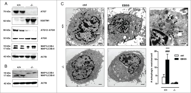
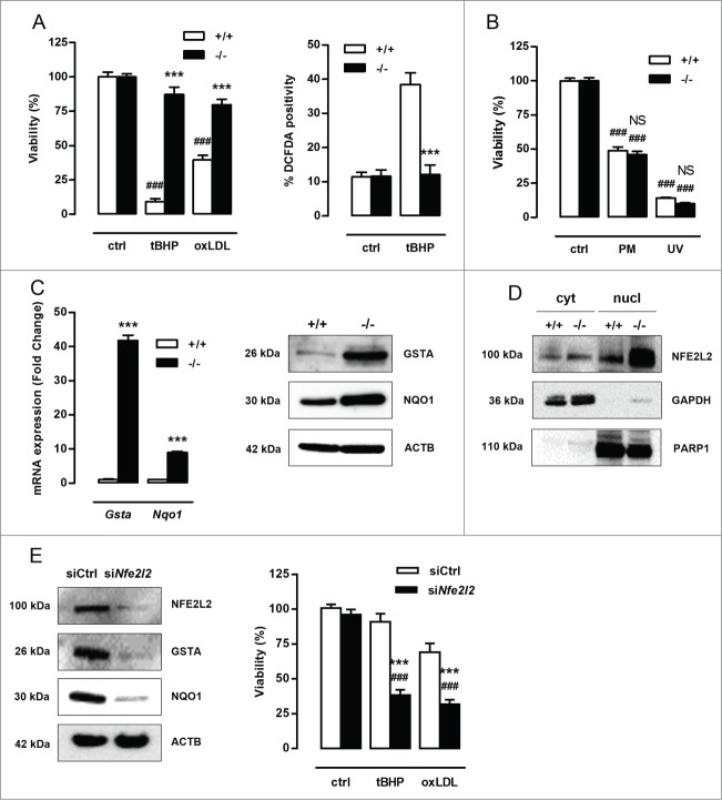
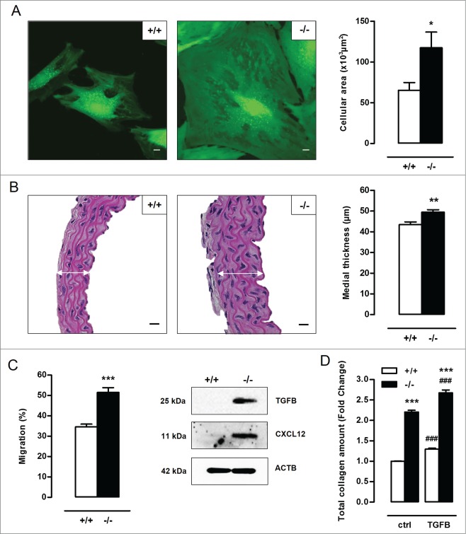
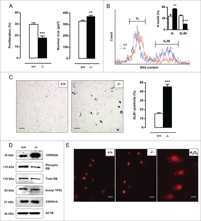
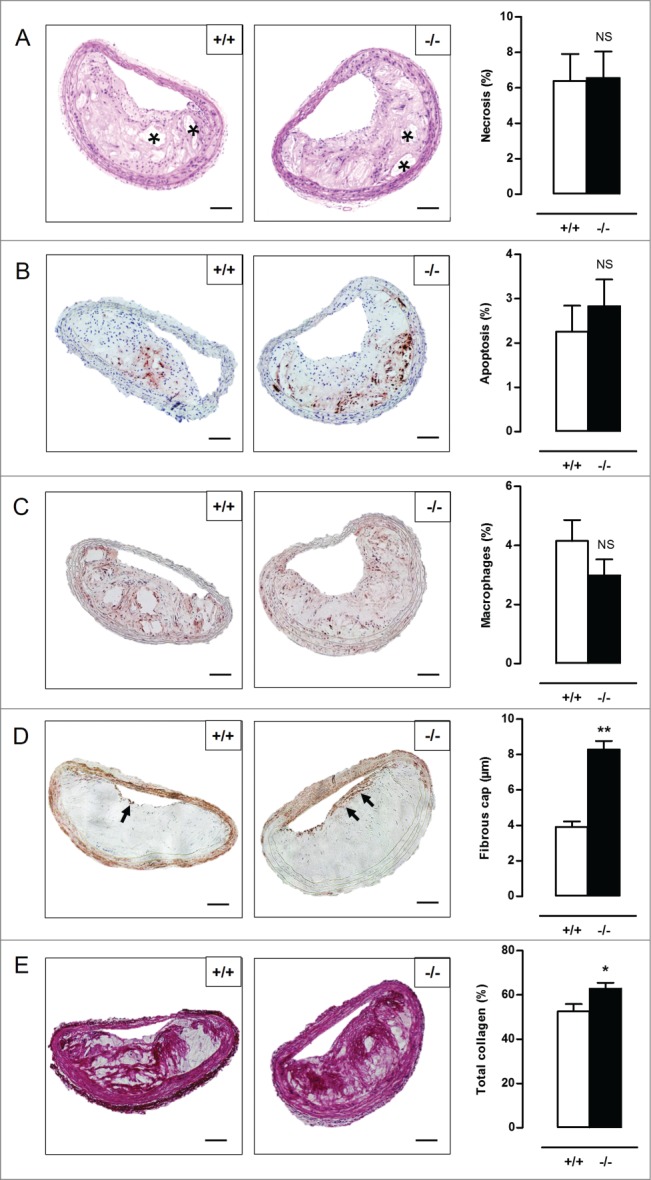
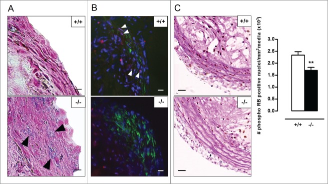
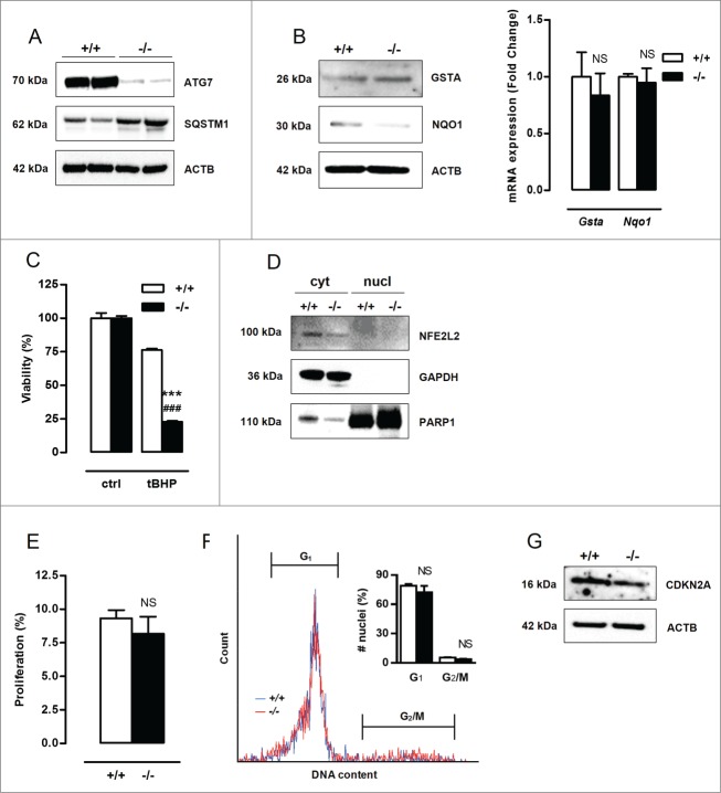
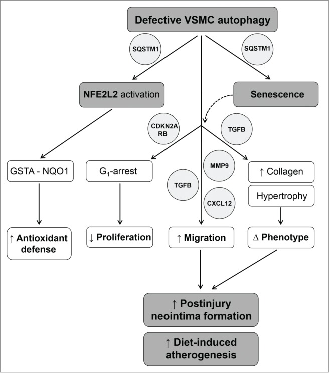
Autophagy is triggered in vascular smooth muscle cells (VSMCs) of diseased arterial vessels. However, the role of VSMC autophagy in cardiovascular disease is poorly understood. Therefore, we investigated the effect of defective autophagy on VSMC survival and phenotype and its significance in the development of postinjury neointima formation and atherosclerosis. Tissue-specific deletion of the essential autophagy gene Atg7 in murine VSMCs (atg7−/− VSMCs) caused accumulation of SQSTM1/p62 and accelerated the development of stress-induced premature senescence as shown by cellular and nuclear hypertrophy, CDKN2A-RB-mediated G1 proliferative arrest and senescence-associated GLB1 activity. Transfection of SQSTM1-encoding plasmid DNA in Atg7+/+ VSMCs induced similar features, suggesting that accumulation of SQSTM1 promotes VSMC senescence. Interestingly, atg7−/− VSMCs were resistant to oxidative stress-induced cell death as compared to controls. This effect was attributed to nuclear translocation of the transcription factor NFE2L2 resulting in upregulation of several antioxidative enzymes. In vivo, defective VSMC autophagy led to upregulation of MMP9, TGFB and CXCL12 and promoted postinjury neointima formation and diet-induced atherogenesis. Lesions of VSMC-specific atg7 knockout mice were characterized by increased total collagen deposition, nuclear hypertrophy, CDKN2A upregulation, RB hypophosphorylation, and GLB1 activity, all features typical of cellular senescence. To conclude, autophagy is crucial for VSMC function, phenotype, and survival. Defective autophagy in VSMCs accelerates senescence and promotes ligation-induced neointima formation and diet-induced atherogenesis, implying that autophagy inhibition as therapeutic strategy in the treatment of neointimal stenosis and atherosclerosis would be unfavorable. Conversely, stimulation of autophagy could be a valuable new strategy in the treatment of arterial disease.
Overview
The claims and statistics of this paper haven’t been checked yet.
Run the check — free, no account needed. It reads the open-access text and checks the sources it cites.
Integrity signals
Registry
Cites
Statistics
Figures
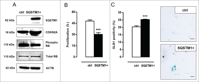
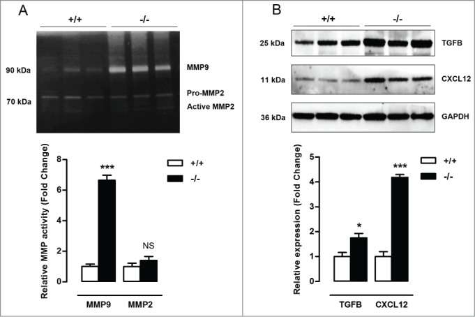
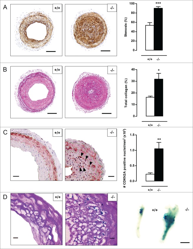
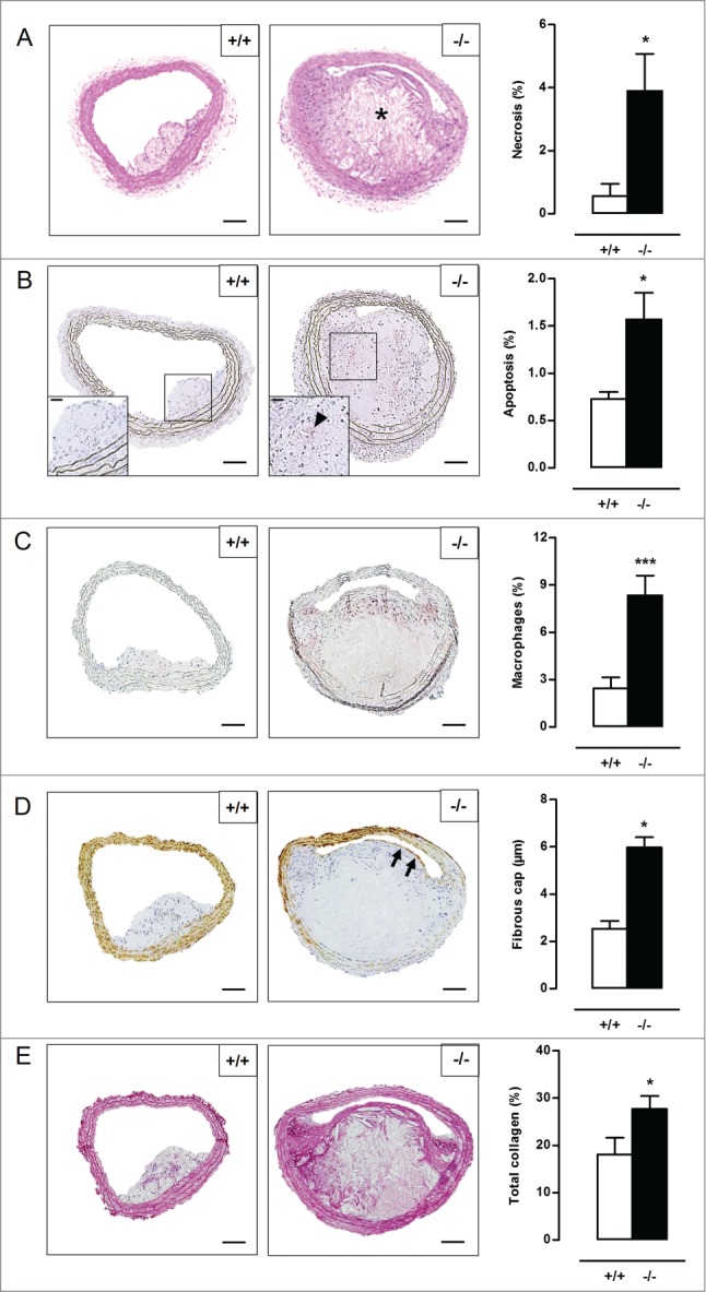
Verification
Claims the paper makes
The claims and statistics of this paper haven’t been checked yet.
Run the check — free, no account needed. It reads the open-access text and checks the sources it cites.
Claims other papers make about this one
Each is checked against this paper’s own text: does it say what the citing paper says it does?
In the citing paper
“Loss of autophagy can also drive VSMC senescence 75 .”
From Vascular smooth muscle cells in atherosclerosis (2019) · cites this paper as [75]
Self-check
Percentages, sums and reported differences re-computed from the paper’s own numbers.
The self-check needs the paper’s full text.
Citations
Cites
Repertoires of Autophagy in the Pathogenesis of Ocular Diseases
Li, Y., Jiang, Q. L., Cao, G.-F., Yao, J., & Yan, B. (2015) · Cellular Physiology and Biochemistry · doi:10.1159/000373980 · Cited by 16,351
Autophagy: Renovation of Cells and Tissues
Mizushima, N., & Komatsu, M. (2011) · Cell · doi:10.1016/j.cell.2011.10.026 · Cited by 6,615 · cited 1×
“amino acids) are recycled in order to replenish nutrient and energy supply.(2) Basal autophagy is therefore an important housekeeping and life-sustaining process.”
BackgroundCellular senescence: when bad things happen to good cells
Campisi, J., & d'Adda di Fagagna, F. (2007) · Nature Reviews Molecular Cell Biology · doi:10.1038/nrm2233 · Cited by 4,441 · cited 3×
“Cellular senescence can be established by the activation of 2 different tumor suppressor pathways: TP53-CDKN1A and CDKN2A-RB.(20,21) The TP53 pathway is triggered by DNA damage and generally associated with CDKN1A upregulation to initiate growth arrest.”
BackgroundLoss of autophagy in the central nervous system causes neurodegeneration in mice
Komatsu, M., Waguri, S., Chiba, T., Murata, S., Iwata, J.-I., Tanida, I., Ueno, T., Koike, M., Uchiyama, Y., Kominami, E., et al. (2006) · Nature · doi:10.1038/nature04723 · Cited by 3,500
Aging, Cellular Senescence, and Cancer
Campisi, J. (2013) · Annual Review of Physiology · doi:10.1146/annurev-physiol-030212-183653 · Cited by 2,954 · cited 3×
“Moreover, atg7¡/¡ VSMCs show an increase in TGFB and CXCL12 expression, which is most likely related to the increase in migration potential and the development of a senescence-associated secretory phenotype.(29,30) Cellular senescence can be established by the activation of 2 different tumor suppressor pathways: TP53-…”
BackgroundImpairment of starvation-induced and constitutive autophagy in Atg7 -deficient mice
Komatsu, M., Waguri, S., Ueno, T., Iwata, J., Murata, S., Tanida, I., Ezaki, J., Mizushima, N., Ohsumi, Y., Uchiyama, Y., et al. (2005) · The Journal of Cell Biology · doi:10.1083/jcb.200412022 · Cited by 2,331
Komatsu, M., Kurokawa, H., Waguri, S., Taguchi, K., Kobayashi, A., Ichimura, Y., Sou, Y.-S., Ueno, I., Sakamoto, A., Tong, K. I., et al. (2010) · Nature Cell Biology · doi:10.1038/ncb2021 · Cited by 2,288 · cited 4×
“SQSTM1 is considered to be a crucial activator of NFE2L2.(19) In the present study, atg7¡/¡ VSMCs showed increased nuclear translocation of NFE2L2.”
BackgroundInfluentialOxidative stress and autophagy: The clash between damage and metabolic needs
Filomeni, G., De Zio, D., & Cecconi, F. (2015) · Cell Death & Differentiation · doi:10.1038/cdd.2014.150 · Cited by 2,195 · cited 1×
“Defective autophagy in VSMCs triggers an antioxidative backup mechanism ROS production, oxidative damage and cell death are major events in cardiovascular disease(15,16) and may be regulated by autophagy.(17,18) To test the role of autophagy in VSMC survival against oxidative stress, Atg7C/C and atg7¡/¡ VSMCs were tre…”
MethodHomeostatic levels of p62 control cytoplasmic inclusion body formation in autophagy-deficient mice.
Komatsu, M., Waguri, S., Koike, M., Sou, Y.-S., Ueno, T., Hara, T., Mizushima, N., Iwata, J.-I., Ezaki, J., Murata, S., et al. (2007) · Cell · doi:10.1016/j.cell.2007.10.035 · Cited by 2,124
Rubinsztein, D. C., Mariño, G., & Kroemer, G. (2011) · Cell · doi:10.1016/j.cell.2011.07.030 · Cited by 2,107 · cited 1×
“Induction of autophagy by caloric restriction, spermidine, resveratrol or rapamycin extends life span in different organisms.(35) For example, the autophagy-inducers rapamycin and resveratrol suppress cellular senescence by partially preventing loss of proliferative potential,(36,37) whereas inhibition of autophagy by…”
Background
Research works printed but not linked: 3
- Grube E, 2004, Rev Cardiovasc Med, 5, S3
- Louis SF, 2010, Exp Clin Cardiol, 15, e75-85
- Seimon TA, 2009, J Clin Invest, 119, 886
Cited by
Sorted by citations
Vascular smooth muscle cells in atherosclerosis
Basatemur, G. L., Jørgensen, H. F., Clarke, M. C. H., Bennett, M. R., & Mallat, Z. (2019) · Nature Reviews Cardiology · doi:10.1038/s41569-019-0227-9 · Cited by 1,099
Vascular smooth muscle cell death, autophagy and senescence in atherosclerosis
Grootaert, M. O. J., Moulis, M., Roth, L., Martinet, W., Vindis, C., Bennett, M. R., & De Meyer, G. R. Y. (2018) · Cardiovascular Research · doi:10.1093/cvr/cvy007 · Cited by 606
Self-citationVascular Calcification—New Insights into Its Mechanism
Lee, S., Lee, I. K., & Jeon, J. (2020) · International Journal of Molecular Sciences · doi:10.3390/ijms21082685 · Cited by 451
Oxidative Stress-Mediated Atherosclerosis: Mechanisms and Therapies
Yang, X., Li, Y., Li, Y., Ren, X., Zhang, X., Hu, D., Gao, Y., Xing, Y., & Shang, H. (2017) · Frontiers in Physiology · doi:10.3389/fphys.2017.00600 · Cited by 418
“Atherosclerotic plaque rupture is a usual reason of the cardiovascular diseases, as stroke and myocardial infarction (Grootaert et al., 2015).”
BackgroundHong, S.-H., & Choi, K. M. (2020) · International Journal of Molecular Sciences · doi:10.3390/ijms21020494 · Cited by 390
“Atherosclerotic plaque rupture can lead to myocardial infarction and stroke [105].”
Background
Metadata
- Authors
- Grootaert, M. O., da Costa Martins, P. A., Bitsch, N., Pintelon, I., De Meyer, G. R., Martinet, W., & Schrijvers, D. M.
- Affiliations
- University of Antwerp (BE)
- Department of Cardiology; Cardiovascular Research Institute Maastricht; Maastricht, The Netherlands
- Maastricht University (NL)
- Laboratory of Physiopharmacology; University of Antwerp; Antwerp, Belgium
- Corresponding author
- Schrijvers, Dorien M
- Venue
- Autophagy
- Publisher
- Informa UK Limited
- Published
- 2015-11-02
- Volume
- 11
- Issue
- 11
- Pages
- 2014-2032
- ISSN
- 1554-8627 (ISSN-L)
- 1554-8635
- PMC
- PMC4824610
- OpenAlex
- W2200751652
- Cited by
- 267
- References
- 73
- Citations by year
- 2026: 8 · 2025: 20 · 2024: 22 · 2023: 28 · 2022: 28 · 2021: 35 · 2020: 30 · 2019: 31 · 2018: 33 · 2017: 19 · 2016: 13
- FWCI
- 17.00
- Type
- journal article
- Language
- en
- Open access
- hybrid
- doi.org
- Licence
- cc-by-nc
- Locations
- Autophagy (publishedVersion, cc-by-nc)
- Europe PMC (PubMed Central) (publishedVersion, cc-by-nc)
- PubMed Central (publishedVersion, cc-by-nc)
- PubMed (publishedVersion)
- Research Publications (Maastricht University) (submittedVersion, other-oa)
- Digital Access to Libraries (submittedVersion, other-oa)
- Funders
- Fonds Wetenschappelijk Onderzoek (G044312N)
- Vlaamse regering
- Topics
- Life Sciences › Biochemistry, Genetics and Molecular Biology › Cell Biology › Autophagy in Disease and Therapy
- Life Sciences › Immunology and Microbiology › Immunology › Atherosclerosis and Cardiovascular Diseases
- Life Sciences › Biochemistry, Genetics and Molecular Biology › Cell Biology › Telomeres, Telomerase, and Senescence
- Keywords
- vascular smooth muscle cells, cellular senescence, neointima formation, atherosclerosis, ATG7, p62, stress-induced premature senescence, Nrf2, oxidative stress resistance, GLB1, MMP-9, CXCL12, collagen deposition
About these checks
Registry, retraction, Cites and statistics results are computed, not judged, with one exception: a printed reference that no index lists is read once by a model before it counts as a citation error. Claims are extracted by a model and kept only when their quote appears verbatim in the text; each cited source is then checked, with a model’s help, for whether it says what the claim relies on. Those verdicts can be wrong — read the claim’s quote and the cited source before relying on one.
Statistics and arithmetic are recomputed, not judged.
Confidence bands are shown only for fields calibrated on our annotated test set. None is calibrated yet, so no band is shown.
What counts as a claim ›How we measure confidence ›How we check this ›
Are you an author, or do you see a problem with this page? Request a review
