

The DOI matches its Crossref record; 72 of the 73 references it cites were found in a registry or an index.
Defective autophagy in vascular smooth muscle cells accelerates senescence and promotes neointima formation and atherogenesis
Open accessAbstract
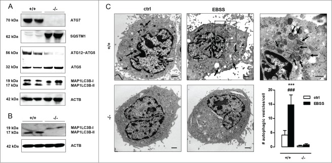
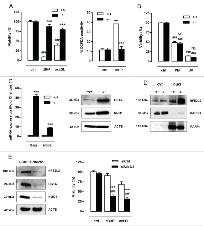
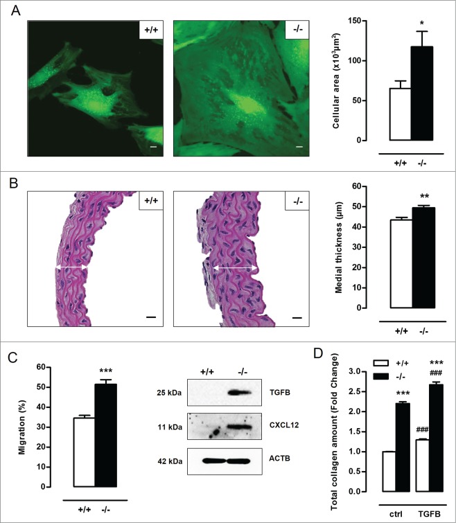
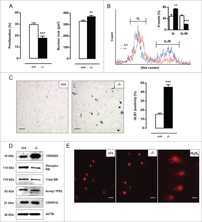
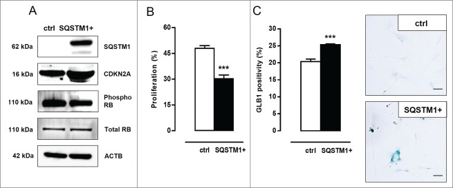
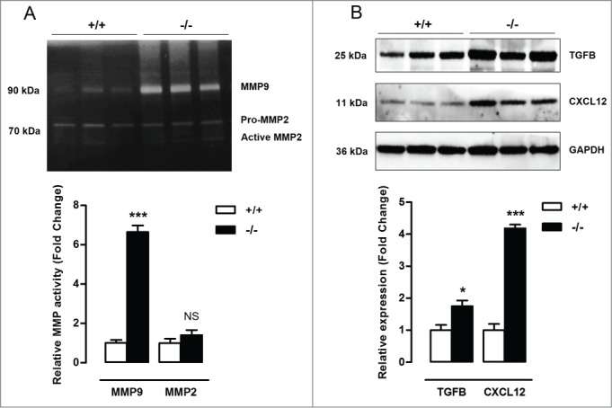
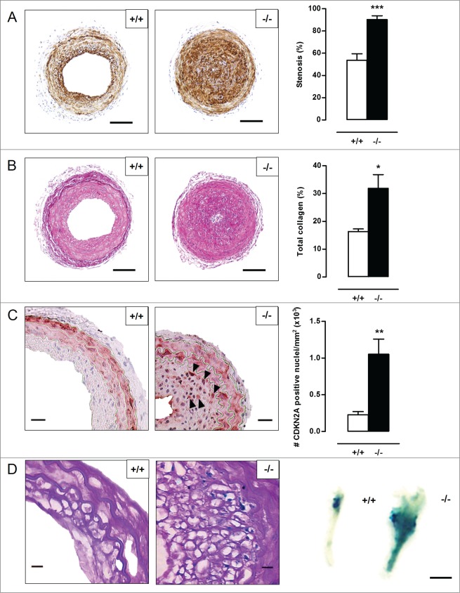
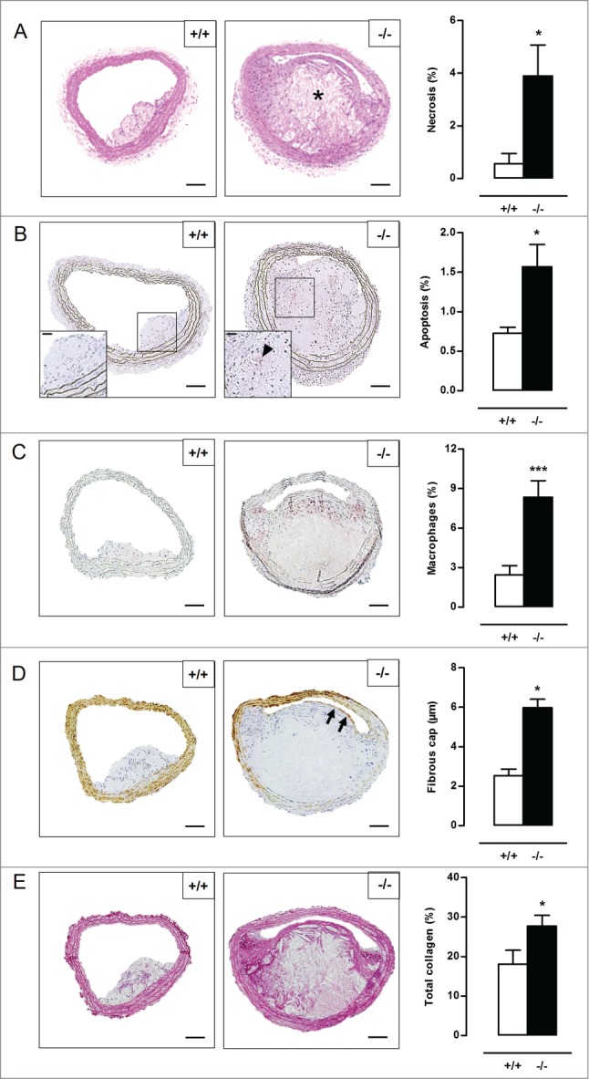
Autophagy is triggered in vascular smooth muscle cells (VSMCs) of diseased arterial vessels. However, the role of VSMC autophagy in cardiovascular disease is poorly understood. Therefore, we investigated the effect of defective autophagy on VSMC survival and phenotype and its significance in the development of postinjury neointima formation and atherosclerosis. Tissue-specific deletion of the essential autophagy gene Atg7 in murine VSMCs (atg7−/− VSMCs) caused accumulation of SQSTM1/p62 and accelerated the development of stress-induced premature senescence as shown by cellular and nuclear hypertrophy, CDKN2A-RB-mediated G1 proliferative arrest and senescence-associated GLB1 activity. Transfection of SQSTM1-encoding plasmid DNA in Atg7+/+ VSMCs induced similar features, suggesting that accumulation of SQSTM1 promotes VSMC senescence. Interestingly, atg7−/− VSMCs were resistant to oxidative stress-induced cell death as compared to controls. This effect was attributed to nuclear translocation of the transcription factor NFE2L2 resulting in upregulation of several antioxidative enzymes. In vivo, defective VSMC autophagy led to upregulation of MMP9, TGFB and CXCL12 and promoted postinjury neointima formation and diet-induced atherogenesis. Lesions of VSMC-specific atg7 knockout mice were characterized by increased total collagen deposition, nuclear hypertrophy, CDKN2A upregulation, RB hypophosphorylation, and GLB1 activity, all features typical of cellular senescence. To conclude, autophagy is crucial for VSMC function, phenotype, and survival. Defective autophagy in VSMCs accelerates senescence and promotes ligation-induced neointima formation and diet-induced atherogenesis, implying that autophagy inhibition as therapeutic strategy in the treatment of neointimal stenosis and atherosclerosis would be unfavorable. Conversely, stimulation of autophagy could be a valuable new strategy in the treatment of arterial disease.
Overview
The claims and statistics of this paper haven’t been checked yet.
Run the check — free, no account needed. It reads the open-access text and checks the sources it cites.
Integrity signals
Registry
Cites
Statistics
Figures
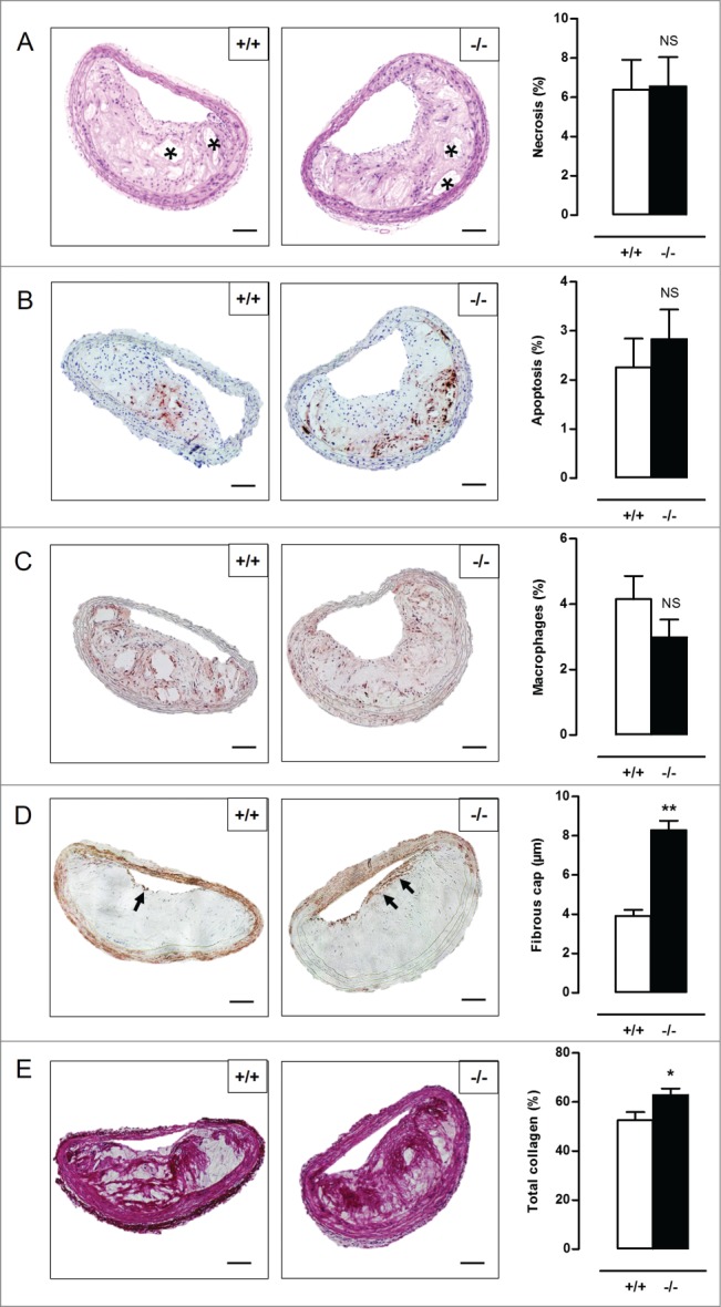
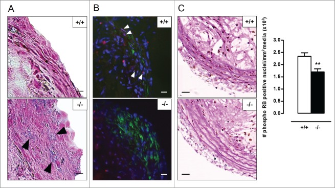
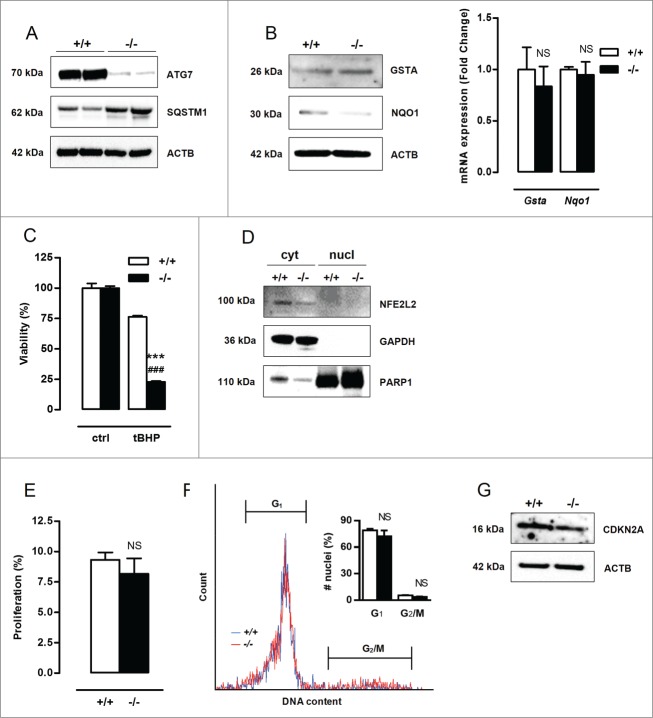
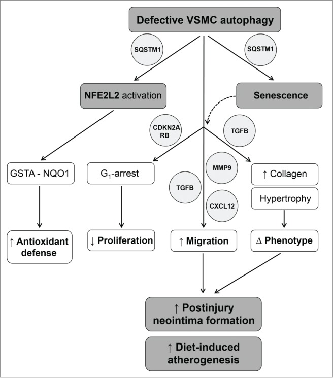
Verification
Claims the paper makes
The claims and statistics of this paper haven’t been checked yet.
Run the check — free, no account needed. It reads the open-access text and checks the sources it cites.
Claims other papers make about this one
Each is checked against this paper’s own text: does it say what the citing paper says it does?
In the citing paper
“Loss of autophagy can also drive VSMC senescence 75 .”
From Vascular smooth muscle cells in atherosclerosis (2019) · cites this paper as [75]
Self-check
Percentages, sums and reported differences re-computed from the paper’s own numbers.
The self-check needs the paper’s full text.
Citations
Cites
Repertoires of Autophagy in the Pathogenesis of Ocular Diseases
Li, Y., Jiang, Q. L., Cao, G.-F., Yao, J., & Yan, B. (2015) · Cellular Physiology and Biochemistry · doi:10.1159/000373980 · Cited by 16,351
Autophagy: Renovation of Cells and Tissues
Mizushima, N., & Komatsu, M. (2011) · Cell · doi:10.1016/j.cell.2011.10.026 · Cited by 6,615 · cited 1×
“amino acids) are recycled in order to replenish nutrient and energy supply.(2) Basal autophagy is therefore an important housekeeping and life-sustaining process.”
BackgroundCellular senescence: when bad things happen to good cells
Campisi, J., & d'Adda di Fagagna, F. (2007) · Nature Reviews Molecular Cell Biology · doi:10.1038/nrm2233 · Cited by 4,441 · cited 3×
“Cellular senescence can be established by the activation of 2 different tumor suppressor pathways: TP53-CDKN1A and CDKN2A-RB.(20,21) The TP53 pathway is triggered by DNA damage and generally associated with CDKN1A upregulation to initiate growth arrest.”
BackgroundLoss of autophagy in the central nervous system causes neurodegeneration in mice
Komatsu, M., Waguri, S., Chiba, T., Murata, S., Iwata, J.-I., Tanida, I., Ueno, T., Koike, M., Uchiyama, Y., Kominami, E., et al. (2006) · Nature · doi:10.1038/nature04723 · Cited by 3,500
Aging, Cellular Senescence, and Cancer
Campisi, J. (2013) · Annual Review of Physiology · doi:10.1146/annurev-physiol-030212-183653 · Cited by 2,954 · cited 3×
“Moreover, atg7¡/¡ VSMCs show an increase in TGFB and CXCL12 expression, which is most likely related to the increase in migration potential and the development of a senescence-associated secretory phenotype.(29,30) Cellular senescence can be established by the activation of 2 different tumor suppressor pathways: TP53-…”
BackgroundImpairment of starvation-induced and constitutive autophagy in Atg7 -deficient mice
Komatsu, M., Waguri, S., Ueno, T., Iwata, J., Murata, S., Tanida, I., Ezaki, J., Mizushima, N., Ohsumi, Y., Uchiyama, Y., et al. (2005) · The Journal of Cell Biology · doi:10.1083/jcb.200412022 · Cited by 2,331
Komatsu, M., Kurokawa, H., Waguri, S., Taguchi, K., Kobayashi, A., Ichimura, Y., Sou, Y.-S., Ueno, I., Sakamoto, A., Tong, K. I., et al. (2010) · Nature Cell Biology · doi:10.1038/ncb2021 · Cited by 2,288 · cited 4×
“SQSTM1 is considered to be a crucial activator of NFE2L2.(19) In the present study, atg7¡/¡ VSMCs showed increased nuclear translocation of NFE2L2.”
BackgroundInfluentialOxidative stress and autophagy: The clash between damage and metabolic needs
Filomeni, G., De Zio, D., & Cecconi, F. (2015) · Cell Death & Differentiation · doi:10.1038/cdd.2014.150 · Cited by 2,195 · cited 1×
“Defective autophagy in VSMCs triggers an antioxidative backup mechanism ROS production, oxidative damage and cell death are major events in cardiovascular disease(15,16) and may be regulated by autophagy.(17,18) To test the role of autophagy in VSMC survival against oxidative stress, Atg7C/C and atg7¡/¡ VSMCs were tre…”
MethodHomeostatic levels of p62 control cytoplasmic inclusion body formation in autophagy-deficient mice.
Komatsu, M., Waguri, S., Koike, M., Sou, Y.-S., Ueno, T., Hara, T., Mizushima, N., Iwata, J.-I., Ezaki, J., Murata, S., et al. (2007) · Cell · doi:10.1016/j.cell.2007.10.035 · Cited by 2,124
Rubinsztein, D. C., Mariño, G., & Kroemer, G. (2011) · Cell · doi:10.1016/j.cell.2011.07.030 · Cited by 2,107 · cited 1×
“Induction of autophagy by caloric restriction, spermidine, resveratrol or rapamycin extends life span in different organisms.(35) For example, the autophagy-inducers rapamycin and resveratrol suppress cellular senescence by partially preventing loss of proliferative potential,(36,37) whereas inhibition of autophagy by…”
BackgroundThe machinery of macroautophagy
Feng, Y., He, D., Yao, Z., & Klionsky, D. J. (2014) · Cell Research · doi:10.1038/cr.2013.168 · Cited by 2,083 · cited 1×
“During this process, fragments of cytoplasmic material are sequestered in double-membrane vesicles, termed autophagosomes, and delivered to lysosomes for degradation.(1) In this way, essential precursor molecules (e.”
BackgroundCellular senescence and the senescent secretory phenotype: therapeutic opportunities
Tchkonia, T., Zhu, Y., van Deursen, J., Campisi, J., & Kirkland, J. L. (2013) · Journal of Clinical Investigation · doi:10.1172/jci64098 · Cited by 1,774 · cited 2×
“Moreover, atg7¡/¡ VSMCs show an increase in TGFB and CXCL12 expression, which is most likely related to the increase in migration potential and the development of a senescence-associated secretory phenotype.(29,30) Cellular senescence can be established by the activation of 2 different tumor suppressor pathways: TP53-…”
BackgroundInk4a/Arf expression is a biomarker of aging
Krishnamurthy, J., Torrice, C., Ramsey, M. R., Kovalev, G. I., Al-Regaiey, K., Su, L., & Sharpless, N. E. (2004) · Journal of Clinical Investigation · doi:10.1172/jci22475 · Cited by 1,486
Angiotensin II induces hypertrophy, not hyperplasia, of cultured rat aortic smooth muscle cells.
Geisterfer, A. A., Peach, M. J., & Owens, G. K. (1988) · Circulation Research · doi:10.1161/01.res.62.4.749 · Cited by 1,059 · cited 1×
“Cell culture VSMCs were isolated from mouse aorta as previously described.(62,63) Briefly, aortas were preincubated in HBSS (Gibco Life Technologies, 14025-050) containing 1 mg/ml collagenase (Worthington, type II CLS, 4176), 1 mg/ml soybean trypsin inhibitor (Worthington, 3571) and 0.”
MethodWang (2012) · doi:10.1161/circresaha.111.261388 · Cited by 957 · cited 1×
“The CDKN2A-RB (retinoblastoma) pathway is engaged when cells are exposed to cellular stress, leading to the development of stress-induced premature senescence (SIPS).(31) During SIPS, the increased expression of CDKN2A leads to hypophosphorylation and activation of RB, resulting in prevention of transcription of proli…”
BackgroundLau, A., Wang, X.-J., Zhao, F. L., Villeneuve, N. F., Wu, T., Jiang, T., Sun, Z., White, E., & Zhang, D. D. (2010) · Molecular and Cellular Biology · doi:10.1128/mcb.00248-10 · Cited by 852 · cited 1×
“However, when SQSTM1 accumulates, it will interfere with the NFE2L2KEAP1 binding complex by sequestrating KEAP1 into aggregates.(19,24) As a result, NFE2L2 is released from its inhibitor and translocated to the nucleus where it promotes transcription of multiple antioxidative genes by binding to the antioxidant respon…”
BackgroundLoss of Autophagy Diminishes Pancreatic β Cell Mass and Function with Resultant Hyperglycemia
Jung, H. S., Chung, K. W., Won Kim, J., Kim, J., Komatsu, M., Tanaka, K., Nguyen, Y. H., Kang, T. M., Yoon, K.-H., Kim, J.-W., et al. (2008) · Cell Metabolism · doi:10.1016/j.cmet.2008.08.013 · Cited by 647
Macrophage Autophagy Plays a Protective Role in Advanced Atherosclerosis
Liao, X., Sluimer, J. C., Wang, Y., Subramanian, M., Brown, K., Pattison, J. S., Robbins, J., Martinez, J., & Tabas, I. (2012) · Cell Metabolism · doi:10.1016/j.cmet.2012.01.022 · Cited by 627 · cited 1×
“atg7¡/¡ macrophages revealed neither NFE2L2 activation nor elevated phase II enzyme expression, which is in line with previous reports showing that various cell types including hepatic cells, neurons, pancreatic b cells, and macrophages are more susceptible to (oxidative stressmediated) cell death under defective auto…”
ResultMatthews, C., Gorenne, I., Scott, S., Figg, N., Kirkpatrick, P., Ritchie, A., Goddard, M., & Bennett, M. (2006) · Circulation Research · doi:10.1161/01.res.0000233315.38086.bc · Cited by 615 · cited 1×
“Different research groups have shown that VSMC senescence is present in atherosclerotic plaques and contributes to the pathogenesis of atherosclerosis.(21,32,53,54) Our observations confirm once more the dual role of VSMCs in atherosclerosis.”
BackgroundVascular Cell Senescence: Contribution to Atherosclerosis
Minamino, T., & Komuro, I. (2007) · Circulation Research · doi:10.1161/01.res.0000256837.40544.4a · Cited by 542
Newby, A. C. (2005) · Cardiovascular Research · doi:10.1016/j.cardiores.2005.08.002 · Cited by 530 · cited 1×
“Matrix metalloproteinases facilitate migration of VSMCs by remodeling the extracellular matrix.(44) In particular MMP9 and MMP2 are upregulated in response to vascular injury and to trigger intimal thickening.”
BackgroundPhagocytosis of apoptotic cells by macrophages is impaired in atherosclerosis
Schrijvers, D. M., De Meyer, G. R. Y., Kockx, M. M., Herman, A. G., & Martinet, W. (2005) · Arteriosclerosis Thrombosis and Vascular Biology · doi:10.1161/01.atv.0000166517.18801.a7 · Cited by 509 · cited 1×
“oxLDL was prepared as previously described.(64) In some experiments, cells were treated with 10 mmol/l puromycin (Sigma Aldrich, P8833) for 12 h or exposed to UV-irradiation for 10 min.”
MethodOwens, G. K., Loeb, A. L., Gordon, D., & Thompson, M. M. (1986) · The Journal of Cell Biology · doi:10.1083/jcb.102.2.343 · Cited by 412 · cited 1×
“Cell culture VSMCs were isolated from mouse aorta as previously described.(62,63) Briefly, aortas were preincubated in HBSS (Gibco Life Technologies, 14025-050) containing 1 mg/ml collagenase (Worthington, type II CLS, 4176), 1 mg/ml soybean trypsin inhibitor (Worthington, 3571) and 0.”
MethodRapamycin decelerates cellular senescence
Demidenko, Z. N., Zubova, S. G., Bukreeva, E. I., Pospelov, V. A., Pospelova, T. V., & Blagosklonny, M. V. (2009) · Cell Cycle · doi:10.4161/cc.8.12.8606 · Cited by 391
Martinet, W., & Meyer, G. R. Y. D. (2009) · Circulation Research · doi:10.1161/circresaha.108.188318 · Cited by 390
Self-citationZernecke, A., Schober, A., Bot, I., von Hundelshausen, P., Liehn, E. A., Moepps, B., Mericskay, M., Gierschik, P., Biessen, E. A. L., & Weber, C. (2005) · Circulation Research · doi:10.1161/01.res.0000162100.52009.38 · Cited by 364 · cited 1×
“CXCL12 is an important chemoattractant that is released by VSMCs after mechanical injury and triggers the recruitment of SMC progenitor cells to the lesion.(51,52) All together, the excessive neointimal thickening in atg7¡/¡ mice could be attributed to a combination of factors: cellular hypertrophy of neointimal VSMCs…”
BackgroundAutophagy in cardiovascular biology
Lavandero, S., Chiong, M., Rothermel, B. A., & Hill, J. A. (2015) · Journal of Clinical Investigation · doi:10.1172/jci73943 · Cited by 331
Jia, G., Cheng, G., Gangahar, D. M., & Agrawal, D. K. (2006) · Immunology and Cell Biology · doi:10.1111/j.1440-1711.2006.01454.x · Cited by 306
Meyer, G. R. Y. D., Grootaert, M. O. J., Michiels, C. F., Kurdi, A., Schrijvers, D. M., & Martinet, W. (2015) · Circulation Research · doi:10.1161/circresaha.116.303804 · Cited by 292
Self-citationCollagen synthesis in atherosclerosis: too much and not enough
Rekhter, M. (1999) · Cardiovascular Research · doi:10.1016/s0008-6363(98)00321-6 · Cited by 280
Autophagy Impairment Induces Premature Senescence in Primary Human Fibroblasts
Kang, H. T., Lee, K. B., Kim, S. Y., Choi, H. R., & Park, S. C. (2011) · PLoS ONE · doi:10.1371/journal.pone.0023367 · Cited by 254 · cited 1×
“For example, the autophagy-inducers rapamycin and resveratrol suppress cellular senescence by partially preventing loss of proliferative potential,(36,37) whereas inhibition of autophagy by knockdown of ATG7 or ATG12 induces premature senescence.(38) In the present study, we dissected out the molecular link between de…”
BackgroundCellular senescence in vivo: Its relevance in ageing and cardiovascular disease
Erusalimsky, J. D., & Kurz, D. J. (2005) · Experimental Gerontology · doi:10.1016/j.exger.2005.04.010 · Cited by 252 · cited 2×
“Different research groups have shown that VSMC senescence is present in atherosclerotic plaques and contributes to the pathogenesis of atherosclerosis.(21,32,53,54) Our observations confirm once more the dual role of VSMCs in atherosclerosis.”
BackgroundFujii, S., Hara, H., Araya, J., Takasaka, N., Kojima, J., Ito, S., Minagawa, S., Yumino, Y., Ishikawa, T., Numata, T., et al. (2012) · OncoImmunology · doi:10.4161/onci.20297 · Cited by 246
Vascular smooth muscle cell sirtuin 1 protects against DNA damage and inhibits atherosclerosis
Gorenne, I., Kumar, S., Gray, K., Figg, N., Yu, H., Mercer, J., & Bennett, M. (2013) · Circulation · doi:10.1161/circulationaha.112.124404 · Cited by 238 · cited 1×
“VSMC-specific deletion of SIRT1 deacetylase in apoe¡/¡ mice promotes atherosclerosis associated with increased DNA damage and senescence.(60) Finally, it is worthwhile to mention that MTOR (mechanistic target of rapamycin [serine/threonine kinase])-inhibitors such as rapamycin (or rapalogs) are able to stimulate autop…”
BackgroundSalabei, J. K., Cummins, T. D., Singh, M., Jones, S. P., Bhatnagar, A., & Hill, B. G. (2013) · Biochemical Journal · doi:10.1042/bj20121344 · Cited by 228
Schober, A., Knarren, S., Lietz, M., Lin, E. A., & Weber, C. (2003) · Circulation · doi:10.1161/01.cir.0000099508.76665.9a · Cited by 198 · cited 1×
“CXCL12 is an important chemoattractant that is released by VSMCs after mechanical injury and triggers the recruitment of SMC progenitor cells to the lesion.(51,52) All together, the excessive neointimal thickening in atg7¡/¡ mice could be attributed to a combination of factors: cellular hypertrophy of neointimal VSMCs…”
BackgroundVascular smooth muscle cell senescence in atherosclerosis
GORENNE, I., KAVURMA, M., SCOTT, S., & BENNETT, M. (2006) · Cardiovascular Research · doi:10.1016/j.cardiores.2006.06.004 · Cited by 196 · cited 1×
“Different research groups have shown that VSMC senescence is present in atherosclerotic plaques and contributes to the pathogenesis of atherosclerosis.(21,32,53,54) Our observations confirm once more the dual role of VSMCs in atherosclerosis.”
BackgroundVascular smooth muscle cell motility: From migration to invasion.
Louis, S. F., & Zahradka, P. C. (2010) · PubMed Central · Cited by 193
Godin, D., Ivan, E., Johnson, C., Magid, R., & Galis, Z. S. (2000) · Circulation · doi:10.1161/01.cir.102.23.2861 · Cited by 192
mTOR inhibition: A promising strategy for stabilization of atherosclerotic plaques
Martinet, W., Loof, H. D., & Meyer, G. R. Y. D. (2014) · Atherosclerosis · doi:10.1016/j.atherosclerosis.2014.01.040 · Cited by 190 · cited 1×
“Finally, it is worthwhile to mention that MTOR (mechanistic target of rapamycin [serine/threonine kinase])-inhibitors such as rapamycin (or rapalogs) are able to stimulate autophagy and have significant promise to treat patients with unstable plaques, particularly in combination with statins or metformin.(61) In concl…”
BackgroundSelf-citationSchrijvers, D. M., Meyer, G. R. Y. D., & Martinet, W. (2011) · Arteriosclerosis Thrombosis and Vascular Biology · doi:10.1161/atvbaha.111.224899 · Cited by 189
Self-citationUnsaturated lipid peroxidation-derived aldehydes activate autophagy in vascular smooth-muscle cells
Hill, B. G., Haberzettl, P., Ahmed, Y., Srivastava, S., & Bhatnagar, A. (2008) · Biochemical Journal · doi:10.1042/bj20071063 · Cited by 187
Takasaka, N., Araya, J., Hara, H., Ito, S., Kobayashi, K., Kurita, Y., Wakui, H., Yoshii, Y., Yumino, Y., Fujii, S., et al. (2013) · The Journal of Immunology · doi:10.4049/jimmunol.1302341 · Cited by 182 · cited 1×
“Indeed, autophagy is an important homeostatic regulator by eliminating damaged intracellular components and may play a critical role in the prevention of cellular senescence.(42) When autophagy is impaired, however, senescence can be engaged as a backup mechanism to protect the cell.”
BackgroundMartinet, W., Knaapen, M., Meyer, G. R. Y. D., Herman, A. G., & Kockx, M. M. (2001) · Circulation Research · doi:10.1161/hh0701.088684 · Cited by 171 · cited 1×
“To detect DNA damage, a comet assay was performed as previously described.(69)”
MethodSelf-citationAutophagy and senescence: A partnership in search of definition
Gewirtz, D. A. (2013) · Autophagy · doi:10.4161/auto.23922 · Cited by 166
Seimon, T. A., Wang, Y., Han, S., Senokuchi, T., Schrijvers, D. M., Kuriakose, G., Tall, A. R., & Tabas, I. A. (2009) · Journal of Clinical Investigation · doi:10.1172/jci37262 · Cited by 155 · cited 1×
“A 3000 mm(2) minimum threshold was implemented in order to avoid counting of regions that likely do not represent substantial areas of necrosis.(70) Lesions were further analyzed by immunohistochemistry with the following primary antibodies: rabbit anti-LAMP2 (BD PharMingen, 553322), rabbit anti-cleaved CASP3 (Cell Si…”
MethodSelf-citationDing, Z., Wang, X., Schnackenberg, L. K., Khaidakov, M., Liu, S., Singla, S., Dai, Y., & Mehta, J. L. (2013) · International Journal of Cardiology · doi:10.1016/j.ijcard.2012.12.045 · Cited by 154
The Role of Oxidative Stress and Autophagy in Atherosclerosis
Perrotta, I. D., & Aquila, S. (2015) · Oxidative Medicine and Cellular Longevity · doi:10.1155/2015/130315 · Cited by 148 · cited 1×
“Defective autophagy in VSMCs triggers an antioxidative backup mechanism ROS production, oxidative damage and cell death are major events in cardiovascular disease(15,16) and may be regulated by autophagy.(17,18) To test the role of autophagy in VSMC survival against oxidative stress, Atg7C/C and atg7¡/¡ VSMCs were tre…”
MethodJohnson, J. L. (2014) · Cardiovascular Research · doi:10.1093/cvr/cvu171 · Cited by 140 · cited 1×
“Although VSMCs are considered beneficial in terms of plaque stability due to the formation of a thick fibrous cap that safeguards plaques from rupturing, VSMCs also play a fundamental role in promoting plaque development.(55) Figure 9.”
BackgroundMouse models of angiogenesis, arterial stenosis, atherosclerosis and hemostasis
Carmeliet, P. (1998) · Cardiovascular Research · doi:10.1016/s0008-6363(98)00108-4 · Cited by 129
Martinet, W., de Bie, M., Schrijvers, D. M., Meyer, G. R. Y. D., Herman, A. G., & Kockx, M. M. (2004) · Arteriosclerosis Thrombosis and Vascular Biology · doi:10.1161/01.atv.0000146266.65820.a1 · Cited by 126
Self-citationAt concentrations that inhibit mTOR, resveratrol suppresses cellular senescence
Demidenko, Z. N., & Blagosklonny, M. V. (2009) · Cell Cycle · doi:10.4161/cc.8.12.8810 · Cited by 124
7-Ketocholesterol Induces Autophagy in Vascular Smooth Muscle Cells through Nox4 and Atg4B
He, C., Zhu, H., Zhang, W., Okon, I. S., Wang, Q., Li, H., Le, Y.-Z., & Xie, Z. (2013) · American Journal Of Pathology · doi:10.1016/j.ajpath.2013.04.028 · Cited by 123
Guo, Z., Mitchell-Raymundo, F., Yang, H., Ikeno, Y., Nelson, J., Diaz, V., Richardson, A., & Reddick, R. (2002) · Mechanisms of Ageing and Development · doi:10.1016/s0047-6374(02)00008-8 · Cited by 121 · cited 1×
“Caloric restriction for example, is a powerful tool to induce autophagy and has both life-span-prolonging and antiatherogenic properties in mice.(59) Also SIRT1 (sirtuin 1) deacetylase, an inducer of autophagy and a negative regulator of cellular aging, plays a protective role in atherosclerosis.”
BackgroundImplications of autophagy for vascular smooth muscle cell function and plasticity
Salabei, J. K., & Hill, B. G. (2013) · Free Radical Biology and Medicine · doi:10.1016/j.freeradbiomed.2013.08.003 · Cited by 108 · cited 1×
“Moreover, plateletderived growth factor-induced autophagy promotes the development of a synthetic and proliferative VSMC phenotype,(8) suggesting that autophagy may regulate VSMC phenotype and function.(10) Because our knowledge of the regulation of autophagy in VSMCs is mainly based on cell culture experiments, we fo…”
BackgroundUnderstanding the role of transforming growth factor-β1 in intimal thickening after vascular injury
KHAN, R., Agrotis, A., & Bobik, A. (2007) · Cardiovascular Research · doi:10.1016/j.cardiores.2007.02.012 · Cited by 104 · cited 1×
“TGFB plays a crucial role in intimal thickening and may stimulate MMP9 and MMP2 expression after arterial injury which in turn may augment the bioavailability of TGFB, thereby creating a positive feedback loop promoting intimal thickening.(47) Given that TGFB is also involved in collagen synthesis, RB-dependent G1-gro…”
BackgroundReactive Oxygen Species and Death
Bennett, M. R. (2001) · Circulation Research · doi:10.1161/hh0701.089955 · Cited by 103
Cell senescence: Hypertrophic arrest beyond the restriction point
Blagosklonny, M. V. (2006) · Journal of Cellular Physiology · doi:10.1002/jcp.20750 · Cited by 97 · cited 1×
“capacity due to a G1-mediated cell cycle arrest and senescenceassociated GLB1 activity, indicative of cellular senescence.(28) Moreover, atg7¡/¡ VSMCs show an increase in TGFB and CXCL12 expression, which is most likely related to the increase in migration potential and the development of a senescence-associated secre…”
BackgroundLöwik, C. W. G. M., Alblas, M. J., Vanderuit, M., Papapoulos, S. E., & Vanderpluijm, G. (1993) · Analytical Biochemistry · doi:10.1006/abio.1993.1442 · Cited by 91 · cited 1×
“Cell viability was evaluated by a neutral red assay.(65) ROS production was determined using the fluorogenic marker carboxy-H2DCFDA (Image-iT LIVE Green Reactive Oxygen Species Detection Kit, Molecular Probes, I36007).”
MethodTGF beta-induced growth inhibition in primary fibroblasts requires the retinoblastoma protein.
Herrera, R. E., Mäkelä, T. P., & Weinberg, R. A. (1996) · Molecular Biology of the Cell · doi:10.1091/mbc.7.9.1335 · Cited by 81
Autophagy plays a protective role in free cholesterol overload-induced death of smooth muscle cells
Xu, K., Yang, Y., Yan, M., Zhan, J., Fu, X., & Zheng, X. (2010) · Journal of Lipid Research · doi:10.1194/jlr.m005702 · Cited by 79
Emerging Roles of Mitochondria ROS in Atherosclerotic Lesions: Causation or Association?
Wang, Y., & Tabas, I. A. (2014) · Journal of Atherosclerosis and Thrombosis · doi:10.5551/jat.23929 · Cited by 71
Vindeløv, L. L., & Christensen, I. J. (1994) · Methods in cell biology · doi:10.1016/s0091-679x(08)61720-3 · Cited by 69 · cited 1×
“Cell cycle analysis was performed by flow cytometry using BD FACScan (BD, Franklin Lakes, NJ, USA) as previously described.(68) Results were analyzed with FCS Express 4 Flow software.”
MethodYu, H., Stoneman, V. E. A., Clarke, M. C. H., Figg, N. L., Xin, H., Kotlikoff, M. I., Littlewood, T. D., & Bennett, M. R. (2011) · Arteriosclerosis Thrombosis and Vascular Biology · doi:10.1161/atvbaha.110.218578 · Cited by 69 · cited 1×
“Total collagen amount was assayed using Sirius red staining as previously described.(67) COL1A1/2 and COL3A1 expression was analyzed by immunofluorescence with rabbit antiCOL1A1/2 antibody (Abcam, ab21286) and anti-rabbit Alexa Fluor 555 secondary antibody (Molecular Probes, A21428), and goat anti-COL3A1 antibody (Bio…”
MethodMethods to Assess Autophagy In Situ—Transmission Electron Microscopy Versus Immunohistochemistry
Martinet, W., Timmermans, J., & Meyer, G. R. Y. D. (2014) · Methods in enzymology on CD-ROM/Methods in enzymology · doi:10.1016/b978-0-12-801329-8.00005-2 · Cited by 64 · cited 1×
“Transmission electron microscopy Samples were prepared for transmission electron microscopy (TEM) as previously described.(66) Sections were examined with a Tecnai microscope (FEI, Eindhoven, The Netherlands) at 120 kV.”
MethodSelf-citationSasaki, M., Miyakoshi, M., Sato, Y., & Nakanuma, Y. (2011) · Liver International · doi:10.1111/j.1478-3231.2011.02656.x · Cited by 59
Debacq‐Chainiaux, F., Pascal, T. G., Boilan, E., Bastin, C., Bauwens, É., & Toussaint, O. (2008) · Free Radical Biology and Medicine · doi:10.1016/j.freeradbiomed.2008.02.001 · Cited by 52
The use of electron microscopy for the detection of autophagy in human atherosclerosis
Perrotta, I. D. (2013) · Micron · doi:10.1016/j.micron.2013.03.007 · Cited by 52
Everolimus for stent-based intracoronary applications.
Grube, E., & Buellesfeld, L. (2004) · PubMed · Cited by 29
Zhao, H. Q., Nikanorov, A., Virmani, R., & Schwartz, L. B. (2012) · Journal of Vascular Surgery · doi:10.1016/j.jvs.2012.04.022 · Cited by 23 · cited 1×
“, everolimus) are essentially used to prevent restenosis.(11,12) Even though autophagy occurs in VSMCs of atherosclerotic plaques,(13,14) the role of VSMC autophagy in atherosclerosis has not been investigated.”
BackgroundAging increases p16INK4a expression in vascular smooth-muscle cells
Rodriguez-Menocal, L., Pham, S. M., Mateu, D., St-Pierre, M., Wei, Y., Pestana, I. A., Aı̈touche, A., & Vázquez-Padrón, R. I. (2009) · Bioscience Reports · doi:10.1042/bsr20080128 · Cited by 20
Research works printed but not linked: 3
- Grube E, 2004, Rev Cardiovasc Med, 5, S3
- Louis SF, 2010, Exp Clin Cardiol, 15, e75-85
- Seimon TA, 2009, J Clin Invest, 119, 886
Cited by
Sorted by citations
Vascular smooth muscle cells in atherosclerosis
Basatemur, G. L., Jørgensen, H. F., Clarke, M. C. H., Bennett, M. R., & Mallat, Z. (2019) · Nature Reviews Cardiology · doi:10.1038/s41569-019-0227-9 · Cited by 1,099
Vascular smooth muscle cell death, autophagy and senescence in atherosclerosis
Grootaert, M. O. J., Moulis, M., Roth, L., Martinet, W., Vindis, C., Bennett, M. R., & De Meyer, G. R. Y. (2018) · Cardiovascular Research · doi:10.1093/cvr/cvy007 · Cited by 606
Self-citationVascular Calcification—New Insights into Its Mechanism
Lee, S., Lee, I. K., & Jeon, J. (2020) · International Journal of Molecular Sciences · doi:10.3390/ijms21082685 · Cited by 451
Oxidative Stress-Mediated Atherosclerosis: Mechanisms and Therapies
Yang, X., Li, Y., Li, Y., Ren, X., Zhang, X., Hu, D., Gao, Y., Xing, Y., & Shang, H. (2017) · Frontiers in Physiology · doi:10.3389/fphys.2017.00600 · Cited by 418
“Atherosclerotic plaque rupture is a usual reason of the cardiovascular diseases, as stroke and myocardial infarction (Grootaert et al., 2015).”
BackgroundHong, S.-H., & Choi, K. M. (2020) · International Journal of Molecular Sciences · doi:10.3390/ijms21020494 · Cited by 390
“Atherosclerotic plaque rupture can lead to myocardial infarction and stroke [105].”
Background
Metadata
- Authors
- Grootaert, M. O., da Costa Martins, P. A., Bitsch, N., Pintelon, I., De Meyer, G. R., Martinet, W., & Schrijvers, D. M.
- Affiliations
- University of Antwerp (BE)
- Department of Cardiology; Cardiovascular Research Institute Maastricht; Maastricht, The Netherlands
- Maastricht University (NL)
- Laboratory of Physiopharmacology; University of Antwerp; Antwerp, Belgium
- Corresponding author
- Schrijvers, Dorien M
- Venue
- Autophagy
- Publisher
- Informa UK Limited
- Published
- 2015-11-02
- Volume
- 11
- Issue
- 11
- Pages
- 2014-2032
- ISSN
- 1554-8627 (ISSN-L)
- 1554-8635
- PMC
- PMC4824610
- OpenAlex
- W2200751652
- Cited by
- 267
- References
- 73
- Citations by year
- 2026: 8 · 2025: 20 · 2024: 22 · 2023: 28 · 2022: 28 · 2021: 35 · 2020: 30 · 2019: 31 · 2018: 33 · 2017: 19 · 2016: 13
- FWCI
- 17.00
- Type
- journal article
- Language
- en
- Open access
- hybrid
- doi.org
- Licence
- cc-by-nc
- Locations
- Autophagy (publishedVersion, cc-by-nc)
- Europe PMC (PubMed Central) (publishedVersion, cc-by-nc)
- PubMed Central (publishedVersion, cc-by-nc)
- PubMed (publishedVersion)
- Research Publications (Maastricht University) (submittedVersion, other-oa)
- Digital Access to Libraries (submittedVersion, other-oa)
- Funders
- Fonds Wetenschappelijk Onderzoek (G044312N)
- Vlaamse regering
- Topics
- Life Sciences › Biochemistry, Genetics and Molecular Biology › Cell Biology › Autophagy in Disease and Therapy
- Life Sciences › Immunology and Microbiology › Immunology › Atherosclerosis and Cardiovascular Diseases
- Life Sciences › Biochemistry, Genetics and Molecular Biology › Cell Biology › Telomeres, Telomerase, and Senescence
- Keywords
- vascular smooth muscle cells, cellular senescence, neointima formation, atherosclerosis, ATG7, p62, stress-induced premature senescence, Nrf2, oxidative stress resistance, GLB1, MMP-9, CXCL12, collagen deposition
About these checks
Registry, retraction, Cites and statistics results are computed, not judged, with one exception: a printed reference that no index lists is read once by a model before it counts as a citation error. Claims are extracted by a model and kept only when their quote appears verbatim in the text; each cited source is then checked, with a model’s help, for whether it says what the claim relies on. Those verdicts can be wrong — read the claim’s quote and the cited source before relying on one.
Statistics and arithmetic are recomputed, not judged.
Confidence bands are shown only for fields calibrated on our annotated test set. None is calibrated yet, so no band is shown.
What counts as a claim ›How we measure confidence ›How we check this ›
Are you an author, or do you see a problem with this page? Request a review
