

The DOI matches its Crossref record; 72 of the 73 references it cites were found in a registry or an index.
Defective autophagy in vascular smooth muscle cells accelerates senescence and promotes neointima formation and atherogenesis
Open accessAbstract
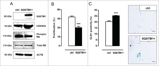
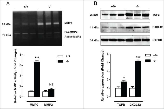
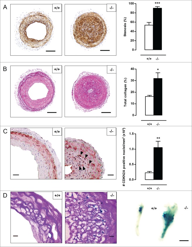
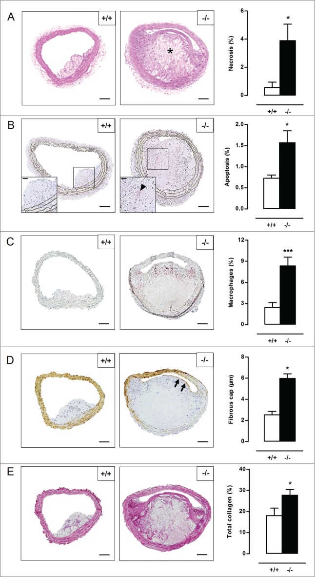
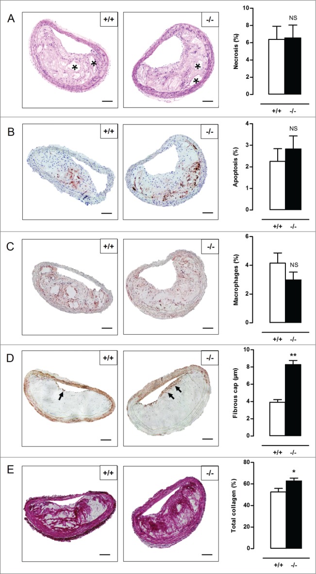
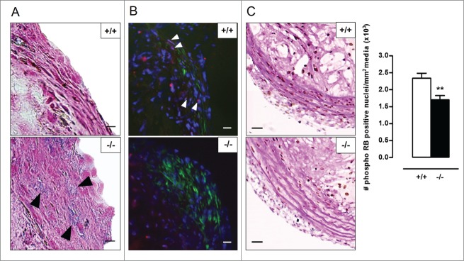
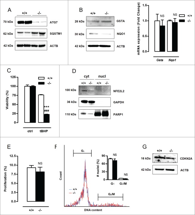
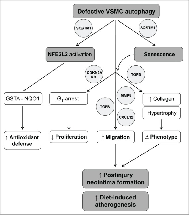
Autophagy is triggered in vascular smooth muscle cells (VSMCs) of diseased arterial vessels. However, the role of VSMC autophagy in cardiovascular disease is poorly understood. Therefore, we investigated the effect of defective autophagy on VSMC survival and phenotype and its significance in the development of postinjury neointima formation and atherosclerosis. Tissue-specific deletion of the essential autophagy gene Atg7 in murine VSMCs (atg7−/− VSMCs) caused accumulation of SQSTM1/p62 and accelerated the development of stress-induced premature senescence as shown by cellular and nuclear hypertrophy, CDKN2A-RB-mediated G1 proliferative arrest and senescence-associated GLB1 activity. Transfection of SQSTM1-encoding plasmid DNA in Atg7+/+ VSMCs induced similar features, suggesting that accumulation of SQSTM1 promotes VSMC senescence. Interestingly, atg7−/− VSMCs were resistant to oxidative stress-induced cell death as compared to controls. This effect was attributed to nuclear translocation of the transcription factor NFE2L2 resulting in upregulation of several antioxidative enzymes. In vivo, defective VSMC autophagy led to upregulation of MMP9, TGFB and CXCL12 and promoted postinjury neointima formation and diet-induced atherogenesis. Lesions of VSMC-specific atg7 knockout mice were characterized by increased total collagen deposition, nuclear hypertrophy, CDKN2A upregulation, RB hypophosphorylation, and GLB1 activity, all features typical of cellular senescence. To conclude, autophagy is crucial for VSMC function, phenotype, and survival. Defective autophagy in VSMCs accelerates senescence and promotes ligation-induced neointima formation and diet-induced atherogenesis, implying that autophagy inhibition as therapeutic strategy in the treatment of neointimal stenosis and atherosclerosis would be unfavorable. Conversely, stimulation of autophagy could be a valuable new strategy in the treatment of arterial disease.
Overview
The claims and statistics of this paper haven’t been checked yet.
Run the check — free, no account needed. It reads the open-access text and checks the sources it cites.
Integrity signals
Registry
Cites
Statistics
Figures
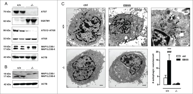
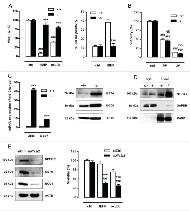
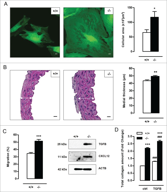
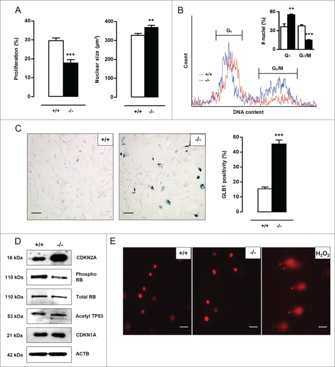
Verification
Claims the paper makes
The claims and statistics of this paper haven’t been checked yet.
Run the check — free, no account needed. It reads the open-access text and checks the sources it cites.
Claims other papers make about this one
Each is checked against this paper’s own text: does it say what the citing paper says it does?
In the citing paper
“Loss of autophagy can also drive VSMC senescence 75 .”
From Vascular smooth muscle cells in atherosclerosis (2019) · cites this paper as [75]
Self-check
Percentages, sums and reported differences re-computed from the paper’s own numbers.
The self-check needs the paper’s full text.
Citations
Cites
Repertoires of Autophagy in the Pathogenesis of Ocular Diseases
Li, Y., Jiang, Q. L., Cao, G.-F., Yao, J., & Yan, B. (2015) · Cellular Physiology and Biochemistry · doi:10.1159/000373980 · Cited by 16,351
Autophagy: Renovation of Cells and Tissues
Mizushima, N., & Komatsu, M. (2011) · Cell · doi:10.1016/j.cell.2011.10.026 · Cited by 6,615 · cited 1×
“amino acids) are recycled in order to replenish nutrient and energy supply.(2) Basal autophagy is therefore an important housekeeping and life-sustaining process.”
BackgroundCellular senescence: when bad things happen to good cells
Campisi, J., & d'Adda di Fagagna, F. (2007) · Nature Reviews Molecular Cell Biology · doi:10.1038/nrm2233 · Cited by 4,441 · cited 3×
“Cellular senescence can be established by the activation of 2 different tumor suppressor pathways: TP53-CDKN1A and CDKN2A-RB.(20,21) The TP53 pathway is triggered by DNA damage and generally associated with CDKN1A upregulation to initiate growth arrest.”
BackgroundLoss of autophagy in the central nervous system causes neurodegeneration in mice
Komatsu, M., Waguri, S., Chiba, T., Murata, S., Iwata, J.-I., Tanida, I., Ueno, T., Koike, M., Uchiyama, Y., Kominami, E., et al. (2006) · Nature · doi:10.1038/nature04723 · Cited by 3,500
Aging, Cellular Senescence, and Cancer
Campisi, J. (2013) · Annual Review of Physiology · doi:10.1146/annurev-physiol-030212-183653 · Cited by 2,954 · cited 3×
“Moreover, atg7¡/¡ VSMCs show an increase in TGFB and CXCL12 expression, which is most likely related to the increase in migration potential and the development of a senescence-associated secretory phenotype.(29,30) Cellular senescence can be established by the activation of 2 different tumor suppressor pathways: TP53-…”
BackgroundImpairment of starvation-induced and constitutive autophagy in Atg7 -deficient mice
Komatsu, M., Waguri, S., Ueno, T., Iwata, J., Murata, S., Tanida, I., Ezaki, J., Mizushima, N., Ohsumi, Y., Uchiyama, Y., et al. (2005) · The Journal of Cell Biology · doi:10.1083/jcb.200412022 · Cited by 2,331
Komatsu, M., Kurokawa, H., Waguri, S., Taguchi, K., Kobayashi, A., Ichimura, Y., Sou, Y.-S., Ueno, I., Sakamoto, A., Tong, K. I., et al. (2010) · Nature Cell Biology · doi:10.1038/ncb2021 · Cited by 2,288 · cited 4×
“SQSTM1 is considered to be a crucial activator of NFE2L2.(19) In the present study, atg7¡/¡ VSMCs showed increased nuclear translocation of NFE2L2.”
BackgroundInfluentialOxidative stress and autophagy: The clash between damage and metabolic needs
Filomeni, G., De Zio, D., & Cecconi, F. (2015) · Cell Death & Differentiation · doi:10.1038/cdd.2014.150 · Cited by 2,195 · cited 1×
“Defective autophagy in VSMCs triggers an antioxidative backup mechanism ROS production, oxidative damage and cell death are major events in cardiovascular disease(15,16) and may be regulated by autophagy.(17,18) To test the role of autophagy in VSMC survival against oxidative stress, Atg7C/C and atg7¡/¡ VSMCs were tre…”
MethodHomeostatic levels of p62 control cytoplasmic inclusion body formation in autophagy-deficient mice.
Komatsu, M., Waguri, S., Koike, M., Sou, Y.-S., Ueno, T., Hara, T., Mizushima, N., Iwata, J.-I., Ezaki, J., Murata, S., et al. (2007) · Cell · doi:10.1016/j.cell.2007.10.035 · Cited by 2,124
Rubinsztein, D. C., Mariño, G., & Kroemer, G. (2011) · Cell · doi:10.1016/j.cell.2011.07.030 · Cited by 2,107 · cited 1×
“Induction of autophagy by caloric restriction, spermidine, resveratrol or rapamycin extends life span in different organisms.(35) For example, the autophagy-inducers rapamycin and resveratrol suppress cellular senescence by partially preventing loss of proliferative potential,(36,37) whereas inhibition of autophagy by…”
Background
Research works printed but not linked: 3
- Grube E, 2004, Rev Cardiovasc Med, 5, S3
- Louis SF, 2010, Exp Clin Cardiol, 15, e75-85
- Seimon TA, 2009, J Clin Invest, 119, 886
Cited by
Sorted by citations
Vascular smooth muscle cells in atherosclerosis
Basatemur, G. L., Jørgensen, H. F., Clarke, M. C. H., Bennett, M. R., & Mallat, Z. (2019) · Nature Reviews Cardiology · doi:10.1038/s41569-019-0227-9 · Cited by 1,099
Vascular smooth muscle cell death, autophagy and senescence in atherosclerosis
Grootaert, M. O. J., Moulis, M., Roth, L., Martinet, W., Vindis, C., Bennett, M. R., & De Meyer, G. R. Y. (2018) · Cardiovascular Research · doi:10.1093/cvr/cvy007 · Cited by 606
Self-citationVascular Calcification—New Insights into Its Mechanism
Lee, S., Lee, I. K., & Jeon, J. (2020) · International Journal of Molecular Sciences · doi:10.3390/ijms21082685 · Cited by 451
Oxidative Stress-Mediated Atherosclerosis: Mechanisms and Therapies
Yang, X., Li, Y., Li, Y., Ren, X., Zhang, X., Hu, D., Gao, Y., Xing, Y., & Shang, H. (2017) · Frontiers in Physiology · doi:10.3389/fphys.2017.00600 · Cited by 418
“Atherosclerotic plaque rupture is a usual reason of the cardiovascular diseases, as stroke and myocardial infarction (Grootaert et al., 2015).”
BackgroundHong, S.-H., & Choi, K. M. (2020) · International Journal of Molecular Sciences · doi:10.3390/ijms21020494 · Cited by 390
“Atherosclerotic plaque rupture can lead to myocardial infarction and stroke [105].”
BackgroundSenescence mechanisms and targets in the heart
Chen, M. S., Lee, R. T., & Garbern, J. C. (2021) · Cardiovascular Research · doi:10.1093/cvr/cvab161 · Cited by 353
ROS signaling and ER stress in cardiovascular disease
Ochoa, C. D., Wu, R.-F., & Terada, L. (2018) · Molecular Aspects of Medicine · doi:10.1016/j.mam.2018.03.002 · Cited by 286
Hallmarks of cardiovascular ageing
Abdellatif, M., Rainer, P. P., Sedej, S., & Kroemer, G. (2023) · Nature Reviews Cardiology · doi:10.1038/s41569-023-00881-3 · Cited by 305
Kitada, M., Ogura, Y., & Koya, D. (2016) · Aging · doi:10.18632/aging.101068 · Cited by 268
Vion, A.-C., Kheloufi, M., Hammoutène, A., Poisson, J., Lasselin, J., Devue, C., Pic, I., Dupont, N., Busse, J., Stark, K., et al. (2017) · Proceedings of the National Academy of Sciences · doi:10.1073/pnas.1702223114 · Cited by 231
Age-Associated Mitochondrial Dysfunction Accelerates Atherogenesis
Tyrrell, D. J., Blin, M. G., Song, J., Wood, S. C., Zhang, M., Beard, D. A., & Goldstein, D. R. (2019) · Circulation Research · doi:10.1161/circresaha.119.315644 · Cited by 230
Macrophage Death as a Pharmacological Target in Atherosclerosis
Martinet, W., Coornaert, I., Puylaert, P., & Meyer, G. R. Y. D. (2019) · Frontiers in Pharmacology · doi:10.3389/fphar.2019.00306 · Cited by 232
“Moreover, NecroX-7 improves important features of plaque stability such as lowering plaque inflammation, reducing oxidative stress and increasing collagen content and fibrous cap thickness (Grootaert et al., 2016).”
BackgroundSelf-citationVascular Smooth Muscle Cells in Aortic Aneurysm: From Genetics to Mechanisms
Lu, H., Du, W., Ren, L., Hamblin, M. H., Becker, R. C., Chen, Y. E., & Fan, Y. (2021) · Journal of the American Heart Association · doi:10.1161/jaha.121.023601 · Cited by 230
Emerging roles of ATG7 in human health and disease
Collier, J. J., Suomi, F., Oláhová, M., McWilliams, T. G., & Taylor, R. W. (2021) · EMBO Molecular Medicine · doi:10.15252/emmm.202114824 · Cited by 201
“…Atg7deficient smooth muscle cells showed diminished survival and proliferation although they demonstrated increased resistance to oxidative stress-induced cell death, reportedly due to increased NRF2 nuclear translocation and antioxidant gene expression (Grootaert et al, 2015; Osonoi et al, 2018).”
BackgroundCao, H., Jia, Q., Li, Y., Chen, C., Xing, S., & Shen, D. (2019) · International Journal of Molecular Sciences · doi:10.3390/ijms20236093 · Cited by 189
SQSTM1/p62 and PPARGC1A/PGC-1alpha at the interface of autophagy and vascular senescence
Salazar, G., Cullen, A. E., Huang, J., Zhao, Y., Serino, A., Hilenski, L. L., Patrushev, N. A., Forouzandeh, F., & Hwang, H. S. (2019) · Autophagy · doi:10.1080/15548627.2019.1659612 · Cited by 186
“In the cardiovascular system, defective autophagy by Atg7 depletion led to the accumulation of SQSTM1 and senescence of VSMCs, and to diet-induced atherosclerosis [12].”
BackgroundVascular Smooth Muscle Cell Plasticity and Autophagy in Dissecting Aortic Aneurysms
Clément, M., Chappell, J., Raffort, J., Lareyre, F., Vandestienne, M., Taylor, A. L., Finigan, A., Harrison, J., Bennett, M. R., Bruneval, P., et al. (2019) · Arteriosclerosis, Thrombosis, and Vascular Biology · doi:10.1161/atvbaha.118.311727 · Cited by 184
Reactive Oxygen Species Generation and Atherosclerosis
Nowak, W. N., Deng, J., Ruan, X. Z., & Xu, Q. (2017) · Arteriosclerosis Thrombosis and Vascular Biology · doi:10.1161/atvbaha.117.309228 · Cited by 188
“In vivo, deletion of Atg7 in SMCs caused enhanced senescence and promoted neointima formation.(73) On the other hand, carotid artery ligation in Sqsmt1 mice caused more neointima formation.”
BackgroundAutophagy, Pyroptosis, and Ferroptosis: New Regulatory Mechanisms for Atherosclerosis
Lin, L., Zhang, M.-X., Zhang, L., Zhang, D., Li, C., & Li, Y. (2022) · Frontiers in Cell and Developmental Biology · doi:10.3389/fcell.2021.809955 · Cited by 166
Apoptosis, autophagy and atherosclerosis: Relationships and the role of Hsp27
Shan, R., Liu, N., Yan, Y., & Liu, B. (2020) · Pharmacological Research · doi:10.1016/j.phrs.2020.105169 · Cited by 169
Guo, F., Wu, Q., Li, P., Zheng, L., Ye, S., Dai, X.-Y., Kang, C., Lu, J., Xu, B., Xu, Y., et al. (2019) · Cell Death and Differentiation · doi:10.1038/s41418-018-0235-z · Cited by 163
Vascular smooth muscle cell senescence and age-related diseases: State of the art
Chen, C., Li, D., Jiang, Y., Tong, J., Fu, H., Wu, Y.-H., & Shen, F. (2018) · Biochimica et Biophysica Acta (BBA) - Molecular Basis of Disease · doi:10.1016/j.bbadis.2018.08.015 · Cited by 161
Defective autophagy in vascular smooth muscle cells enhances cell death and atherosclerosis
Osonoi, Y., Mita, T., Azuma, K., Nakajima, K., Masuyama, A., Goto, H., Nishida, Y., Miyatsuka, T., Fujitani, Y., Koike, M., et al. (2018) · Autophagy · doi:10.1080/15548627.2018.1501132 · Cited by 149
“Similar to a previous study [18], SMCs from Atg7cKO mice were more resistant to exogenous high oxidative stress-induced cell death compared with control Atg7 mice (Figure 1(H)).”
BackgroundResultInfluentialAutophagy in health and disease: focus on the cardiovascular system
Mialet‐Perez, J., & Vindis, C. (2017) · Essays in Biochemistry · doi:10.1042/ebc20170022 · Cited by 151
Defective autophagy in atherosclerosis: to die or to senesce?
Grootaert, M. O. J., Roth, L., Schrijvers, D. M., De Meyer, G. R. Y., & Martinet, W. (2018) · Oxidative Medicine and Cellular Longevity · doi:10.1155/2018/7687083 · Cited by 147
“It is important to note that accumulation of p62 also leads to activation of the nuclear factor-erythroid 2-related factor 2 (Nrf2) and antioxidant responsive element (ARE) pathway in Atg7-deficient VSMCs, rendering them more resistant to oxidative stress-induced cell death as compared to autophagy-competent VSMCs [16…”
BackgroundInfluentialSelf-citationCellular senescence links aging and diabetes in cardiovascular disease
Shakeri, H., Lemmens, K., Gevaert, A. B., De Meyer, G. R. Y., & Segers, V. F. M. (2018) · American Journal of Physiology-Heart and Circulatory Physiology · doi:10.1152/ajpheart.00287.2018 · Cited by 134
Zhang, X., Ramírez, C. M., Aryal, B., Madrigal‐Matute, J., Liu, X., Díaz, A., Torrecilla-Parra, M., Suárez, Y., Cuervo, A. M., Sessa, W. C., et al. (2020) · Arteriosclerosis Thrombosis and Vascular Biology · doi:10.1161/atvbaha.120.314291 · Cited by 130
Swiader, A., Nahapetyan, H., Faccini, J., D’Angelo, R., Mucher, E., Elbaz, M., Boya, P., & Vindis, C. (2016) · Oncotarget · doi:10.18632/oncotarget.8936 · Cited by 129
The role of autophagy in cardiovascular pathology
Gatica, D., Chiong, M., Lavandero, S., & Klionsky, D. J. (2021) · Cardiovascular Research · doi:10.1093/cvr/cvab158 · Cited by 123
Peng, S., Xu, L., Che, X., Xiao, Q., Pu, J., Shao, Q., & He, B. (2018) · Frontiers in Pharmacology · doi:10.3389/fphar.2018.00438 · Cited by 107
“In response to oxidative stress-inducing stimuli, which prevail in atherosclerotic lesions (Wang and Bennett, 2012), the VSMC has mainly three choices, either fight, adapt or die, basically through autophagy, senescence or apoptosis (Grootaert et al., 2018).”
BackgroundMitophagy in Cardiovascular Diseases
Morciano, G., Patergnani, S., Bonora, M., Pedriali, G., Tarocco, A., Bouhamida, E., Marchi, S., Ancora, G., Anania, G., Wieckowski, M. R., et al. (2020) · Journal of Clinical Medicine · doi:10.3390/jcm9030892 · Cited by 110
“A dysfunction in the autophagic process, caused by either autophagy related 7 (ATG7) deletion in VSMCs [49,50] or a macrophage-specific ATG5-null mutant mouse model [46], has been linked to atheroma development in AS.”
BackgroundInfluentialProtective role of chaperone-mediated autophagy against atherosclerosis
Madrigal-Matute, J., de Bruijn, J., van Kuijk, K., Riascos-Bernal, D. F., Diaz, A., Tasset, I., Martín-Segura, A., Gijbels, M. J. J., Sander, B., Kaushik, S., et al. (2022) · Proceedings of the National Academy of Sciences · doi:10.1073/pnas.2121133119 · Cited by 107
“Preclinical studies have shown that VSMCs up-regulate macroautophagy in response to several proatherogenic stimuli, such as lipids, proinflammatory cytokines, growth factors, and/or oxidative stress (50), whereas autophagic malfunction leads to VSMC phenotype switch and foam cell formation (15, 51, 52).”
BackgroundJiang, Y., Kou, J., Han, X. B., Li, X., Zhong, Z., Liu, Z., Zheng, Y., Tian, Y., & Yang, L. M. (2017) · Oxidative Medicine and Cellular Longevity · doi:10.1155/2017/8519169 · Cited by 99
Michiels, C. F., Kurdi, A., Timmermans, J., Meyer, G. R. Y. D., & Martinet, W. (2016) · Atherosclerosis · doi:10.1016/j.atherosclerosis.2016.07.899 · Cited by 102
“Lack of ATG7 expression in isolated Atg7 F/F SM22αCre + and Atg7 F/F LysM-Cre + cells is associated with several hallmarks of impaired autophagy such as SQSTM1/p62 accumulation and absence of LC3-II formation, as previously described [25, 26].”
BackgroundMethodSelf-citationMitochondrial Dynamics and Mitophagy in Cardiometabolic Disease
Lin, J., Duan, J., Wang, Q., Xu, S., Zhou, S., & Yao, K. (2022) · Frontiers in Cardiovascular Medicine · doi:10.3389/fcvm.2022.917135 · Cited by 103
Song, J., Zhang, Y., Frieler, R. A., Andren, A. C., Wood, S. C., Tyrrell, D. J., Sajjakulnukit, P., Deng, J. C., Lyssiotis, C. A., Mortensen, R. M., et al. (2023) · Journal of Clinical Investigation · doi:10.1172/jci173034 · Cited by 100
“We further examined Nrf2 responses in BMDMs exposed to oxidized low-density lipoprotein cholesterol (oxLDL), a major activator of atherogenesis (58–60), and/or OI.”
DNA Damage: A Main Determinant of Vascular Aging
Bautista‐Niño, P. K., Portilla-Fernández, E., Vaughan, D. E., Danser, A. H. J., & Roks, A. J. M. (2016) · International Journal of Molecular Sciences · doi:10.3390/ijms17050748 · Cited by 95
“Importantly, genetic ablation of Atg7, an important mediator of autophagy, causes an accelerated appearance of vascular aging hallmarks in mice [136].”
BackgroundRole of autophagy in atherosclerosis: foe or friend?
Hassanpour, M., Rahbarghazi, R., Nouri, M., Aghamohammadzadeh, N., Safaei, N., & Ahmadi, M. (2019) · Journal of Inflammation · doi:10.1186/s12950-019-0212-4 · Cited by 94
Metformin suppresses vascular smooth muscle cell senescence by promoting autophagic flux
Tai, S., Sun, J., Zhou, Y., Zhu, Z., He, Y., Chen, M., Yang, H., Xiao, Y., Tu, T., Tang, L., et al. (2021) · Journal of Advanced Research · doi:10.1016/j.jare.2021.12.009 · Cited by 99
“Recent studies found that the accumulation of senescent VSMCs can result from defective autophagy [18,19], and blockage of autophagic flux can enhance senescence in VSMCs [20], suggesting that reduced autophagy might induce senescence.”
BackgroundAutophagy as a novel therapeutic target in vascular calcification
Phadwal, K., Feng, D., Zhu, D., & MacRae, V. E. (2019) · Pharmacology & Therapeutics · doi:10.1016/j.pharmthera.2019.107430 · Cited by 90
The roles of autophagy in vascular smooth muscle cells
Tai, S., Hu, X., Peng, D., Zhou, S., & Zheng, X. (2016) · International Journal of Cardiology · doi:10.1016/j.ijcard.2016.02.128 · Cited by 88
Wu, H., Song, A., Hu, W., & Dai, M. (2018) · Frontiers in Pharmacology · doi:10.3389/fphar.2017.00948 · Cited by 88
“Defective autophagy in VSMCs disturbed cell homeostasis and induced cell proliferation, which finally led to and even accelerated atherogenesis (Grootaert et al., 2015).”
BackgroundPawłowska, E., Szczepańska, J., Wiśniewski, K., Tokarz, P., Jaskólski, D. J., & Błasiak, J. (2018) · International Journal of Molecular Sciences · doi:10.3390/ijms19041245 · Cited by 85
Targeting senescent cells to attenuate cardiovascular disease progression
Song, P., Zhao, Q., & Zou, M. (2020) · Ageing Research Reviews · doi:10.1016/j.arr.2020.101072 · Cited by 81
“ATG7 deletion in VSMCs of ApoE−/− mice promotes ligation-induced neointima formation and Western diet-induced atherogenesis in mice (Grootaert et al., 2015).”
BackgroundXu, J., Kitada, M., Ogura, Y., & Koya, D. (2021) · Frontiers in Cell and Developmental Biology · doi:10.3389/fcell.2021.641852 · Cited by 75
Lázaro, I., Lopez‐Sanz, L., Bernal, S., Oguiza, A., Recio, C., Melgar, A., Jiménez-Castilla, L., Egido, J., Madrigal‐Matute, J., & Gómez‐Guerrero, C. (2018) · Frontiers in Pharmacology · doi:10.3389/fphar.2018.00819 · Cited by 75
“Indeed, tBHQ treatment suppresses ischemia and reperfusion injury in brain (Shih et al., 2005; Hou et al., 2018; Xu et al., 2018) and kidney (Guerrero-Beltrán et al., 2012).”
BackgroundUryga, A. K., Grootaert, M. O. J., Garrido, A. M., Oc, S., Foote, K., Chappell, J., Finigan, A., Rossiello, F., d’Adda di Fagagna, F., Aravani, D., et al. (2021) · Communications Biology · doi:10.1038/s42003-021-02123-z · Cited by 73
Self-citationLuo, Z., Xu, W., Ma, S., Qiao, H., Gao, L., Zhang, R., Yang, B. Y., Qiu, Y. F., Chen, J., Zhang, M., et al. (2017) · Oxidative Medicine and Cellular Longevity · doi:10.1155/2017/3018190 · Cited by 73
Nakagawa, K., & Nakashima, Y. (2018) · Atherosclerosis · doi:10.1016/j.atherosclerosis.2018.03.039 · Cited by 70
Yang, J., Yu, J., Li, D., Yu, S., Jingbin, K., Wang, L., Wang, Y., Qiu, Y., Gao, X., Zhang, J., et al. (2016) · Autophagy · doi:10.1080/15548627.2016.1245261 · Cited by 67
Nahapetyan, H., Moulis, M., Grousset, E., Faccini, J., Grazide, M.-H., Mucher, E., Elbaz, M., Martinet, W., & Vindis, C. (2019) · Cell Death and Disease · doi:10.1038/s41419-019-1400-0 · Cited by 70
Self-citationHe, L., Chen, Q., Wang, L., Pu, Y., Huang, J., Cheng, C. K., Luo, J., Kang, L., Lin, X., Li, X., et al. (2024) · Redox Biology · doi:10.1016/j.redox.2024.103229 · Cited by 69
“Recent studies have also highlighted the role of Nrf2 in connecting autophagy and vascular senescence in atherosclerosis [38,39].”
Role of AMPK in atherosclerosis via autophagy regulation
Ou, H.-X., Liu, C., Feng, W., Xiao, X., Tang, S., & Mo, Z. (2018) · Science China Life Sciences · doi:10.1007/s11427-017-9240-2 · Cited by 62
Ma, C., Wu, H., Yang, G., Xiang, J., Feng, K., Zhang, J., Hua, Y., Kang, L., Fan, G., & Yang, S. (2021) · British Journal of Pharmacology · doi:10.1111/bph.15720 · Cited by 61
“KLF2 is a Krüppel-like transcription factor that contains highly conserved DNA-binding zinc finger domains and can regulate gene expression after nuclear translocation (Guixé-Muntet et al., 2017).”
BackgroundLysosome (Dys)function in Atherosclerosis—A Big Weight on the Shoulders of a Small Organelle
Marques, A. R. A., Ramos, C., Machado-Oliveira, G., & Vieira, O. V. (2021) · Frontiers in Cell and Developmental Biology · doi:10.3389/fcell.2021.658995 · Cited by 53
Galindo, C. L., Khan, S. R., Zhang, X., Yeh, Y., Liu, Z., & Razani, B. (2023) · Expert Opinion on Therapeutic Targets · doi:10.1080/14728222.2023.2288272 · Cited by 56
“…[134,147], retards atheroprogression [154], protects against apoptosis [134], and importantly is not macrophage-specific, since VSMCs and other neighboring cell types express the same mechanistic machinery and respond similarly to endogenous and exogenous autophagic regulators [129,130,132,145].”
BackgroundRole of Autophagy in Proteostasis: Friend and Foe in Cardiac Diseases
Li, J., Zhang, D., Wiersma, M., & Brundel, B. J. J. M. (2018) · Cells · doi:10.3390/cells7120279 · Cited by 59
The Long Non-coding Road to Atherosclerosis
Josefs, T., & Boon, R. A. (2020) · Current Atherosclerosis Reports · doi:10.1007/s11883-020-00872-6 · Cited by 52
Quercetin has a protective effect on atherosclerosis via enhancement of autophagy in ApoE‑/‑ mice
Cao, H., Jia, Q., Shen, D., Li, Y., Chen, C., & Xing, S. (2019) · Experimental and Therapeutic Medicine · doi:10.3892/etm.2019.7851 · Cited by 51
“It has been demonstrated that the presence of senescent VSMCs in plaques is associated with AS development, and impaired autophagy may increase the senescence process of VSMCs to drive the formation of plaque and the development AS (25).”
Lysosomes in the immunometabolic reprogramming of immune cells in atherosclerosis
Bonacina, F., Zhang, X., Manel, N., Yvan‐Charvet, L., Razani, B., & Norata, G. D. (2024) · Nature Reviews Cardiology · doi:10.1038/s41569-024-01072-4 · Cited by 53
“As the vascular lesion progresses, the autophagic process in macrophages and vascular cells worsens, leading to cell senescence and death, which contributes to atherosclerosis progression 129 .”
Cell senescence in cardiometabolic diseases
Grootaert, M. O. J. (2024) · npj Aging · doi:10.1038/s41514-024-00170-4 · Cited by 56
Self-citationFang, Z., Zhang, S., Luo, H., Jiang, D., Huo, B., Zhong, X., Feng, X., Cheng, W., Chen, Y., Feng, G., et al. (2022) · Cell Proliferation · doi:10.1111/cpr.13386 · Cited by 46
“For example, Grootaert et al.(16) demonstrated that defective autophagy in VSMCs accelerated senescence and promoted neointima formation, whereas Ouyang et al.”
BackgroundGarcía-Miguel, M., Riquelme, J. A., Norambuena‐Soto, I., Morales, P. E., Sanhueza‐Olivares, F., Núñez-Soto, C., Mondaca‐Ruff, D., Cancino-Arenas, N., Martín, A. S., & Chiong, M. (2018) · PLoS ONE · doi:10.1371/journal.pone.0197210 · Cited by 46
“Furthermore, defective autophagic activity in VSMC induces G1-cell cycle arrest, increases migration and up-regulates MMP-9, transforming growth factor β (TGF- β), chemokine (C-X-C) motif ligand 12 (CXCL12) and elicits post-injury neointima formation [29].”
BackgroundAutophagy in Metabolic Age-Related Human Diseases
Moulis, M., & Vindis, C. (2018) · Cells · doi:10.3390/cells7100149 · Cited by 48
“The importance of autophagy in atherosclerosis progression was further demonstrated in ApoE-null mice with specific VSMC deletion of Atg7 that exhibited accelerated atherosclerotic plaque development after 10 weeks of HFD, as shown by increased plaque cell death, inflammation, and fibrous cap thinning [120,121].”
BackgroundClement, M., Raffort, J., Lareyre, F., Tsiantoulas, D., Newland, S., Lu, Y., Masters, L., Harrison, J., Saveljeva, S., Ma, M. K. L., et al. (2019) · Circulation Research · doi:10.1161/circresaha.119.315248 · Cited by 44
“…autophagy in atherosclerosis promotes apoptosis and senescence of endothelial cells, 5 premature senescence of vascular smooth muscle cells (SMCs), 6 disturbs the cholesterol efflux pathway 7 and activates NLRP3 (NOD [nucleotide-binding oligomerization domain]-like receptor family, pyrin domain…”
Smooth muscle-specific HuR knockout induces defective autophagy and atherosclerosis
Liu, S., Jiang, X., Cui, X., Wang, J., Liu, S., Li, H., Yang, J., Zhang, C., & Zhang, W. (2021) · Cell Death and Disease · doi:10.1038/s41419-021-03671-2 · Cited by 43
You, G., Long, X., Song, F., Huang, J., Tian, M., Xiao, Y., Deng, S., & Wu, Q. (2020) · Drug Design Development and Therapy · doi:10.2147/dddt.s233932 · Cited by 44
The Role of SGLT2 Inhibitors in Vascular Aging
Liu, L., Ni, Y., Zhan, J., & Liu, Y. (2021) · Aging and Disease · doi:10.14336/ad.2020.1229 · Cited by 44
A new perspective on intervertebral disc calcification—from bench to bedside
Novais, E. J., Narayanan, R., Canseco, J. A., van de Wetering, K., Kepler, C. K., Hilibrand, A. S., Vaccaro, A. R., & Risbud, M. V. (2024) · Bone Research · doi:10.1038/s41413-023-00307-3 · Cited by 43
“87,88 For example, vascular calci fi cation shows the involvement of activated PERK-eIF2-ATF4-CHOP complex in different mouse models, which further promotes osteogenic differentiation and mineralization through BMP and RUNX2 pathways.”
BackgroundSubstrate stiffness differentially impacts autophagy of endothelial cells and smooth muscle cells
Hu, M., Jia, F., Huang, W., Xu, L. L., Hu, D., Wang, J., Ren, K., Fu, G., Wang, Y., & Ji, J. (2020) · Bioactive Materials · doi:10.1016/j.bioactmat.2020.10.013 · Cited by 44
“For instance, defective autophagy of VSMCs was found to accelerate cellular senescence, promoting neointima formation and atherogenesis [13,14].”
BackgroundRamadan, A., Singh, K. K., Quan, A., Plant, P. J., Al-Omran, M. A., Teoh, H., & Verma, S. (2017) · Journal of Vascular Surgery · doi:10.1016/j.jvs.2017.08.086 · Cited by 41
Wang, Y., Li, X., Chen, J., McConnell, B. K., Chen, L., Li, P., Chen, Y., & Zhang, Y. (2019) · Cell Death and Disease · doi:10.1038/s41419-019-1931-4 · Cited by 41
Atherosclerosis and Coenzyme Q10
Suarez-Rivero, J. M., Pastor‐Maldonado, C. J., de la Mata, M., Paz, M. V., Povea-Cabello, S., Álvarez-Córdoba, M., Villalón-García, I., Suárez-Carrillo, A., Talaverón-Rey, M., Munuera-Cabeza, M., et al. (2019) · International Journal of Molecular Sciences · doi:10.3390/ijms20205195 · Cited by 44
Caspase‐3 Deletion Promotes Necrosis in Atherosclerotic Plaques of ApoE Knockout Mice
Grootaert, M. O. J., Schrijvers, D. M., Hermans, M., Hoof, V. O. V., Meyer, G. R. Y. D., & Martinet, W. (2016) · Oxidative Medicine and Cellular Longevity · doi:10.1155/2016/3087469 · Cited by 39
“…University Hospital, 2610 Antwerp, Belgium Correspondence should be addressed to Mandy O. J. Grootaert; mandy.grootaert@uantwerpen.be Received 2 June 2016; Revised 23 September 2016; Accepted 29 September 2016 Academic Editor: Chris Kevil Copyright © 2016 Mandy O. J. Grootaert et al. This is an…”
BackgroundSelf-citationWang, X., Li, D., Chen, H., Wei, X., & Xu, X. (2019) · Medical Science Monitor · doi:10.12659/msm.915681 · Cited by 39
“In addition, the expression of MMP2 and MMP9 [18,19], which are important regulators of VSMC migration, was significantly increased by LIPCAR upregulation.”
BackgroundInfluentialSaha, S., Profumo, E., Togna, A. R., Riganò, R., Saso, L., & Buttari, B. (2020) · Oxidative Medicine and Cellular Longevity · doi:10.1155/2020/1232816 · Cited by 39
The putative role of autophagy in the pathogenesis of abdominal aortic aneurysms
Ramadan, A., Al-Omran, M. A., & Verma, S. (2017) · Atherosclerosis · doi:10.1016/j.atherosclerosis.2017.01.017 · Cited by 38
“To investigate the role of autophagy deficiency in VSMC survival, viability was measured in ATG7 deficient and WT murine VSMCs treated with oxidative stressors oxLDL and tert-Butyl hydroperoxide [35].”
BackgroundMethodInfluentialProfumo, E., Maggi, E., Arese, M., Cristofano, C. D., Salvati, B., Saso, L., Businaro, R., & Buttari, B. (2022) · International Journal of Molecular Sciences · doi:10.3390/ijms232113009 · Cited by 37
Liu, Q., Wang, Y., & Gu, H. (2020) · Clinica Chimica Acta · doi:10.1016/j.cca.2020.10.025 · Cited by 38
Programmed Cell Death: Complex Regulatory Networks in Cardiovascular Disease
Zhou, L., Sun, J., Gu, L., Wang, S., Yang, T., Wei, T., Shan, T., Wang, H., & Wang, L. (2021) · Frontiers in Cell and Developmental Biology · doi:10.3389/fcell.2021.794879 · Cited by 37
Chronic Stress A Potential Suspect Zero of Atherosclerosis: A Systematic Review
Meng, L., Zhang, Y.-M., Luo, Y., Gong, T., & Liu, D. (2021) · Frontiers in Cardiovascular Medicine · doi:10.3389/fcvm.2021.738654 · Cited by 38
Ho‐Tin‐Noé, B., Vo, S., Bayles, R., Ferrière, S., Ladjal, H., Toumi, S., Deschildre, C., Ollivier, V., & Michel, J. (2016) · The Journal of Pathology · doi:10.1002/path.4873 · Cited by 38
Komaravolu, R. K., Waltmann, M. D., Konaniah, E. S., Jaeschke, A., & Hui, D. Y. (2019) · Arteriosclerosis Thrombosis and Vascular Biology · doi:10.1161/atvbaha.119.313194 · Cited by 38
Chen, Y., Su, X., Qin, Q., Yu, Y., Jia, M., Zhang, H., Li, H., & Pei, L. (2019) · Biomedicine & Pharmacotherapy · doi:10.1016/j.biopha.2019.109758 · Cited by 37
Jarad, S., Gill, G. S., Amadi, P. U., Gu, H.-M., & Zhang, D. (2025) · Pharmacological Research · doi:10.1016/j.phrs.2025.107833 · Cited by 40
Perpetual change: autophagy, the endothelium, and response to vascular injury
Sachdev, U., & Lotze, M. T. (2017) · Journal of Leukocyte Biology · doi:10.1189/jlb.3ru1116-484rr · Cited by 35
Small-Vessel Vasculopathy Due to Aberrant Autophagy in LAMP-2 Deficiency
Nguyen, H. T., Noguchi, S., Sugie, K., Matsuo, Y., Nguyen, C. T. H., Koito, H., Shiojima, I., Nishino, I., & Tsukaguchi, H. (2018) · Scientific Reports · doi:10.1038/s41598-018-21602-8 · Cited by 35
Metabolic Alterations in Cardiopulmonary Vascular Dysfunction
Smolders, V. F. E. D., Zodda, E., Quax, P. H. A., Carini, M., Barberà, J. A., Thomson, T. M., Tura-Ceide, O., & Cascante, M. (2019) · Frontiers in Molecular Biosciences · doi:10.3389/fmolb.2018.00120 · Cited by 37
“Atherogenesis is the result of complex sequences of events associated with processes such as endothelial dysfunction, neovascularization, vascular proliferation, apoptosis, matrix degradation, inflammation, oxidative stress, and thrombosis (Hansson, 2005).”
BackgroundCelastrol ameliorates vascular neointimal hyperplasia through Wnt5a-involved autophagy
Shi, Y., Liu, L., Deng, C., Zhao, T., Shi, Z., Yan, J., Gong, Y.-Z., Liao, D., & Li, Q. (2021) · International Journal of Biological Sciences · doi:10.7150/ijbs.58715 · Cited by 36
Gkaliagkousi, Ε., Lazaridis, A. A., Doğan, S., Fraenkel, E., Tuna, B. G., Mozos, I. M., VUKIĆEVIĆ, M., Yalçın, Ö., & Gopčević, K. (2022) · International Journal of Molecular Sciences · doi:10.3390/ijms23158672 · Cited by 35
“Furthermore, defective autophagy has been implicated in angiogenesis, calcification of the vessel wall and atherosclerosis [11,212].”
BackgroundMa, M., Li, F., Li, W., Zhao, H., Jiang, M., Yu, Y., Dong, Y., Zhang, Y., Li, P., Bu, W., et al. (2022) · British Journal of Pharmacology · doi:10.1111/bph.15949 · Cited by 35
Xu, X.-J., Zhao, W., Feng, S., Sun, C., Chen, Q., Ni, B., & Hu, H. (2017) · Molecular Medicine Reports · doi:10.3892/mmr.2017.7533 · Cited by 34
“Recent studies have suggested that autophagy is relevant to human diseases (20,26).”
BackgroundInfluentialLi, D., Chen, A., Lan, T., Zou, Y., Zhao, L., Yang, P., Qu, H., Li, W., Varghese, Z., Moorhead, J. F., et al. (2018) · The FASEB Journal · doi:10.1096/fj.201800975rrr · Cited by 34
“atherogenesis and that up-regulated autophagy could prevent the development of atherosclerosis, especially in the formation of foam cells (26).”
BackgroundPharmacological modulation of vascular ageing: A review from VascAgeNet
Roth, L., Doğan, S., Tuna, B. G., Arányi, T., Benı́tez, S., Borrell-Pagés, M., Bozaykut, P., Meyer, G. R. Y. D., Duca, L., Durmuş, N., et al. (2023) · Ageing Research Reviews · doi:10.1016/j.arr.2023.102122 · Cited by 38
“Defective auto-phagy in VSMCs, that were isolated from Atg7 F/F SM22 α -Cre + mice, triggers stress-induced premature senescence (Grootaert et al., 2015).”
BackgroundSelf-citationZhang, P., Guan, Y., Chen, J., Li, X., McConnell, B. K., Zhou, W. J., Boini, K. M., & Zhang, Y. (2018) · Cell Death and Disease · doi:10.1038/s41419-018-1197-2 · Cited by 33
Xiao, Q., Che, X., Cai, B., Tao, Z., Zhang, H., Shao, Q., & Pu, J. (2019) · Journal of Cellular and Molecular Medicine · doi:10.1111/jcmm.14715 · Cited by 33
Ouyang, C., Mu, J., Lu, Q., Li, J., Zhu, H., Wang, Q., Zou, M., & Xie, Z. (2019) · Autophagy · doi:10.1080/15548627.2019.1707488 · Cited by 32
Lin, J., Chen, R., Yang, L., Gong, M., Du, M.-Y., Mu, S.-Q., Jiang, Z.-A., Li, H., Yang, Y., Wang, X., et al. (2023) · Journal of Advanced Research · doi:10.1016/j.jare.2023.07.010 · Cited by 31
Ouyang, C., Li, J., Zheng, X., Mu, J., Torres, G., Wang, Q., Zou, M., & Xie, Z. (2021) · Autophagy · doi:10.1080/15548627.2021.1911018 · Cited by 33
“However, VSMC-specific deletion of Atg7 leads to VSMC senescence, enhances cell migration, and promotes ligation-induced neointima formation and diet-induced atherogenesis [7].”
BackgroundNakagawa, K., Tanaka, M., Hahm, T., Nguyen, H.-N., Matsui, T., Chen, Y.-X., & Nakashima, Y. (2021) · Arteriosclerosis Thrombosis and Vascular Biology · doi:10.1161/atvbaha.121.316154 · Cited by 32
Metadata
- Authors
- Grootaert, M. O., da Costa Martins, P. A., Bitsch, N., Pintelon, I., De Meyer, G. R., Martinet, W., & Schrijvers, D. M.
- Affiliations
- University of Antwerp (BE)
- Department of Cardiology; Cardiovascular Research Institute Maastricht; Maastricht, The Netherlands
- Maastricht University (NL)
- Laboratory of Physiopharmacology; University of Antwerp; Antwerp, Belgium
- Corresponding author
- Schrijvers, Dorien M
- Venue
- Autophagy
- Publisher
- Informa UK Limited
- Published
- 2015-11-02
- Volume
- 11
- Issue
- 11
- Pages
- 2014-2032
- ISSN
- 1554-8627 (ISSN-L)
- 1554-8635
- PMC
- PMC4824610
- OpenAlex
- W2200751652
- Cited by
- 267
- References
- 73
- Citations by year
- 2026: 8 · 2025: 20 · 2024: 22 · 2023: 28 · 2022: 28 · 2021: 35 · 2020: 30 · 2019: 31 · 2018: 33 · 2017: 19 · 2016: 13
- FWCI
- 17.00
- Type
- journal article
- Language
- en
- Open access
- hybrid
- doi.org
- Licence
- cc-by-nc
- Locations
- Autophagy (publishedVersion, cc-by-nc)
- Europe PMC (PubMed Central) (publishedVersion, cc-by-nc)
- PubMed Central (publishedVersion, cc-by-nc)
- PubMed (publishedVersion)
- Research Publications (Maastricht University) (submittedVersion, other-oa)
- Digital Access to Libraries (submittedVersion, other-oa)
- Funders
- Fonds Wetenschappelijk Onderzoek (G044312N)
- Vlaamse regering
- Topics
- Life Sciences › Biochemistry, Genetics and Molecular Biology › Cell Biology › Autophagy in Disease and Therapy
- Life Sciences › Immunology and Microbiology › Immunology › Atherosclerosis and Cardiovascular Diseases
- Life Sciences › Biochemistry, Genetics and Molecular Biology › Cell Biology › Telomeres, Telomerase, and Senescence
- Keywords
- vascular smooth muscle cells, cellular senescence, neointima formation, atherosclerosis, ATG7, p62, stress-induced premature senescence, Nrf2, oxidative stress resistance, GLB1, MMP-9, CXCL12, collagen deposition
About these checks
Registry, retraction, Cites and statistics results are computed, not judged, with one exception: a printed reference that no index lists is read once by a model before it counts as a citation error. Claims are extracted by a model and kept only when their quote appears verbatim in the text; each cited source is then checked, with a model’s help, for whether it says what the claim relies on. Those verdicts can be wrong — read the claim’s quote and the cited source before relying on one.
Statistics and arithmetic are recomputed, not judged.
Confidence bands are shown only for fields calibrated on our annotated test set. None is calibrated yet, so no band is shown.
What counts as a claim ›How we measure confidence ›How we check this ›
Are you an author, or do you see a problem with this page? Request a review
