

The DOI matches its Crossref record; 243 of the 243 references it cites were found in a registry or an index.
Brain temperature and its role in physiology and pathophysiology: Lessons from 20 years of thermorecording
Open accessAbstract
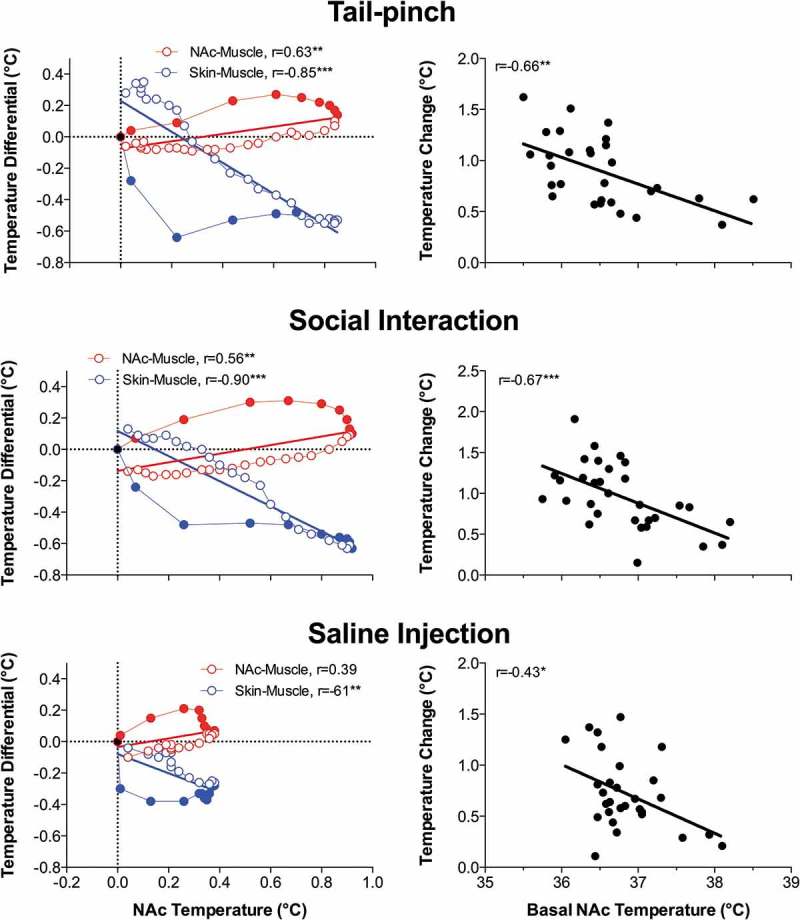
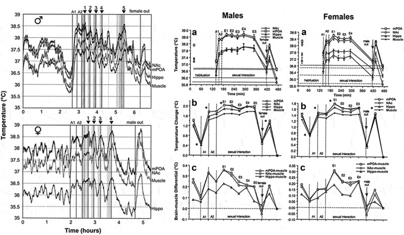
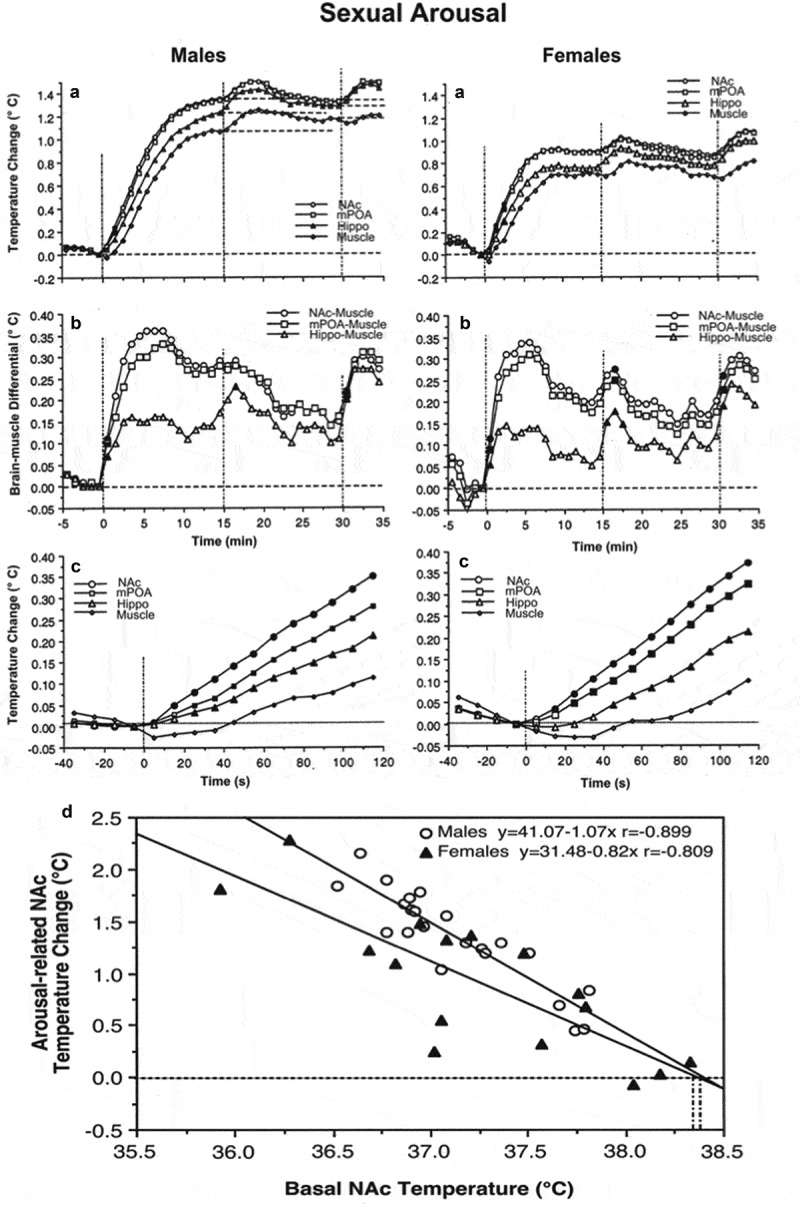
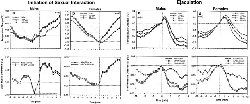
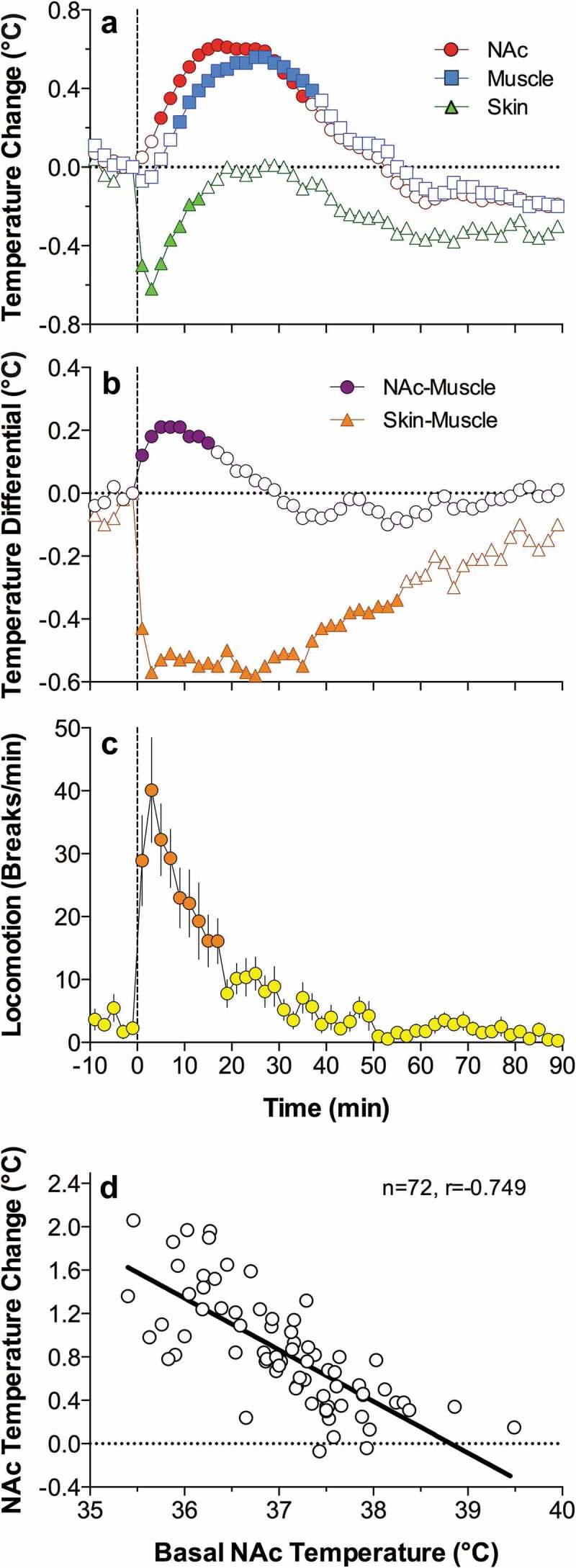
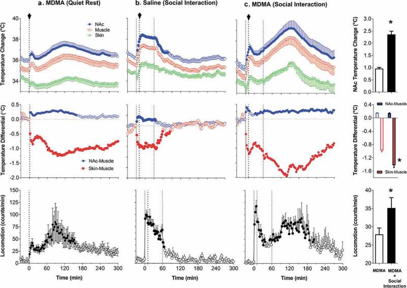
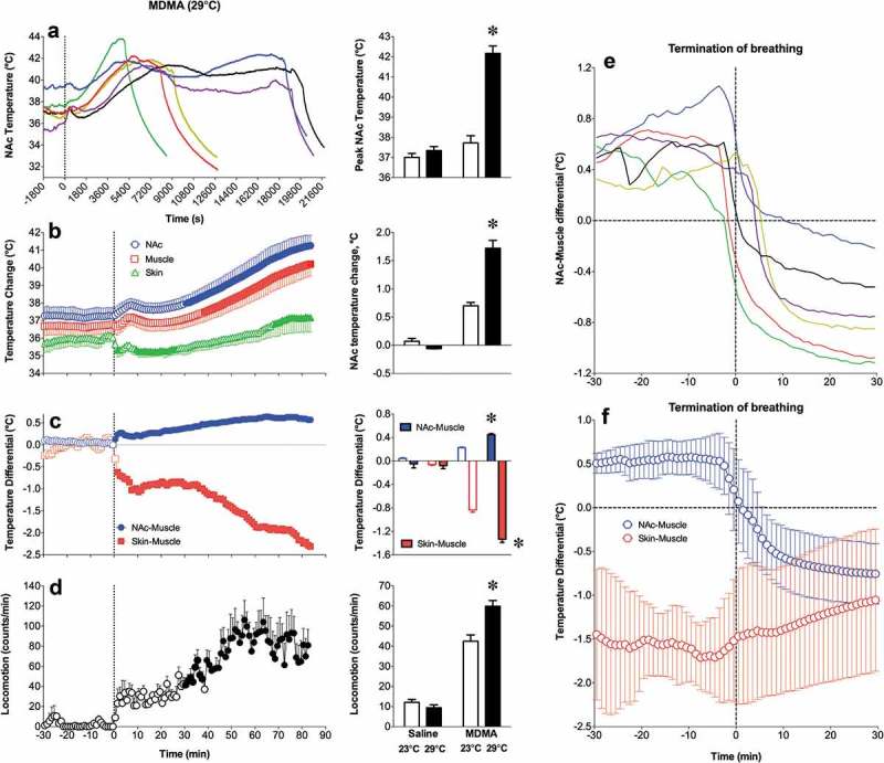
It is well known that temperature affects the dynamics of all physicochemical processes governing neural activity. It is also known that the brain has high levels of metabolic activity, and all energy used for brain metabolism is finally transformed into heat. However, the issue of brain temperature as a factor reflecting neural activity and affecting various neural functions remains in the shadow and is usually ignored by most physiologists and neuroscientists. Data presented in this review demonstrate that brain temperature is not stable, showing relatively large fluctuations (2-4°C) within the normal physiological and behavioral continuum. I consider the mechanisms underlying these fluctuations and discuss brain thermorecording as an important tool to assess basic changes in neural activity associated with different natural (sexual, drinking, eating) and drug-induced motivated behaviors. I also consider how naturally occurring changes in brain temperature affect neural activity, various homeostatic parameters, and the structural integrity of brain cells as well as the results of neurochemical evaluations conducted in awake animals. While physiological hyperthermia appears to be adaptive, enhancing the efficiency of neural functions, under specific environmental conditions and following exposure to certain psychoactive drugs, brain temperature could exceed its upper limits, resulting in multiple brain abnormalities and life-threatening health complications.
Overview
The claims and statistics of this paper haven’t been checked yet.
Run the check — free, no account needed. It reads the open-access text and checks the sources it cites.
Integrity signals
Registry
Cites
Statistics
Figures
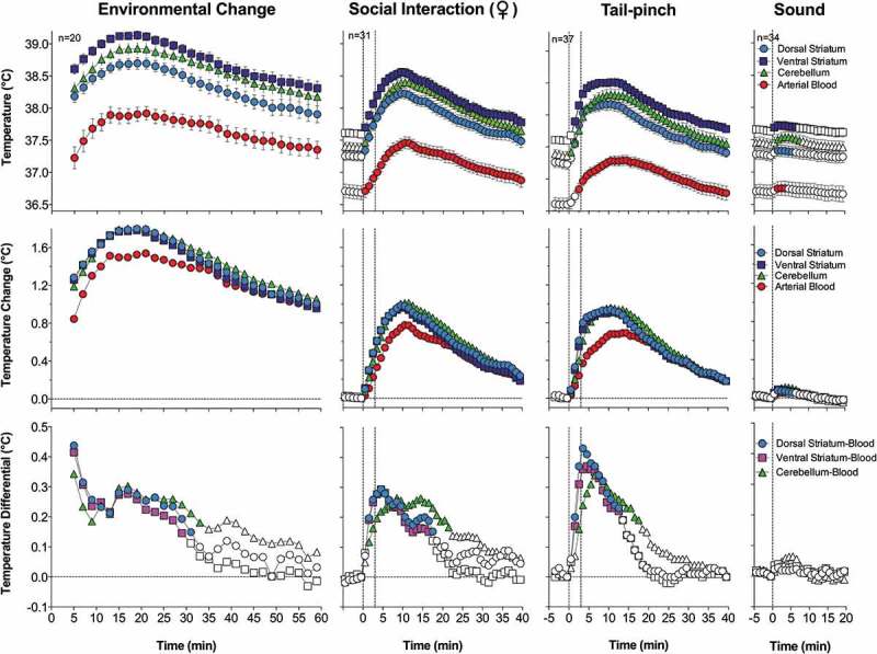
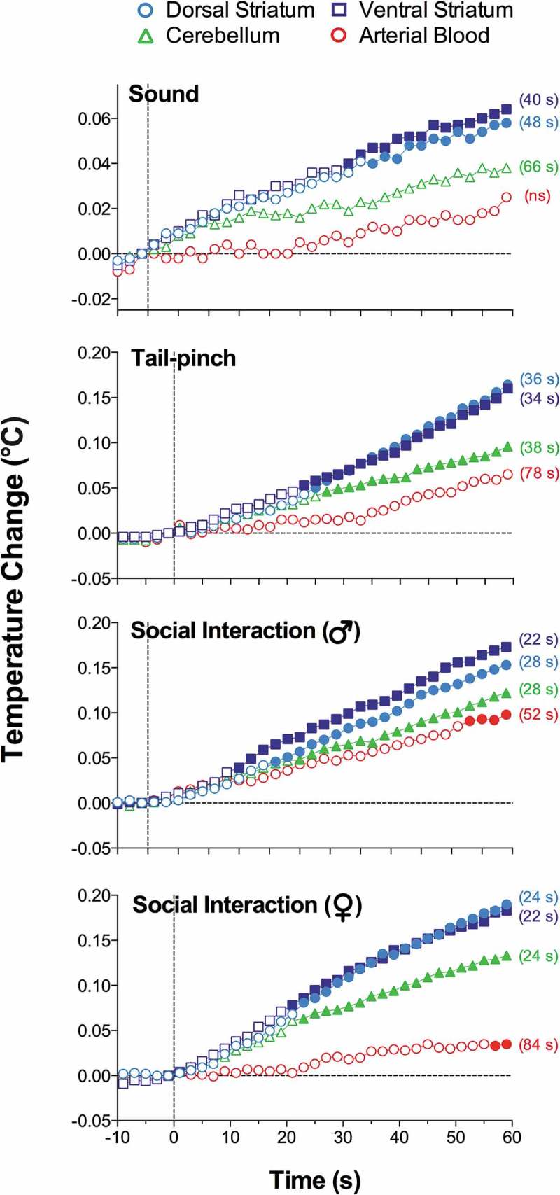
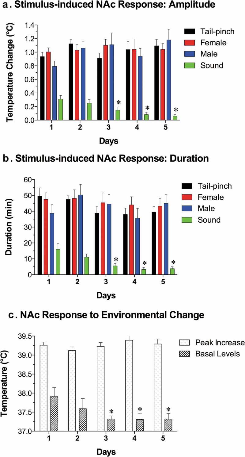
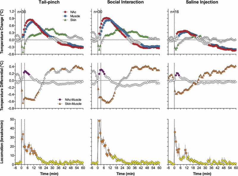
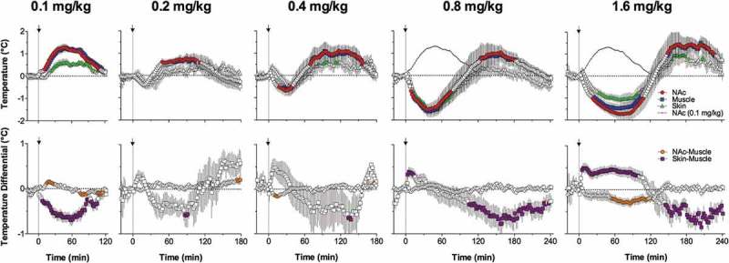
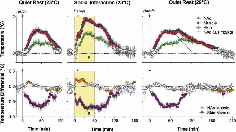
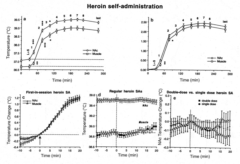
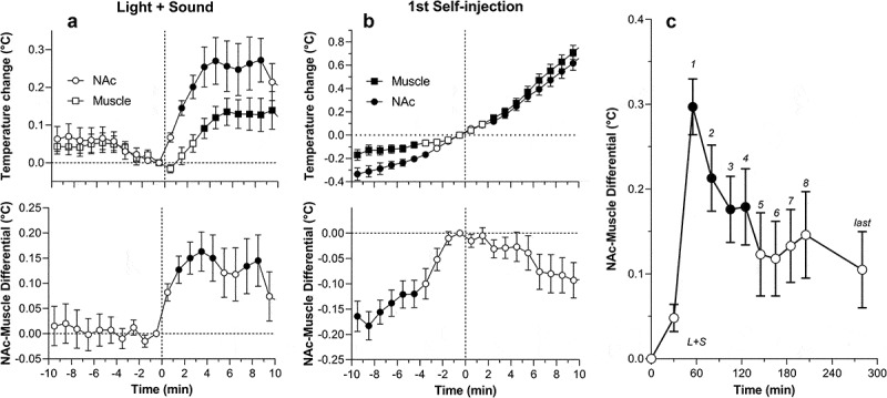
Verification
Claims the paper makes
The claims and statistics of this paper haven’t been checked yet.
Run the check — free, no account needed. It reads the open-access text and checks the sources it cites.
Claims other papers make about this one
Each is checked against this paper’s own text: does it say what the citing paper says it does?
The cited source says what the claim attributes to it.
In the citing paper
“however, they further discuss it in the light of chemically induced temperature changes such as those caused by drugs which can exceed beyond its upper physiological limit disrupting thermal dynamics leading to maladaptive neural activity and life-threatening complications (Kiyatkin, 2019).”
From Comparative analysis of energy transfer mechanisms for neural implants (2024) · cites this paper as Kiyatkin, 2019
The cited source says what the claim attributes to it.
In the citing paper
“Furthermore, physiological hyperthermia in the rat brain appears to be adaptive under normal conditions, enhancing neural functions (Kiyatkin, 2019).”
From Comparative analysis of energy transfer mechanisms for neural implants (2024) · cites this paper as Kiyatkin, 2019
The cited source says what the claim attributes to it.
In the citing paper
“Studies on rat models reveal that brain temperature is inherently unstable, fluctuating between 2 and 4°C under normal physiological and behavioral conditions.”
From Comparative analysis of energy transfer mechanisms for neural implants (2024) · cites this paper as Kiyatkin, 2019
Self-check
Percentages, sums and reported differences re-computed from the paper’s own numbers.
The self-check needs the paper’s full text.
Citations
Cites
ELECTRODE SYSTEMS FOR CONTINUOUS MONITORING IN CARDIOVASCULAR SURGERY
Clark, L. C., & Lyons, C. (1962) · Annals of the New York Academy of Sciences · doi:10.1111/j.1749-6632.1962.tb13623.x · Cited by 3,945
The Blood-Brain Barrier in Health and Chronic Neurodegenerative Disorders
Zlokovic, B. V. (2008) · Neuron · doi:10.1016/j.neuron.2008.01.003 · Cited by 3,280
Masters, W. H., & Johnson, V. E. (1966) · Medical Entomology and Zoology · Cited by 3,080
A psychomotor stimulant theory of addiction.
Wise, R. A., & Bozarth, M. A. (1987) · Psychological Review · doi:10.1037/0033-295x.94.4.469 · Cited by 2,747
Glial and neuronal control of brain blood flow
Attwell, D., Buchan, A. M., Charpak, S., Lauritzen, M., MacVicar, B. A., & Newman, E. A. (2010) · Nature · doi:10.1038/nature09613 · Cited by 2,511
From motivation to action: Functional interface between the limbic system and the motor system
MOGENSON, G., JONES, D., & YIM, C. (1980) · Progress in Neurobiology · doi:10.1016/0301-0082(80)90018-0 · Cited by 2,331
Fox, P. T., & Raichle, M. E. (1986) · Proceedings of the National Academy of Sciences · doi:10.1073/pnas.83.4.1140 · Cited by 2,056
On the Regulation of the Blood‐supply of the Brain
Roy, C., & Sherrington, C. (1890) · The Journal of Physiology · doi:10.1113/jphysiol.1890.sp000321 · Cited by 1,834
Nonoxidative Glucose Consumption During Focal Physiologic Neural Activity
Fox, P. T., Raichle, M. E., Mintun, M. A., & Dence, C. (1988) · Science · doi:10.1126/science.3260686 · Cited by 1,796
Busto, R., Dietrich, W. D., Globus, M. Y. ‐., Valdés, I., Scheinberg, P., & Ginsberg, M. D. (1987) · Journal of Cerebral Blood Flow & Metabolism · doi:10.1038/jcbfm.1987.127 · Cited by 1,786
Siesjö, B. K. (1978) · Medical Entomology and Zoology · Cited by 1,426
The Pharmacology and Clinical Pharmacology of 3,4-Methylenedioxymethamphetamine (MDMA, “Ecstasy”)
Green, A. R., Mechan, A. O., Elliott, J. M., O’Shea, E., & Colado, M. I. (2003) · Pharmacological Reviews · doi:10.1124/pr.55.3.3 · Cited by 1,272
Schmidt‐Nielsen, K. (1997) · Cambridge University Press eBooks · doi:10.1017/9780511801822 · Cited by 1,162
Skin blood flow in adult human thermoregulation: How it works, when it does not, and why
Charkoudian, N. (2003) · Mayo Clinic Proceedings · doi:10.4065/78.5.603 · Cited by 1,041
The dopamine hypothesis of the reinforcing properties of cocaine
Kuhar, M. J., Ritz, M. C., & Boja, J. W. (1991) · Trends in Neurosciences · doi:10.1016/0166-2236(91)90141-g · Cited by 1,005
Nucleus accumbens shell and core dopamine: differential role in behavior and addiction
Di Chiara, G. (2002) · Behavioural Brain Research · doi:10.1016/s0166-4328(02)00286-3 · Cited by 996
Blood-Brain Barrier in Physiology and Medicine
Rapoport, S. I. (1976) · Medical Entomology and Zoology · Cited by 889
Wise, R. A. (2002) · Neuron · doi:10.1016/s0896-6273(02)00965-0 · Cited by 881
Temperature Monitoring and Perioperative Thermoregulation
Sessler, D. I., Warner, D. S., & Warner, M. A. (2008) · Anesthesiology · doi:10.1097/aln.0b013e31817f6d76 · Cited by 758
James, W. (1892) · Macmillan eBooks · doi:10.1037/11630-000 · Cited by 725
Substance Abuse: A Comprehensive Textbook
Liskow, B. I. (1998) · JAMA · doi:10.1001/jama.279.10.802-a · Cited by 713
The pharmacology of cocaine related to its abuse.
Johanson, C. E., & Fischman, M. W. (1989) · Pharmacological Reviews · doi:10.1016/s0031-6997(25)00022-5 · Cited by 694
Margaria, R., Cerretelli, P., Aghemo, P., & Sassi, G. (1963) · Journal of Applied Physiology · doi:10.1152/jappl.1963.18.2.367 · Cited by 666
Welsh, F. A., & Shapiro, H. M. (1979) · Anesthesiology · doi:10.1097/00000542-197904000-00027 · Cited by 647
Pfaff, D. W. (1980) · Springer eBooks · doi:10.1007/978-1-4613-8084-9 · Cited by 631
Hyperthermia and central fatigue during prolonged exercise in humans
Nybo, L., & Nielsen, B. (2001) · Journal of Applied Physiology · doi:10.1152/jappl.2001.91.3.1055 · Cited by 623
The pharmacology and toxicology of "ecstasy" (MDMA) and related drugs.
Kalant, H. (2001) · Europe PMC (PubMed Central) · Cited by 604
Energetic basis of brain activity: implications for neuroimaging
Shulman, R. G., Rothman, D. L., Behar, K. L., & Hyder, F. (2004) · Trends in Neurosciences · doi:10.1016/j.tins.2004.06.005 · Cited by 590
The Conduction of the Nervous Impulse.
Lucas, K., & Adrian, E. D. A. (1918) · JAMA · doi:10.1001/jama.1918.02600110061026 · Cited by 583
Role of the preoptic-anterior hypothalamus in thermoregulation and fever
Boulant, J. A. (2000) · Clinical Infectious Diseases · doi:10.1086/317521 · Cited by 562
The Conduction of the Nervous Impulse
Landau, W. M. (1965) · Neurology · doi:10.1212/wnl.15.6.595 · Cited by 540
Mapping of human brown adipose tissue in lean and obese young men
Leitner, B. P., Huang, S., Brychta, R. J., Duckworth, C. J., Baskin, A. S., McGehee, S., Tal, I., Dieckmann, W., Gupta, G., Kolodny, G. M., et al. (2017) · Proceedings of the National Academy of Sciences · doi:10.1073/pnas.1705287114 · Cited by 530 · cited 2×
“[52] Leitner BP, Huang S, Brychta RJ, et al.”
BackgroundBenveniste, H. (1989) · Journal of Neurochemistry · doi:10.1111/j.1471-4159.1989.tb07243.x · Cited by 526
Neural Organization and Evolution of Thermal Regulation in Mammals
Satinoff, E. (1978) · Science · doi:10.1126/science.351802 · Cited by 512
Silver, I. A., & Erecińska, M. (1994) · Journal of Neuroscience · doi:10.1523/jneurosci.14-08-05068.1994 · Cited by 489
Heikkila, R. E., Orlansky, H., & Cohen, G. (1975) · Biochemical Pharmacology · doi:10.1016/0006-2952(75)90152-5 · Cited by 468
Central Neural Regulation of Brown Adipose Tissue Thermogenesis and Energy Expenditure
Morrison, S. F., Madden, C. J., & Tupone, D. (2014) · Cell Metabolism · doi:10.1016/j.cmet.2014.02.007 · Cited by 437
Blood–Brain Barrier in Physiology and Medicine
Michenfelder, J. D. (1977) · Anesthesiology · doi:10.1097/00000542-197701000-00021 · Cited by 433
Energetics of Functional Activation in Neural Tissues
Sokoloff, L. (1999) · Neurochemical Research · doi:10.1023/a:1022534709672 · Cited by 380
Damsma, G., Pfaus, J. G., Wenkstern, D., Phillips, A. G., & al, E. (1992) · Behavioral Neuroscience · doi:10.1037//0735-7044.106.1.181 · Cited by 370
Electrochemical Methods for Neuroscience
Michael, A. C., & Borland, L. M. (2006) · CRC Press eBooks · doi:10.1201/9781420005868 · Cited by 369
Prolonged presence of glutamate during excitatory synaptic transmission to cerebellar Purkinje cells
Barbour, B. (1994) · Neuron · doi:10.1016/0896-6273(94)90448-0 · Cited by 363
Reference values for resting blood flow to organs of man
Williams, L. R., & Leggett, R. W. (1989) · Clinical Physics and Physiological Measurement · doi:10.1088/0143-0815/10/3/001 · Cited by 358
The effect of temperature on the synaptic delay at the neuromuscular junction.
Katz, B., & Miledi, R. (1965) · The Journal of Physiology · doi:10.1113/jphysiol.1965.sp007790 · Cited by 356
Regulation of Body Temperature
WHITTOW, G. C. (1986) · Springer eBooks · doi:10.1007/978-1-4612-4862-0_9 · Cited by 353
Romanovsky, A. A., Ivanov, A. I., & Shimansky, Y. P. (2002) · Journal of Applied Physiology · doi:10.1152/japplphysiol.01173.2001 · Cited by 351
Yablonskiy, D. A., Sukstanskii, A. L., Leawoods, J. C., Gierada, D. S., Bretthorst, G. L., Lefrak, S. S., Cooper, J. D., & Conradi, M. S. (2002) · Proceedings of the National Academy of Sciences · doi:10.1073/pnas.052594699 · Cited by 346
Brain temperature exceeds systemic temperature in head-injured patients
Rumana, C. S., Gopinath, S. P., Uzura, M., Valadka, A. B., & Robertson, C. S. (1998) · Critical Care Medicine · doi:10.1097/00003246-199803000-00032 · Cited by 346
THE LAW OF INITIAL VALUE IN NEUROLOGY AND PSYCHIATRY: Facts and Problems1
WILDER, J. (1957) · The Journal of Nervous and Mental Disease · doi:10.1097/00005053-195701000-00009 · Cited by 339
Esposito, P., Gheorghe, D., Kandere‐Grzybowska, K., Pang, X., Connolly, R. J., Jacobson, S., & Theoharides, T. C. (2001) · Brain Research · doi:10.1016/s0006-8993(00)03026-2 · Cited by 339
Overview of Brain Microdialysis
Chefer, V. I., Thompson, A. C., Zapata, A. G., & Shippenberg, T. S. (2009) · Current Protocols in Neuroscience · doi:10.1002/0471142301.ns0701s47 · Cited by 332
Inadequate heat release from the human brain during prolonged exercise with hyperthermia
Nybo, L., Secher, N. H., & Nielsen, B. (2002) · The Journal of Physiology · doi:10.1113/jphysiol.2002.030023 · Cited by 316
Electrochemical Analysis of Neurotransmitters
Bucher, E. S., & Wightman, R. M. (2015) · Annual Review of Analytical Chemistry · doi:10.1146/annurev-anchem-071114-040426 · Cited by 307
Thompson, S., Masukawa, L., & Prince, D. (1985) · Journal of Neuroscience · doi:10.1523/jneurosci.05-03-00817.1985 · Cited by 297
Thermoregulation and Marathon Running
Cheuvront, S. N., & Haymes, E. M. (2001) · Sports Medicine · doi:10.2165/00007256-200131100-00004 · Cited by 285
Cardiovascular aspects of human thermoregulation.
Rowell, L. B. (1983) · Circulation Research · doi:10.1161/01.res.52.4.367 · Cited by 279
Chiara, G. D., Loddo, P., & Tanda, G. (1999) · Biological Psychiatry · doi:10.1016/s0006-3223(99)00236-x · Cited by 262
Rougé‐Pont, F., Piazza, P. V., Kharouby, M., Moal, M. L., & Simon, H. (1993) · Brain Research · doi:10.1016/0006-8993(93)90260-t · Cited by 260
CEREBRAL CIRCULATION AND METABOLISM DURING THIOPENTAL ANESTHESIA AND HYPERVENTILATION IN MAN*
Pierce, E. C., Lambertsen, C. J., Deutsch, S., Chase, P. E., Linde, H. W., Dripps, R. D., & Price, H. L. (1962) · Journal of Clinical Investigation · doi:10.1172/jci104623 · Cited by 259
Opioids for hedonic experience and dopamine to get ready for it
Barbano, M. F., & Cador, M. (2006) · Psychopharmacology · doi:10.1007/s00213-006-0521-1 · Cited by 257
Albers, D. S., & Sonsalla, P. K. (1995) · Journal of Pharmacology and Experimental Therapeutics · doi:10.1016/s0022-3565(25)12160-5 · Cited by 256
15O PET Measurement of Blood Flow and Oxygen Consumption in Cold-Activated Human Brown Fat
Muzik, O., Mangner, T. J., Leonard, W. R., Kumar, A., Janisse, J. J., & Granneman, J. G. (2013) · Journal of Nuclear Medicine · doi:10.2967/jnumed.112.111336 · Cited by 250
Permeability of the blood–brain barrier depends on brain temperature
Kiyatkin, E. A., & Sharma, H. S. (2009) · Neuroscience · doi:10.1016/j.neuroscience.2009.04.004 · Cited by 247
Self-citationRole of cerebral arterial blood in the regulation of brain temperature in the monkey
Hayward, J. N., & Baker, M. (1968) · American Journal of Physiology-Legacy Content · doi:10.1152/ajplegacy.1968.215.2.389 · Cited by 245
Kiyatkin, E. A., & Rebec, G. V. (1996) · Journal of Neurophysiology · doi:10.1152/jn.1996.75.1.142 · Cited by 243
Self-citationSexual activity increases dopamine transmission in the nucleus accumbens and striatum of female rats
Pfaus, J. G., Damsma, G., Wenkstern, D., & Fibiger, H. C. (1995) · Brain Research · doi:10.1016/0006-8993(95)00679-k · Cited by 236
Mechan, A. O., Esteban, B. M., O’Shea, E., Elliott, J. M., Colado, M. I., & Green, A. R. (2002) · British Journal of Pharmacology · doi:10.1038/sj.bjp.0704442 · Cited by 221
Effects of 3,4-methylenedioxymethamphetamine on autonomic thermoregulatory responses of the rat
Gordon, C. J., Watkinson, W. P., O'Callaghan, J. P., & Miller, D. B. (1991) · Pharmacology Biochemistry and Behavior · doi:10.1016/0091-3057(91)90288-d · Cited by 220
Everitt, B. J., & Stacey, P. (1987) · Journal of comparative psychology · doi:10.1037/0735-7036.101.4.407 · Cited by 215
Natural selective cooling of the human brain: evidence of its occurrence and magnitude.
Cabanac, M., & Caputa, M. (1979) · The Journal of Physiology · doi:10.1113/jphysiol.1979.sp012617 · Cited by 214
Normal male sexual function: emphasis on orgasm and ejaculation
Alwaal, A., Breyer, B. N., & Lue, T. F. (2015) · Fertility and Sterility · doi:10.1016/j.fertnstert.2015.08.033 · Cited by 209
McNay, E. C., McCarty, R. C., & Gold, P. E. (2001) · Neurobiology of Learning and Memory · doi:10.1006/nlme.2000.3976 · Cited by 200
Brain temperature monitoring and modulation in patients with severe MCA infarction
Schwab, S., Spranger, M., Aschoff, A., Steiner, T., & Hacke, W. (1997) · Neurology · doi:10.1212/wnl.48.3.762 · Cited by 199
Non‐synaptic receptors and transporters involved in brain functions and targets of drug treatment
Vizi, E. S., Fekete, Á., Károly, R., & Mike, Á. (2010) · British Journal of Pharmacology · doi:10.1111/j.1476-5381.2009.00624.x · Cited by 193
Heart Rate, Rate-Pressure Product, and Oxygen Uptake During Four Sexual Activities
Bohlen, J. G. (1984) · Archives of Internal Medicine · doi:10.1001/archinte.1984.00350210057007 · Cited by 190
The thermoregulation system and how it works
(2018) · Handbook of Clinical Neurology · doi:10.1016/b978-0-444-63912-7.00001-1 · Cited by 186 · cited 2×
“It is well known that the brain plays a crucial role in the regulation of body temperature by detecting changes in environmental temperatures, integrating sensory information, and adjusting, via effector mechanisms, heat production and/or heat loss to the external environment [4,5].”
BackgroundMethodRapid Changes in Local Extracellular Rat Brain Glucose Observed with an In Vivo Glucose Sensor
Hu, Y., & Wilson, G. S. (1997) · Journal of Neurochemistry · doi:10.1046/j.1471-4159.1997.68041745.x · Cited by 186
Brain temperature fluctuation: a reflection of functional neural activation
Kiyatkin, E. A., Brown, P. L., & Wise, R. A. (2002) · European Journal of Neuroscience · doi:10.1046/j.1460-9568.2002.02066.x · Cited by 185
Self-citationRougé‐Pont, F., Deroche‐Gamonet, V., Moal, M. L., & Piazza, P. V. (1998) · European Journal of Neuroscience · doi:10.1046/j.1460-9568.1998.00438.x · Cited by 184
Horvath, T. L., Warden, C. H., Hajós, M., Lombardi, A., Goglia, F., & Diano, S. (1999) · Journal of Neuroscience · doi:10.1523/jneurosci.19-23-10417.1999 · Cited by 181
Fever: Causes and consequences
Moltz, H. (1993) · Neuroscience & Biobehavioral Reviews · doi:10.1016/s0149-7634(05)80009-0 · Cited by 180
Fellows, L. K., Boutelle, M. G., & Fillenz, M. (1993) · Journal of Neurochemistry · doi:10.1111/j.1471-4159.1993.tb03285.x · Cited by 176
Brain temperature and hippocampal function
Andersen, P. N., & Moser, E. I. (1995) · Hippocampus · doi:10.1002/hipo.450050602 · Cited by 176
CYTOLOGICAL RESPONSES OF BROWN FAT TISSUE IN COLD-EXPOSED RATS
Cameron, I. L., & Smith, R. E. (1964) · The Journal of Cell Biology · doi:10.1083/jcb.23.1.89 · Cited by 163
Bowyer, J. F., & Ali, S. F. (2006) · Synapse · doi:10.1002/syn.20324 · Cited by 162
The onset of fever: new insights into its mechanism
Blatteis, C. M. (2007) · Progress in brain research · doi:10.1016/s0079-6123(06)62001-3 · Cited by 157
Alterations in Blood–Brain Barrier Function by Morphine and Methamphetamine
Sharma, H. S., & Ali, S. F. (2006) · Annals of the New York Academy of Sciences · doi:10.1196/annals.1369.020 · Cited by 153
Microdialysis of GABA and glutamate: Analysis, interpretation and comparison with microsensors
van der Zeyden, M., Oldenziel, W. H., Rea, K., Cremers, T. I. F. H., & Westerink, B. H. C. (2007) · Pharmacology Biochemistry and Behavior · doi:10.1016/j.pbb.2007.09.004 · Cited by 152
Dose dependent reduction of glucose utilization by pentobarbital in rat brain.
Crane, P. D., Braun, L. D., Cornford, E. M., Cremer, J. E., Glass, J. M., & Oldendorf, W. H. (1978) · Stroke · doi:10.1161/01.str.9.1.12 · Cited by 152
Modern Psychophysiology and the Law of Initial Value
Wilder, J. (1958) · American Journal of Psychotherapy · doi:10.1176/appi.psychotherapy.1958.12.2.199 · Cited by 152
Fever and hypothermia in systemic inflammation
Garami, A., Steiner, A. A., & Romanovsky, A. A. (2018) · Handbook of clinical neurology · doi:10.1016/b978-0-444-64074-1.00034-3 · Cited by 148
Persistently active, pacemaker-like neurons in neocortex
Bon-Jégo, M. L. (2007) · Frontiers in Neuroscience · doi:10.3389/neuro.01.1.1.009.2007 · Cited by 145
Relationship among Brain and Blood Glucose Levels and Spontaneous and Glucoprivic Feeding
Dunn-Meynell, A. A., Sanders, N. M., Compton, D. S., Becker, T. C., Eiki, J., Zhang, B. B., & Levin, B. E. (2009) · Journal of Neuroscience · doi:10.1523/jneurosci.0334-09.2009 · Cited by 143
Kiyatkin, E. A., & Gratton, A. P. (1994) · Brain Research · doi:10.1016/0006-8993(94)90231-3 · Cited by 143
Self-citationSelective brain cooling in humans: “fancy” or fact?
Cabanac, M. (1993) · The FASEB Journal · doi:10.1096/fasebj.7.12.8375612 · Cited by 140
SHARMA, H. S., & Dey, P. K. (1986) · Journal of the Neurological Sciences · doi:10.1016/0022-510x(86)90036-5 · Cited by 139
Expression of aquaporin 1 and aquaporin 4 water channels in rat choroid plexus
Speake, T., Freeman, L. J., & Brown, P. D. N. (2002) · Biochimica et Biophysica Acta (BBA) - Biomembranes · doi:10.1016/s0005-2736(02)00658-2 · Cited by 136
LOCALIZED THERMAL CHANGES IN THE CAT'S BRAIN
Serota, H. M., & Gerard, R. W. (1938) · Journal of Neurophysiology · doi:10.1152/jn.1938.1.2.115 · Cited by 131
Kiyatkin, E. A., Brown, P. L., & Sharma, H. S. (2007) · European Journal of Neuroscience · doi:10.1111/j.1460-9568.2007.05741.x · Cited by 130
Self-citationHeart rate and blood pressure responses during sexual activity in normal males
Nemec, E. D., Mansfield, L., & Kennedy, J. W. (1976) · American Heart Journal · doi:10.1016/s0002-8703(76)80106-8 · Cited by 130
Thermoregulatory effects of 3,4-methylenedioxymethamphetamine (MDMA) in humans
Freedman, R. R., Johanson, C., & Tancer, M. E. (2005) · Psychopharmacology · doi:10.1007/s00213-005-0149-6 · Cited by 129
Hypothermia as a potential treatment for cerebral ischemia.
Maher, J., & Hachinski, V. (1993) · PubMed · Cited by 129
Li, Y.-Q., Chen, P., Jain, V., Reilly, R. M., & Wong, C. S. (2004) · Radiation Research · doi:10.1667/rr3117 · Cited by 126
Hypothalamic temperature in unanesthetized albino rats during feeding and sleeping
Abrams, R. M., & Hammel, H. T. (1964) · American Journal of Physiology-Legacy Content · doi:10.1152/ajplegacy.1964.206.3.641 · Cited by 124
Donaldson, G. C., Keatinge, W. R., & Saunders, R. D. (2003) · International Journal of Hyperthermia · doi:10.1080/0265673021000058357 · Cited by 123
Striatal dopamine uptake in the rat: In vivo analysis by fast cyclic voltammetry
Stamford, J. A., Kruk, Z. L., Millar, J., & Wightman, R. M. (1984) · Neuroscience Letters · doi:10.1016/0304-3940(84)90274-x · Cited by 122
Brain hyperthermia as physiological and pathological phenomena
Kiyatkin, E. A. (2005) · Brain Research Reviews · doi:10.1016/j.brainresrev.2005.04.001 · Cited by 120
Self-citationPedersen, N. P., & Blessing, W. W. (2001) · Journal of Neuroscience · doi:10.1523/jneurosci.21-21-08648.2001 · Cited by 120
Intracerebral temperatures in free-moving cats
Jm, D., & Hanai, T. (1966) · American Journal of Physiology-Legacy Content · doi:10.1152/ajplegacy.1966.211.3.755 · Cited by 119
SHARMA, H. S., & Kiyatkin, E. A. (2008) · Journal of Chemical Neuroanatomy · doi:10.1016/j.jchemneu.2008.08.002 · Cited by 118
Self-citationRichie, J. M. (1973) · Progress in Biophysics and Molecular Biology · doi:10.1016/0079-6107(73)90019-9 · Cited by 118
Synaptic transmission in the neocortex during reversible cooling
Volgushev, M., Vidyasagar, T. R., Chistiakova, M., & Eysel, U. T. (2000) · Neuroscience · doi:10.1016/s0306-4522(00)00109-3 · Cited by 117
Direct cooling of the human brain by heat loss from the upper respiratory tract
Mariak, Z. D., White, M. D., Lewko, J., Łysoń, T., & Piekarski, P. (1999) · Journal of Applied Physiology · doi:10.1152/jappl.1999.87.5.1609 · Cited by 116
Spinal nerve lesion alters blood–spinal cord barrier function and activates astrocytes in the rat
Gordh, T., Chu, H., & Sharma, H. S. (2006) · Pain · doi:10.1016/j.pain.2006.05.020 · Cited by 115
Medial preoptic area/anterior hypothalamus and sexual motivation
Paredes, R. G. (2003) · Scandinavian Journal of Psychology · doi:10.1111/1467-9450.00337 · Cited by 115
Kiyatkin, E. A., Wise, R. A., & Gratton, A. P. (1993) · Synapse · doi:10.1002/syn.890140109 · Cited by 112
Self-citationPhillips, P. E. M., Robinson, D. L., Stuber, G. D., Carelli, R. M., & Wightman, R. M. (2003) · Humana Press eBooks · doi:10.1385/1-59259-358-5:443 · Cited by 112
Blood-brain barrier integrity may be threatened by exercise in a warm environment
Watson, P., Shirreffs, S. M., & Maughan, R. J. (2005) · American Journal of Physiology-Regulatory, Integrative and Comparative Physiology · doi:10.1152/ajpregu.00676.2004 · Cited by 110
Theoretical model of temperature regulation in the brain during changes in functional activity
Sukstanskii, A. L., & Yablonskiy, D. A. (2006) · Proceedings of the National Academy of Sciences · doi:10.1073/pnas.0604376103 · Cited by 110
Guatteo, E., Chung, K. K. H., Bowala, T. K., Bernardi, G., Mercuri, N. B., & Lipski, J. (2005) · Journal of Neurophysiology · doi:10.1152/jn.00066.2005 · Cited by 109
Popular lectures on scientific subjects
Helmholtz, H. L. F., & Atkinson, E. (1873) · D. Appleton eBooks · doi:10.5962/bhl.title.29497 · Cited by 108
Specialized brain cooling in humans?
Brengelmann, G. L. (1993) · The FASEB Journal · doi:10.1096/fasebj.7.12.8375613 · Cited by 108
Kiyatkin, E. A., & Rebec, G. V. (1998) · Neuroscience · doi:10.1016/s0306-4522(98)00054-2 · Cited by 107
Self-citationThe Effect of anesthesia on body temperature control
Lenhardt, R. (2010) · Frontiers in Bioscience-Scholar · doi:10.2741/s123 · Cited by 104
Brain hyperthermia induced by MDMA (‘ecstasy’): modulation by environmental conditions
Brown, P. L., & Kiyatkin, E. A. (2004) · European Journal of Neuroscience · doi:10.1111/j.0953-816x.2004.03453.x · Cited by 104
Self-citationLocalized thermal changes evoked in the brain by visual and auditory stimulation
McElligott, J. G., & Melzack, R. (1967) · Experimental Neurology · doi:10.1016/0014-4886(67)90108-2 · Cited by 104
Shimura, T., Yamamoto, T., & Shimokochi, M. (1994) · Brain Research · doi:10.1016/0006-8993(94)91875-9 · Cited by 102
de Groote, L., & Linthorst, A. C. E. (2007) · Neuroscience · doi:10.1016/j.neuroscience.2007.06.030 · Cited by 100
DIRECT ARTERIAL PRESSURE, HEART RATE AND ELECTROCARDIOGRAM DURING HUMAN COITUS
Littler, W. A., Honour, A. J., & Sleight, P. (1974) · Reproduction · doi:10.1530/jrf.0.0400321 · Cited by 99
Doblin, R. (2002) · Journal of Psychoactive Drugs · doi:10.1080/02791072.2002.10399952 · Cited by 99
Sessa, B. (2017) · Neuroscience Letters · doi:10.1016/j.neulet.2016.07.004 · Cited by 97
Effect of anesthesia on glucose production and utilization in rats
Pénicaud, L., Ferré, P., Kandé, J., Leturque, A., Issad, T., & Girard, J. R. (1987) · American Journal of Physiology-Endocrinology and Metabolism · doi:10.1152/ajpendo.1987.252.3.e365 · Cited by 96
Kiyatkin, E. A., & Brown, P. L. (2005) · Physiology & Behavior · doi:10.1016/j.physbeh.2005.02.002 · Cited by 96
Self-citationFellows, L. K., & Boutelle, M. G. (1993) · Brain Research · doi:10.1016/0006-8993(93)90373-u · Cited by 94
Brain Hyperthermia Is Induced by Methamphetamine and Exacerbated by Social Interaction
Brown, P. L., Wise, R. A., & Kiyatkin, E. A. (2003) · Journal of Neuroscience · doi:10.1523/jneurosci.23-09-03924.2003 · Cited by 93
Self-citationTHERMOREGULATION AND BIOENERGETICS
Johnson, R. H. (1976) · Journal of Neurology Neurosurgery & Psychiatry · doi:10.1136/jnnp.39.5.514-a · Cited by 89
Epidural temperature and possible intracerebral temperature gradients in man
Mellergård, P., & Nordström, C. (1990) · British Journal of Neurosurgery · doi:10.3109/02688699009000679 · Cited by 89
Hyperthermia impairs liver mitochondrial function in vitro
Willis, W. T., Jackman, M. R., Bizeau, M. E., Pagliassotti, M. J., & Hazel, J. R. (2000) · American Journal of Physiology-Regulatory, Integrative and Comparative Physiology · doi:10.1152/ajpregu.2000.278.5.r1240 · Cited by 86
Kiyatkin, E. A., & Stein, E. A. (1995) · Neuroscience · doi:10.1016/0306-4522(94)00436-9 · Cited by 80
Self-citationCentral control of sexual behavior
Oomura, Y., Aou, S., Koyama, Y., Fujita, I., & Yoshimatsu, H. (1988) · Brain Research Bulletin · doi:10.1016/0361-9230(88)90103-7 · Cited by 77
An amperometric enzyme electrode for monitoring brain glucose in the freely moving rat
Boutelle, M. G., Stanford, S. C., Fillenz, M., Albery, W. J., & Bartlett, P. (1986) · Neuroscience Letters · doi:10.1016/0304-3940(86)90527-6 · Cited by 77
Depletion of mesolimbic dopamine during behavioral despair: Partial reversal by chronic imipramine
Rossetti, Z. L., Lai, M., Hmaidan, Y., & Gessa, G. L. (1993) · European Journal of Pharmacology · doi:10.1016/0014-2999(93)90257-i · Cited by 76
Autonomic Basis for the Rise in Brain Temperature during Paradoxical Sleep
Baker, M. A., & Hayward, J. N. (1967) · Science · doi:10.1126/science.157.3796.1586 · Cited by 72
Cardiovascular response to sexual activity
Stein, R. A. (2000) · The American Journal of Cardiology · doi:10.1016/s0002-9149(00)00888-2 · Cited by 70
Variability of skin temperature in the waking monkey
Baker, M., Cronin, M., & Mountjoy, D. G. (1976) · American Journal of Physiology-Legacy Content · doi:10.1152/ajplegacy.1976.230.2.449 · Cited by 68
Lin, P.-S., Quamo, S., Ho, K.-C., & Gladding, J. (1991) · Radiation Research · doi:10.2307/3578169 · Cited by 68
Kiyatkin, E. A., & Rebec, G. V. (1999) · Brain Research · doi:10.1016/s0006-8993(99)01093-8 · Cited by 67
Self-citationIwagami, Y. (1996) · PubMed · Cited by 67
Regional Temperature Changes in the Brain during Somatosensory Stimulation
Trübel, H., Sacolick, L. I., & Hyder, F. (2005) · Journal of Cerebral Blood Flow & Metabolism · doi:10.1038/sj.jcbfm.9600164 · Cited by 62
Tympanic temperatures during hemiface cooling
Cabanac, M., Germain, M., & Brinnel, H. (1987) · European Journal of Applied Physiology · doi:10.1007/bf00635366 · Cited by 62
BLOOD PRESSURE AND RESPIRATORY PATTERNS DURING HUMAN COITUS
FOX, C. A., & FOX, B. (1969) · Reproduction · doi:10.1530/jrf.0.0190405 · Cited by 62
Lee, J. C. F., Callaway, J. C., & Foehring, R. C. (2004) · Journal of Neurophysiology · doi:10.1152/jn.01017.2004 · Cited by 62
Localization of GTP cyclohydrolase in monoaminergic but not nitric oxide-producing cells
Hwang, O., Baker, H. A., Gross, S. S., & JOH, T. H. (1998) · Synapse · doi:10.1002/(sici)1098-2396(199802)28:2 · Cited by 62
Popular lectures on scientific subjects
Herschel, J. F. W. (1895) · doi:10.5962/bhl.title.32855 · Cited by 61
Enhanced brain protection during passive hyperthermia in humans
Brinnel, H., Nagasaka, T., & Cabanac, M. (1987) · European Journal of Applied Physiology · doi:10.1007/bf00635367 · Cited by 61
Kiaytkin, E. A. (1988) · International Journal of Neuroscience · doi:10.3109/00207458808985756 · Cited by 59
Self-citationTemperature Sensitivity of the Suprachiasmatic Nucleus of Ground Squirrels and Rats in vitro
Ruby, N. F., & Heller, H. C. (1996) · Journal of Biological Rhythms · doi:10.1177/074873049601100205 · Cited by 58
Physiologic Responses During Coitus
Bartlett, R. G. (1956) · Journal of Applied Physiology · doi:10.1152/jappl.1956.9.3.469 · Cited by 57
Food restriction affects energy metabolism in rat liver mitochondria
Dumas, J. (2003) · Biochimica et Biophysica Acta (BBA) - General Subjects · doi:10.1016/j.bbagen.2003.11.002 · Cited by 56
Hyperthermia Modulates Respiratory Pacemaker Bursting Properties
Tryba, A. K., & Ramirez, J. (2004) · Journal of Neurophysiology · doi:10.1152/jn.00752.2003 · Cited by 55
TEMPERATURE CHANGES IN THE CORTEX AND HYPOTHALAMUS DURING SLEEP
Serota, H. M. (1939) · Journal of Neurophysiology · doi:10.1152/jn.1939.2.1.42 · Cited by 54
Linthorst, A. C. E., Flachskamm, C., & Reul, J. M. H. M. (2008) · Stress · doi:10.1080/10253890701533231 · Cited by 53
Temperature-sensitive properties of rat suprachiasmatic nucleus neurons
Burgoon, P. W., & Boulant, J. A. (2001) · American Journal of Physiology-Regulatory, Integrative and Comparative Physiology · doi:10.1152/ajpregu.2001.281.3.r706 · Cited by 53
Central Leptin Infusion Attenuates the Cardiovascular and Metabolic Effects of Fasting in Rats
Overton, J. M., Williams, T. D., Chambers, J. B., & Rashotte, M. E. (2001) · Hypertension · doi:10.1161/01.hyp.37.2.663 · Cited by 51
Sharma, H. S., & Johanson, C. E. (2007) · Annals of the New York Academy of Sciences · doi:10.1196/annals.1403.008 · Cited by 51
Kiyatkin, E. A., & Lenoir, M. (2012) · Journal of Neurophysiology · doi:10.1152/jn.00521.2012 · Cited by 50
Self-citationHeart Rate and Blood Pressure Response in Adult Men and Women During Exercise and Sexual Activity
Palmeri, S. T., Kostis, J. B., Casazza, L., Sleeper, L. A., Lü, M., Nezgoda, J. T., & Rosen, R. S. (2007) · The American Journal of Cardiology · doi:10.1016/j.amjcard.2007.07.040 · Cited by 50
Behavioral and neurochemical effects of acute swim stress are due to hypothermia
Stone, E. A. (1970) · Life Sciences · doi:10.1016/0024-3205(70)90050-0 · Cited by 48
Brain and arterial blood temperatures of free-ranging oryx (Oryx gazella)
Maloney, S. K., Fuller, A., Mitchell, G., & Mitchell, D. (2002) · Pflügers Archiv - European Journal of Physiology · doi:10.1007/s004240100704 · Cited by 48
Kiyatkin, E. A., Kim, A. H., Wakabayashi, K. T., Baumann, M. H., & Shaham, Y. (2014) · Journal of Neuroscience · doi:10.1523/jneurosci.0506-14.2014 · Cited by 47
Self-citationBrain temperature variations during natural sleep and arousal in white rats
Kovalzon, V. M. (1973) · Physiology & Behavior · doi:10.1016/0031-9384(73)90141-8 · Cited by 47
Resting oxygen consumption in rats during food restriction, starvation and refeeding
Munch, I. C., Markussen, N. H., & Øritsland, N. A. (1993) · Acta Physiologica Scandinavica · doi:10.1111/j.1748-1716.1993.tb09564.x · Cited by 47
Kiyatkin, E. A., & Mitchum, R. D. (2003) · Neuroscience · doi:10.1016/s0306-4522(03)00222-7 · Cited by 46
Self-citationSevere brain hypothermia as a factor underlying behavioral immobility during cold-water forced swim
Taltavull, J., Chefer, V. I., Shippenberg, T. S., & Kiyatkin, E. A. (2003) · Brain Research · doi:10.1016/s0006-8993(03)02695-7 · Cited by 46
Self-citationRapid Dopamine Release in Freely Moving Rats
Robinson, D. L., & Wightman, R. M. (2006) · Frontiers in neuroengineering series/Frontiers in neuroengineering · doi:10.1201/9781420005868.ch2 · Cited by 44
Does the preoptic anterior hypothalamus receive thermoafferent information?
Berner, N. J., & Heller, H. C. (1998) · American Journal of Physiology-Regulatory, Integrative and Comparative Physiology · doi:10.1152/ajpregu.1998.274.1.r9 · Cited by 44
Davis, W. M., Hatoum, H. T., & Waters, I. W. (1987) · PubMed · Cited by 44
On the role of anesthesia on the body/brain temperature differential in rats
Zhu, M., Nehra, D., Ackerman, J. J. H., & Yablonskiy, D. A. (2004) · Journal of Thermal Biology · doi:10.1016/j.jtherbio.2004.08.029 · Cited by 43
Kiyatkin, E. A., Wakabayashi, K. T., & Lenoir, M. (2013) · ACS Chemical Neuroscience · doi:10.1021/cn300232m · Cited by 42
Self-citationOvadia, H., Abramsky, O., Feldman, S., & Weidenfeld, J. (2001) · Brain Research · doi:10.1016/s0006-8993(01)02361-7 · Cited by 41
Relationship between neuronal activity and brain temperature in rats
Moser, E. I., & Mathiesen, L. (1996) · Neuroreport · doi:10.1097/00001756-199607290-00038 · Cited by 41
Neuronal Responses in the Nucleus Accumbens Shell during Sexual Behavior in Male Rats
Matsumoto, J., Urakawa, S., Hori, E., de Araújo, M. F. P., Sakuma, Y., Ono, T., & Nishijo, H. (2012) · Journal of Neuroscience · doi:10.1523/jneurosci.5140-11.2012 · Cited by 40
Hypothalamic temperature and deep body temperature during copulation in the male rat
Blumberg, M. S., Mennella, J. A., & Moltz, H. (1987) · Physiology & Behavior · doi:10.1016/0031-9384(87)90236-8 · Cited by 39
Temperature Gradients Between Arterial Blood and Brain in the Monkey.
Hayward, J. N., Smith, E., & Stuart, D. G. (1966) · Experimental Biology and Medicine · doi:10.3181/00379727-121-30827 · Cited by 39
Wakabayashi, K. T., & Kiyatkin, E. A. (2012) · Journal of Neurophysiology · doi:10.1152/jn.01167.2011 · Cited by 38
Self-citationPhysiological Roles of Aquaporins in the Choroid Plexus
Boassa, D., & Yool, A. J. (2005) · Current topics in developmental biology/Current Topics in Developmental Biology · doi:10.1016/s0070-2153(05)67005-6 · Cited by 37
Altschule, M. D. (1951) · Circulation · doi:10.1161/01.cir.3.3.444 · Cited by 36
Lowry, J., Demestre, M., & Fillenz, M. (1998) · Developmental Neuroscience · doi:10.1159/000017298 · Cited by 34
Kiyatkin, E. A., & Brown, P. L. (2003) · Neuroscience · doi:10.1016/s0306-4522(02)00711-x · Cited by 34
Self-citationLucas, J. H., Emery, D. G., WANG, G., Rosenberg-Schaffer, L. J., Jordan, R. S., & Gross, G. W. (1994) · Journal of Neurotrauma · doi:10.1089/neu.1994.11.35 · Cited by 34
Brain and Body Hyperthermia Associated with Heroin Self-Administration in Rats
Kiyatkin, E. A., & Wise, R. A. (2002) · Journal of Neuroscience · doi:10.1523/jneurosci.22-03-01072.2002 · Cited by 32
Self-citationPeripheral Vasoconstriction Induced By Emotional Stress in Rats
Solomon, G. F., Moos, R. H., Stone, G. C., & Fessel, W. J. (1964) · Angiology · doi:10.1177/000331976401500806 · Cited by 32
Erickson, K. M., & Lanier, W. L. (2003) · Anesthesia & Analgesia · doi:10.1213/01.ane.0000061221.23197.ce · Cited by 32
Fuller, A., Kamerman, P. R., Maloney, S. K., Mitchell, G., & Mitchell, D. (2003) · Journal of Experimental Biology · doi:10.1242/jeb.00230 · Cited by 32
No Specific Brain Protection Against Thermal Stress in Fever
Mariak, Z. D., Jadeszko, M., Lewko, J., Wj, Ł., & Łysoń, T. (1998) · Acta Neurochirurgica · doi:10.1007/s007010050144 · Cited by 31
Cerebral Blood Flow Changes During Localized Hyperthermia
Moriyama, E. (1990) · Neurologia medico-chirurgica · doi:10.2176/nmc.30.923 · Cited by 29
In vitro thermosensitivity of the midline thalamus
Travis, K. A., Bockholt, H. J., Zardetto‐Smith, A. M., & Johnson, A. K. (1995) · Brain Research · doi:10.1016/0006-8993(95)00428-s · Cited by 29
Arai, H., Uto, A., Ogawa, Y., & Sato, K. (1993) · Neuroscience Letters · doi:10.1016/0304-3940(93)90363-p · Cited by 28
Parsing Glucose Entry into the Brain: Novel Findings Obtained with Enzyme-Based Glucose Biosensors
Kiyatkin, E. A., & Wakabayashi, K. T. (2014) · ACS Chemical Neuroscience · doi:10.1021/cn5002304 · Cited by 28
Self-citationBrown, P. L., & Kiyatkin, E. A. (2005) · Psychopharmacology · doi:10.1007/s00213-005-2244-0 · Cited by 28
Self-citation
Research works printed but not linked: 28
- Schiff M, 1870, Achieves De Physiologue
- Bernard C, 1872, Revue Des Deux Mondes (1829–1971), 98, 373
- Amidon R, 1880, Med Rec (1866–1922), 18, 706
- Francois-Franck, 1880, Le Progres Medical, 8, 456
- Donhoffer S, 1967, Acta Physiol Acad Sci Hung, 32, 53
- Szekely M, 1973, Acta Physiol Acad Sci Hung, 43, 85
- Le Bon-Jego M, 2007, Neurosci, 1, 123
- Oobu K, 1993, Fukuoka Igaku Zasshi, 84, 497
- Uda M, 1990, Gan No Rinsho, 36, 2362
- 10.1037/0735-7044.106.1.181 · DOI · Paper page
- Pavlov IP, 1927, Conditioned reflexes
- Mariak Z, 2000, Neurol Neurochir Pol, 34, 509
- Berner NJ, 1998, Am J Physiol, 274, R9
- Nadel E, 2003, Medical physiology. Philadelphia: Saunders, 1231
- Iwagami Y, 1996, Hum Cell, 9, 353
- Chen YZ, 2003, Di Yi Jun Yi Da Xue Xue Bao, 23, 21
- Oifa AI, 1985, Zh Nevropatol Psikhiatr Im S S Korsakova, 85, 1016
- Penicaud L, 1987, Am J Physiol, 252, E365
- Phillips PE, 2003, Methods Mol Med, 79, 443
- Zhang HY, 2009, Zhejiang Da Xue Xue Bao Yi Xue Ban, 38, 271
- Maher J, 1993, Cerebrovasc Brain Metab Rev, 5, 277
- Johanson CE, 1989, Pharmacol Rev, 41, 3
- Jaffe J, 1997, Substance abuse: a comprehensive textbook
- Kalant H, 2001, CMAJ, 165, 917
- Albers DS, 1995, J Pharmacol Exp Ther, 275, 1104
- Davis WM, 1987, Alcohol Drug Res, 7, 123
- Lyson T, 2006, Neurol Neurochir Pol, 40, 269
- Brown P, 2005, Int J Neuroprot Neuroregener, 1, 86
Cited by
Sorted by citations
Climate change and neurodegenerative diseases
Bongioanni, P., Carratore, R. D., Corbianco, S., Diana, A., Cavallini, G., Masciandaro, S. M., Dini, M., & Buizza, R. (2021) · Environmental Research · doi:10.1016/j.envres.2021.111511 · Cited by 120
Futile cycles: Emerging utility from apparent futility
Sharma, A. K., Khandelwal, R., & Wolfrum, C. (2024) · Cell Metabolism · doi:10.1016/j.cmet.2024.03.008 · Cited by 91
Lang, X., Wargocki, P. L., & Liu, W. (2021) · Indoor Air · doi:10.1111/ina.12941 · Cited by 65
The effects of extreme high indoor temperature on EEG during a low intensity activity
Lang, X., Wang, Z., Tian, X., Wu, Y., Zhu, S., & Liu, W. (2022) · Building and Environment · doi:10.1016/j.buildenv.2022.109225 · Cited by 46
Irwin, M. R., Curay, C. M., Choi, S., & Kiyatkin, E. A. (2023) · Brain Research · doi:10.1016/j.brainres.2023.148251 · Cited by 38
Self-citation
Metadata
- Authors
- Kiyatkin, E. A.
- Affiliations
- National Institutes of Health (US)
- Corresponding author
- Kiyatkin, Eugene A.
- ORCID
- Kiyatkin, Eugene A.
- Venue
- Temperature
- Publisher
- Informa UK Limited
- Published
- 2019-10-02
- Volume
- 6
- Issue
- 4
- Pages
- 271-333
- ISSN
- 2332-8940 (ISSN-L)
- 2332-8959
- PMC
- PMC6949027
- OpenAlex
- W2994590621
- Cited by
- 122
- References
- 243
- Citations by year
- 2026: 23 · 2025: 24 · 2024: 23 · 2023: 19 · 2022: 18 · 2021: 12 · 2020: 3
- FWCI
- 1.48
- Type
- journal article
- Language
- en
- Open access
- hybrid
- doi.org
- Licence
- public-domain
- creativecommons.org/publicdomain/mark/1.0/ (vor)
- Locations
- Temperature (publishedVersion, public-domain)
- Europe PMC (PubMed Central) (publishedVersion, public-domain)
- PubMed Central (publishedVersion, public-domain)
- PubMed (publishedVersion)
- Funders
- National Institutes of Health
- National Institute on Drug Abuse (ZIADA000445)
- Topics
- Health Sciences › Medicine › Critical Care and Intensive Care Medicine › Thermal Regulation in Medicine
- Health Sciences › Medicine › Physiology (medical) › Thermoregulation and physiological responses
- Life Sciences › Neuroscience › Cognitive Neuroscience › Neural dynamics and brain function
- Keywords
- brain temperature, neuronal activity, brain metabolism, hyperthermia, motivated behavior, psychoactive substances, homeostasis, neurochemistry, neurophysiology
About these checks
Registry, retraction, Cites and statistics results are computed, not judged, with one exception: a printed reference that no index lists is read once by a model before it counts as a citation error. Claims are extracted by a model and kept only when their quote appears verbatim in the text; each cited source is then checked, with a model’s help, for whether it says what the claim relies on. Those verdicts can be wrong — read the claim’s quote and the cited source before relying on one.
Statistics and arithmetic are recomputed, not judged.
Confidence bands are shown only for fields calibrated on our annotated test set. None is calibrated yet, so no band is shown.
What counts as a claim ›How we measure confidence ›How we check this ›
Are you an author, or do you see a problem with this page? Request a review
