

The DOI matches its Crossref record; 35 of the 36 references it cites were found in a registry or an index.
Detection of a Novel Intraneuronal Pool of Insoluble Amyloid β Protein that Accumulates with Time in Culture
Open accessAbstract
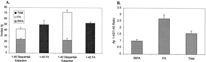
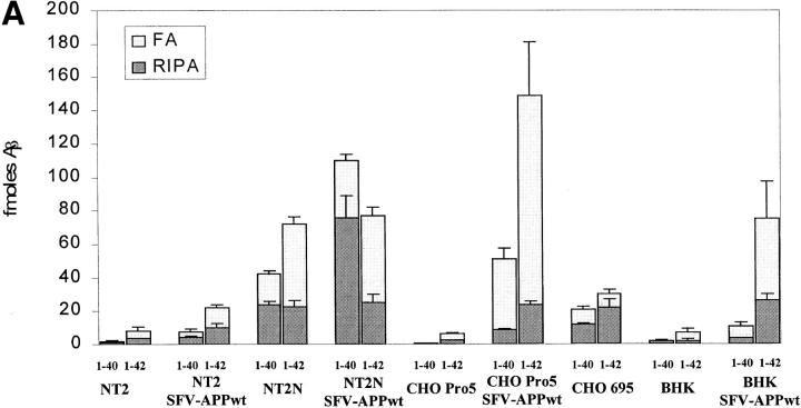
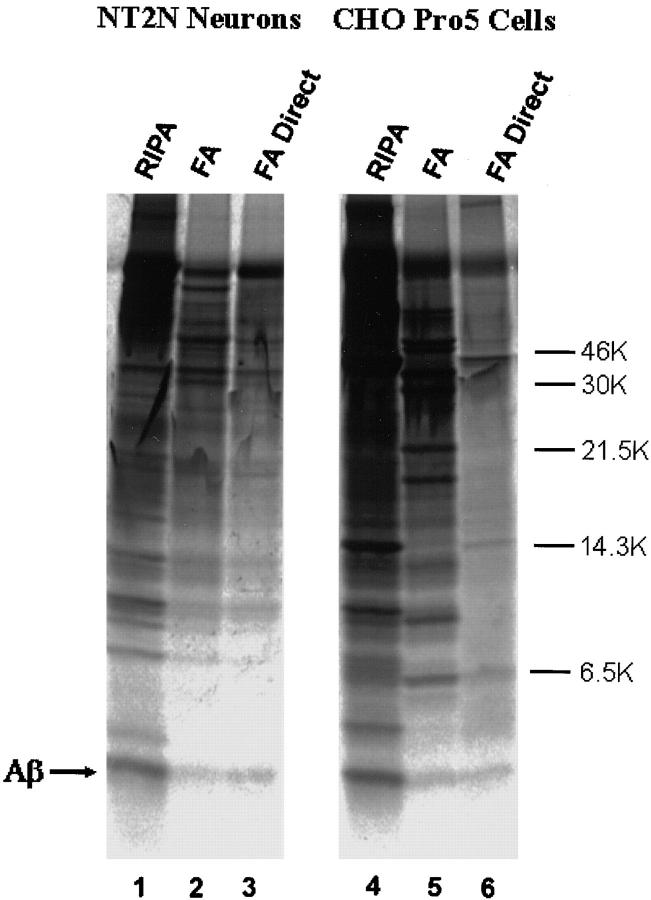
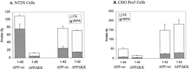
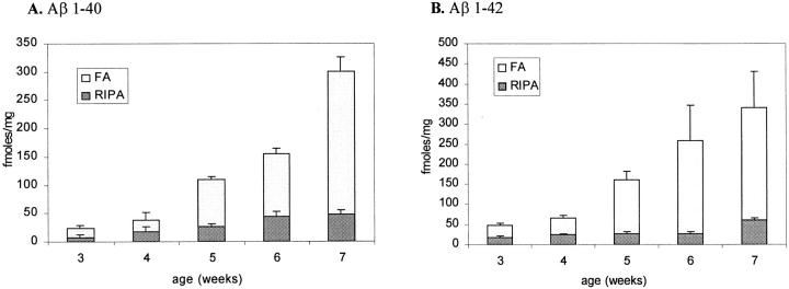
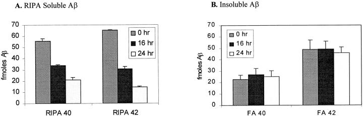
The amyloid-β peptide (Aβ) is produced at several sites within cultured human NT2N neurons with Aβ1-42 specifically generated in the endoplasmic reticulum/intermediate compartment. Since Aβ is found as insoluble deposits in senile plaques of the AD brain, and the Aβ peptide can polymerize into insoluble fibrils in vitro, we examined the possibility that Aβ1-40, and particularly the more highly amyloidogenic Aβ1-42, accumulate in an insoluble pool within NT2N neurons. Remarkably, we found that formic acid extraction of the NT2N cells solubilized a pool of previously undetectable Aβ that accounted for over half of the total intracellular Aβ. Aβ1-42 was more abundant than Aβ1-40 in this pool, and most of the insoluble Aβ1-42 was generated in the endoplasmic reticulum/intermediate compartment pathway. High levels of insoluble Aβ were also detected in several nonneuronal cell lines engineered to overexpress the amyloid-β precursor protein. This insoluble intracellular pool of Aβ was exceptionally stable, and accumulated in NT2N neurons in a time-dependent manner, increasing 12-fold over a 7-wk period in culture. These novel findings suggest that Aβ amyloidogenesis may be initiated within living neurons rather than in the extracellular space. Thus, the data presented here require a reexamination of the prevailing view about the pathogenesis of Aβ deposition in the AD brain.
Overview
The claims and statistics of this paper haven’t been checked yet.
Run the check — free, no account needed. It reads the open-access text and checks the sources it cites.
Integrity signals
Registry
Cites
Statistics
Figures
Verification
Claims the paper makes
The claims and statistics of this paper haven’t been checked yet.
Run the check — free, no account needed. It reads the open-access text and checks the sources it cites.
Claims other papers make about this one
Each is checked against this paper’s own text: does it say what the citing paper says it does?
The source backs part of the claim, not all of it — or backs it for a different population, condition or comparison.
In the citing paper
“The amyloid cascade hypothesis predicts that a gradual increase of A-(1-42) levels in brain interstitial fluid (3,4) may lead to A oligomerization and eventually to A fibrillization (5).”
From Internalization of β-Amyloid Peptide by Primary Neurons in the Absence of Apolipoprotein E (2007) · cites this paper as [3]
Experimental — accuracy not yet measuredIn the citing paper
“Subsequent work showed that particularly the disease-linked Aβ42 peptide, rather than the more abundantly secreted Aβ40 peptide, increased with differentiation and time in culture within clonal NT2 cells that can develop a neuron-like phenotype [174, 195].”
From Intraneuronal β-amyloid accumulation and synapse pathology in Alzheimer’s disease (2010) · cites this paper as [174]
Self-check
Percentages, sums and reported differences re-computed from the paper’s own numbers.
The self-check needs the paper’s full text.
Citations
Cites
Correlative Memory Deficits, Aβ Elevation, and Amyloid Plaques in Transgenic Mice
Hsiao, K., Chapman, P., Nilsen, S., Eckman, C., Harigaya, Y., Younkin, S., Yang, F., & Cole, G. (1996) · Science · doi:10.1126/science.274.5284.99 · Cited by 4,210
Cloning of a gene bearing missense mutations in early-onset familial Alzheimer's disease
Sherrington, R., Rogaev, E. I., Liang, Y., Rogaeva, E. A., Levesque, G., Ikeda, M., Chi, H., Lin, C., Li, G., Holman, K., et al. (1995) · Nature · doi:10.1038/375754a0 · Cited by 4,051
Scheuner, D., Eckman, C., Jensen, M., Song, X., Citron, M., Suzuki, N., Bird, T. D., Hardy, J., Hutton, M., Kukull, W., et al. (1996) · Nature Medicine · doi:10.1038/nm0896-864 · Cited by 2,599
Candidate gene for the chromosome 1 familial Alzheimer’s disease locus
Levy-Lahad, E., Wasco, W., Poorkaj, P., Romano, D. M., Oshima, J., Pettingell, W. H., Yu, C.-E., Jondro, P. D., Schmidt, S. D., Wang, K., et al. (1995) · Science · doi:10.1126/science.7638622 · Cited by 2,577
Alzheimer-type neuropathology in transgenic mice overexpressing V717F β-amyloid precursor protein
Games, D., Adams, D., Alessandrini, R., Barbour, R., Borthelette, P., Blackwell, C., Carr, T., Clemens, J., Donaldson, T., Gillespie, F., et al. (1995) · Nature · doi:10.1038/373523a0 · Cited by 2,516
Jarrett, J. T., & Lansbury, P. T. (1993) · Cell · doi:10.1016/0092-8674(93)90635-4 · Cited by 2,178
Jarrett, J. T., Berger, E. P., & Lansbury, P. T. (1993) · Biochemistry · doi:10.1021/bi00069a001 · Cited by 2,022
Amyloid β-peptide is produced by cultured cells during normal metabolism
Haass, C., Schlossmacher, M. G., Hung, A. Y., Vigo-Pelfrey, C., Mellon, A., Ostaszewski, B. L., Lieberburg, I., Koo, E. H., Schenk, D., Teplow, D. B., et al. (1992) · Nature · doi:10.1038/359322a0 · Cited by 1,921
Iwatsubo, T., Odaka, A., Suzuki, N., Mizusawa, H., Nukina, N., & Ihara, Y. (1994) · Neuron · doi:10.1016/0896-6273(94)90458-8 · Cited by 1,774
Citron, M., Oltersdorf, T., Haass, C., McConlogue, L., Hung, A. Y., Seubert, P., Vigo-Pelfrey, C., Lieberburg, I., & Selkoe, D. J. (1992) · Nature · doi:10.1038/360672a0 · Cited by 1,757
Increased amyloid-β42(43) in brains of mice expressing mutant presenilin 1
Duff, K., Eckman, C., Zehr, C., Yu, X., Prada, C.-M., Pérez‐Tur, J., Hutton, M., Buée, L., Harigaya, Y., Yager, D., et al. (1996) · Nature · doi:10.1038/383710a0 · Cited by 1,486
Borchelt, D. R., Thinakaran, G., Eckman, C. B., Lee, M. K., Davenport, F., Ratovitsky, T., Prada, C.-M., Kim, G., Seekins, S., Yager, D., et al. (1996) · Neuron · doi:10.1016/s0896-6273(00)80230-5 · Cited by 1,478
Suzuki, N., Cheung, T. T., Cai, X.-D., Odaka, A., Otvos, L., Eckman, C., Golde, T. E., & Younkin, S. G. (1994) · Science · doi:10.1126/science.8191290 · Cited by 1,471
Production of the Alzheimer Amyloid β Protein by Normal Proteolytic Processing
Shoji, M., Golde, T. E., Ghiso, J., Cheung, T. T., Estus, S., Shaffer, L. M., Cai, X.-D., McKay, D. M., Tintner, R., Frangione, B., et al. (1992) · Science · doi:10.1126/science.1439760 · Cited by 1,447
Assembly and aggregation properties of synthetic Alzheimer's A4/beta amyloid peptide analogs.
Burdick, D., Soreghan, B., Kwon, M., Kosmoski, J., Knauer, M., Henschen, A., Yates, J., Cotman, C., & Glabe, C. (1992) · Journal of Biological Chemistry · doi:10.1016/s0021-9258(18)48529-8 · Cited by 1,014
Evidence that production and release of amyloid beta-protein involves the endocytic pathway
Koo, E. H., & Squazzo, S. L. (1994) · Journal of Biological Chemistry · doi:10.1016/s0021-9258(17)32449-3 · Cited by 829
Distinct sites of intracellular production for Alzheimer's disease Aβ40/42 amyloid peptides
Hartmann, T., Bieger, S. C., Brühl, B., Tienari, P. J., Ida, N., Allsop, D., Roberts, G. W., Masters, C. L., Dotti, C. G., Unsicker, K., et al. (1997) · Nature Medicine · doi:10.1038/nm0997-1016 · Cited by 719
Pleasure, S., Page, C., & Lee, V. (1992) · Journal of Neuroscience · doi:10.1523/jneurosci.12-05-01802.1992 · Cited by 607
Self-citationKovacs, D. M., Fausett, H. J., Page, K. J., Kim, T., Moir, R. D., Merriam, D. E., Hollister, R. D., Hallmark, O. G., Mancini, R., Felsenstein, K. M., et al. (1996) · Nature Medicine · doi:10.1038/nm0296-224 · Cited by 552
Isolation of Low‐Molecular‐Weight Proteins from Amyloid Plaque Fibers in Alzheimer's Disease
Selkoe, D. J., Abraham, C. R., Podlisny, M. B., & Duffy, L. K. (1986) · Journal of Neurochemistry · doi:10.1111/j.1471-4159.1986.tb08501.x · Cited by 452
Cook, D. G., Forman, M. S., Sung, J. C., Leight, S., Kolson, D. L., Iwatsubo, T., Lee, V. M.-Y., & Doms, R. W. (1997) · Nature Medicine · doi:10.1038/nm0997-1021 · Cited by 444
Self-citationPleasure, S. J., & LEE, V. M.-Y. (1993) · Journal of Neuroscience Research · doi:10.1002/jnr.490350603 · Cited by 437
Self-citationMasliah, E., Sisk, A., Mallory, M. E., Mucke, L., Schenk, D., & Games, D. (1996) · Journal of Neuroscience · doi:10.1523/jneurosci.16-18-05795.1996 · Cited by 389
The Molecular and Genetic Basis of Neurological Disease
Trojanowski, J. Q. (1993) · Journal of Neuropathology & Experimental Neurology · doi:10.1097/00005072-199309000-00013 · Cited by 341
Intracellular Generation and Accumulation of Amyloid β-Peptide Terminating at Amino Acid 42
Wild-Bode, C., Yamazaki, T., Capell, A., Leimer, U., Steiner, H., Ihara, Y., & Haass, C. (1997) · Journal of Biological Chemistry · doi:10.1074/jbc.272.26.16085 · Cited by 303
Xu, H., Sweeney, D. A., Wang, R., Thinakaran, G., Lo, A., Sisodia, S. S., Greengard, P., & Gandy, S. (1997) · Proceedings of the National Academy of Sciences · doi:10.1073/pnas.94.8.3748 · Cited by 281
Intracellular accumulation and resistance to degradation of the Alzheimer amyloid A4/beta protein.
Knauer, M. F., Soreghan, B., Burdick, D., Kosmoski, J., & Glabe, C. G. (1992) · Proceedings of the National Academy of Sciences · doi:10.1073/pnas.89.16.7437 · Cited by 233
Wertkin, A. M., Turner, R. S., Pleasure, S. J., Golde, T. E., Younkin, S. G., Trojanowski, J. Q., & Lee, V. M. (1993) · Proceedings of the National Academy of Sciences · doi:10.1073/pnas.90.20.9513 · Cited by 231
Self-citationCook, D. G., Sung, J. C., Golde, T. E., Felsenstein, K. M., Wójczyk, B. S., Tanzi, R. E., Trojanowski, J. Q., Lee, V. M., & Doms, R. W. (1996) · Proceedings of the National Academy of Sciences · doi:10.1073/pnas.93.17.9223 · Cited by 202
Self-citationTurner, R. S., Suzuki, N., Chyung, A. S. C., Younkin, S. G., & Lee, V. M.-Y. (1996) · Journal of Biological Chemistry · doi:10.1074/jbc.271.15.8966 · Cited by 161
Self-citationAmyloid β protein in plasma from patients with sporadic Alzheimer's disease
Tamaoka, A., Fukushima, T., Sawamura, N., Ishikawa, K., Oguni, E., Komatsuzaki, Y., & Shoji, S. (1996) · Journal of the Neurological Sciences · doi:10.1016/0022-510x(96)00143-8 · Cited by 154
Chyung, A. S. C., Greenberg, B., Cook, D. G., Doms, R. W., & Lee, V. M. ‐. (1997) · The Journal of Cell Biology · doi:10.1083/jcb.138.3.671 · Cited by 136
Self-citationYang, A. J., Knauer, M. F., Burdick, D., & Glabe, C. G. (1995) · Journal of Biological Chemistry · doi:10.1074/jbc.270.24.14786 · Cited by 126
Forman, M. S., Cook, D. G., Leight, S., Doms, R. W., & Lee, V. M. ‐. (1997) · Journal of Biological Chemistry · doi:10.1074/jbc.272.51.32247 · Cited by 57
Self-citationHarigaya, Y., Shoji, M., Kawarabayashi, T., Kanai, M., Nakamura, T., Iizuka, T., Igeta, Y., Saido, T. C., Sahara, N., Mori, H., et al. (1995) · Biochemical and Biophysical Research Communications · doi:10.1006/bbrc.1995.1912 · Cited by 46
Research works printed but not linked: 1
- Kim, 1988, Production and characterization of monoclonal antibodies reactive to synthetic cerebrovascular amyloid peptide, Neurosci Res Commun, 2, 121
Cited by
Sorted by citations
Neuropathological alterations in Alzheimer disease
Serrano-Pozo, A., Frosch, M. P., Masliah, E., & Hyman, B. T. (2011) · Cold Spring Harbor Perspectives in Medicine · doi:10.1101/cshperspect.a006189 · Cited by 3,345
Intracellular amyloid-β in Alzheimer's disease
LaFerla, F. M., Green, K. N., & Oddo, S. (2007) · Nature Reviews Neuroscience · doi:10.1038/nrn2168 · Cited by 1,942
Translating cell biology into therapeutic advances in Alzheimer's disease
Selkoe, D. J. (1999) · Nature · doi:10.1038/399a023 · Cited by 1,166
Deciphering the Molecular Basis of Memory Failure in Alzheimer's Disease
Walsh, D. M., & Selkoe, D. J. (2004) · Neuron · doi:10.1016/j.neuron.2004.09.010 · Cited by 1,207
Metadata
- Authors
- Skovronsky, D. M., Doms, R. W., & Lee, V. M.-Y.
- Affiliations
- University of Pennsylvania (US)
- ORCID
- Lee, Virginia M.-Y.
- Venue
- The Journal of Cell Biology
- Publisher
- Rockefeller University Press
- Published
- 1998-05-18
- Volume
- 141
- Issue
- 4
- Pages
- 1031-1039
- ISSN
- 0021-9525 (ISSN-L)
- 1540-8140
- PMC
- PMC2132781
- OpenAlex
- W2130792655
- Cited by
- 290
- References
- 36
- Citations by year
- 2026: 2 · 2025: 4 · 2024: 4 · 2023: 4 · 2022: 6 · 2021: 3 · 2019: 8 · 2018: 2 · 2017: 4 · 2016: 1 · 2015: 6 · 2014: 3 · 2013: 8 · 2012: 8
- FWCI
- 15.16
- Type
- journal article
- Language
- en
- Open access
- green
- www.ncbi.nlm.nih.gov
- Licence
- cc-by-nc-sa
- Locations
- The Journal of Cell Biology (publishedVersion)
- Europe PMC (PubMed Central) (publishedVersion, cc-by-nc-sa)
- PubMed Central (publishedVersion, cc-by-nc-sa)
- PubMed (publishedVersion)
- Topics
- Life Sciences › Neuroscience › Cellular and Molecular Neuroscience › Alzheimer's disease research and treatments
- Health Sciences › Medicine › Psychiatry and Mental health › Dementia and Cognitive Impairment Research
- Life Sciences › Neuroscience › Neurology › Neuroinflammation and Neurodegeneration Mechanisms
- Keywords
- amyloid-beta, amyloid-beta 42, Aβ40, endoplasmic reticulum, amyloidogenesis, Alzheimer's disease, amyloid plaques, amyloid precursor protein overexpression, formic acid extraction
- MeSH terms
- Alzheimer Disease, Animals, Brain, Cell Line, Endoplasmic Reticulum, Genetic Vectors, Cricetinae, Humans, Neurons, Peptide Fragments, Recombinant Proteins, Semliki forest virus, Solubility, Subcellular Fractions, Transfection, Tumor Cells, Cultured, Amyloid beta-Peptides, CHO Cells, Carcinoma, Embryonal
About these checks
Registry, retraction, Cites and statistics results are computed, not judged, with one exception: a printed reference that no index lists is read once by a model before it counts as a citation error. Claims are extracted by a model and kept only when their quote appears verbatim in the text; each cited source is then checked, with a model’s help, for whether it says what the claim relies on. Those verdicts can be wrong — read the claim’s quote and the cited source before relying on one.
Statistics and arithmetic are recomputed, not judged.
Confidence bands are shown only for fields calibrated on our annotated test set. None is calibrated yet, so no band is shown.
What counts as a claim ›How we measure confidence ›How we check this ›
Are you an author, or do you see a problem with this page? Request a review
