

The DOI matches its Crossref record; 58 of the 58 references it cites were found in a registry or an index.
Assembly of Myelin by Association of Proteolipid Protein with Cholesterol- and Galactosylceramide-Rich Membrane Domains
Open accessAbstract
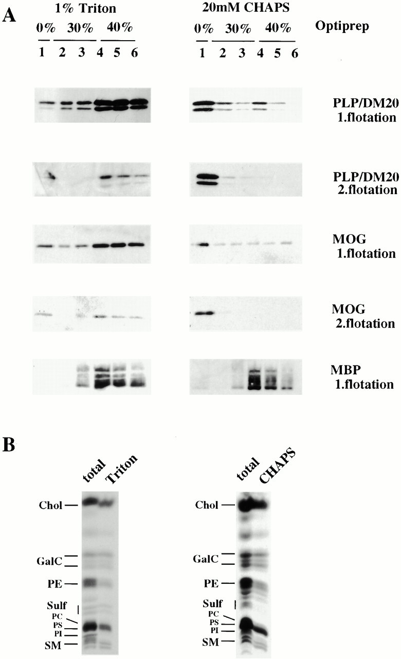
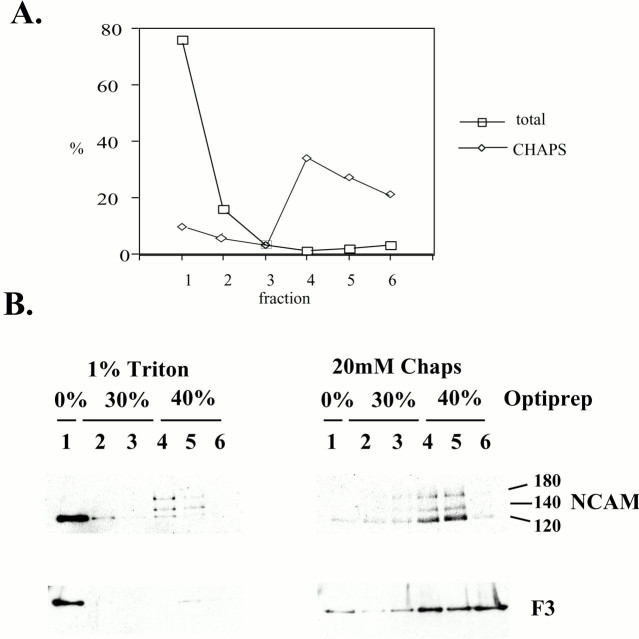
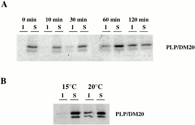
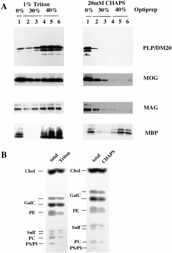
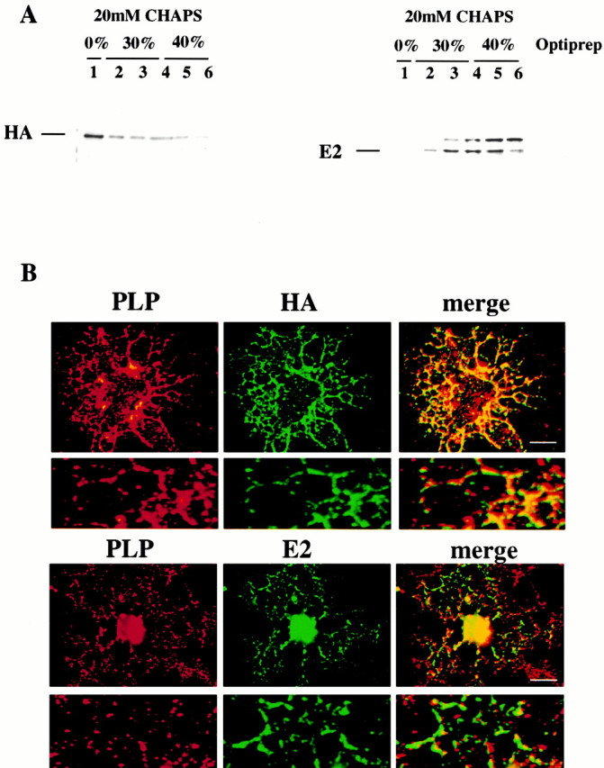
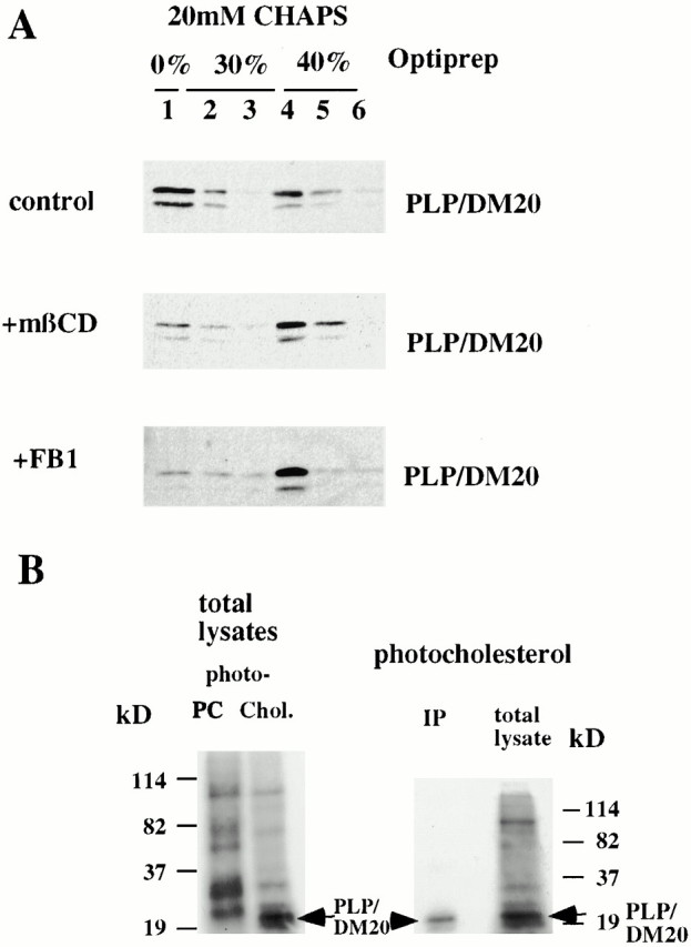
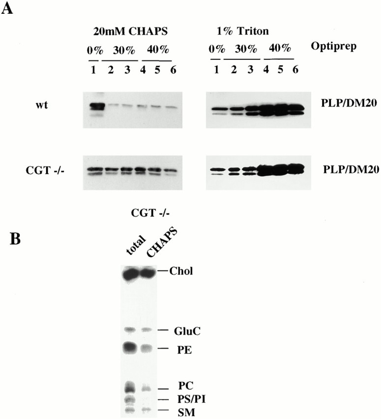
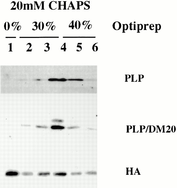
Myelin is a specialized membrane enriched in glycosphingolipids and cholesterol that contains a limited spectrum of proteins. We investigated the assembly of myelin components by oligodendrocytes and analyzed the role of lipid–protein interactions in this process. Proteolipid protein (PLP), the major myelin protein, was recovered from cultured oligodendrocytes from a low-density CHAPS-insoluble membrane fraction (CIMF) enriched in myelin lipids. PLP associated with the CIMF after leaving the endoplasmic reticulum but before exiting the Golgi apparatus, suggesting that myelin lipid and protein components assemble in the Golgi complex. The specific association of PLP with myelin lipids in CIMF was supported by the finding that it was efficiently cross-linked to photoactivable cholesterol, but not to phosphatidylcholine, which is underrepresented in both myelin and CIMF. Furthermore, depletion of cholesterol or inhibition of sphingolipid synthesis in oligodendrocytes abolished the association of PLP with CIMF. Thus, PLP may be recruited to myelin rafts, represented by CIMF, via lipid–protein interactions. In contrast to oligodendrocytes, after transfection in BHK cells, PLP is absent from isolated CIMF, suggesting that PLP requires specific lipids for raft association. In mice deficient in the enzyme ceramide galactosyl transferase, which cannot synthesize the main myelin glycosphingolipids, a large fraction of PLP no longer associates with rafts. Formation of a cholesterol- and galactosylceramide-rich membrane domain (myelin rafts) may be critical for the sorting of PLP and assembly of myelin in oligodendrocytes.
Overview
The claims and statistics of this paper haven’t been checked yet.
Run the check — free, no account needed. It reads the open-access text and checks the sources it cites.
Integrity signals
Registry
Cites
Statistics
Figures
Verification
Claims the paper makes
The claims and statistics of this paper haven’t been checked yet.
Run the check — free, no account needed. It reads the open-access text and checks the sources it cites.
Claims other papers make about this one
Each is checked against this paper’s own text: does it say what the citing paper says it does?
The source backs part of the claim, not all of it — or backs it for a different population, condition or comparison.
In the citing paper
“The extra cholesterol supply is not simply the building blocks for lipid content in the myelin sheet, but also the key for proper myelin protein assembly and sorting (Simons et al. 2000).”
From Apolipoprotein E ε4 disrupts oligodendrocyte differentiation by interfering with astrocyte‐derived lipid transport (2023) · cites this paper as Simons et al. 2000
Experimental — accuracy not yet measured
Self-check
Percentages, sums and reported differences re-computed from the paper’s own numbers.
The self-check needs the paper’s full text.
Citations
Cites
A RAPID METHOD OF TOTAL LIPID EXTRACTION AND PURIFICATION
Bligh, E. G., & Dyer, W. J. (1959) · Canadian Journal of Biochemistry and Physiology · doi:10.1139/o59-099 · Cited by 54,751
Functional rafts in cell membranes
Simons, K., & Ikonen, E. (1997) · Nature · doi:10.1038/42408 · Cited by 9,757
Srimaroeng, C., Cecile, J. P., Walden, R., & Pritchard, J. B. (2013) · Cellular Physiology and Biochemistry · doi:10.1159/000350077 · Cited by 2,989
Brown, D. A., & Rose, J. K. (1992) · Cell · doi:10.1016/0092-8674(92)90189-j · Cited by 2,921
Structure and Function of Sphingolipid- and Cholesterol-rich Membrane Rafts
Brown, D. A., & London, E. (2000) · Journal of Biological Chemistry · doi:10.1074/jbc.r000005200 · Cited by 2,344
Adams, R. D., & Victor, M. (1977) · Medical Entomology and Zoology · Cited by 1,718
ENDOCYTOSIS AND MOLECULAR SORTING
Mellman, I. (1996) · Annual Review of Cell and Developmental Biology · doi:10.1146/annurev.cellbio.12.1.575 · Cited by 1,552
Lipid sorting in epithelial cells
Simons, K., & Gerrit van, M. E. E. R. (1988) · Biochemistry · doi:10.1021/bi00417a001 · Cited by 1,373
GPI-anchored proteins are organized in submicron domains at the cell surface
Varma, R., & Mayor, S. (1998) · Nature · doi:10.1038/29563 · Cited by 1,209
Lipid Domain Structure of the Plasma Membrane Revealed by Patching of Membrane Components
Harder, T., Scheiffele, P., Verkade, P., & Simons, K. (1998) · The Journal of Cell Biology · doi:10.1083/jcb.141.4.929 · Cited by 1,186
Cited by
Sorted by citations
Items 101–200 of 292
Schweitzer, J., Becker, T., Schachner, M., Nave, K., & Werner, H. B. (2005) · Molecular and Cellular Neuroscience · doi:10.1016/j.mcn.2005.10.007 · Cited by 63
Boggs, J. M., Gao, W., Zhao, J., Park, H.-J., Liu, Y., & Basu, A. (2009) · FEBS Letters · doi:10.1016/j.febslet.2009.11.074 · Cited by 63
Lüders, K. A., Nessler, S., Kusch, K., Patzig, J., Jung, R. B., Möbius, W., Nave, K., & Werner, H. B. (2019) · Glia · doi:10.1002/glia.23549 · Cited by 62
Protein-sphingolipid interactions within cellular membranes
Haberkant, P., Schmitt, O., Contreras, F. ‐., Thiele, C., Hanada, K., Sprong, H., Reinhard, C., Wieland, F., & Brügger, B. (2007) · Journal of Lipid Research · doi:10.1194/jlr.d700023-jlr200 · Cited by 62
Self-citationBoggs, J., Gao, W., & Hirahara, Y. (2008) · Biochimica et Biophysica Acta · doi:10.1016/j.bbagen.2007.10.015 · Cited by 70
Myelin: Methods for Purification and Proteome Analysis
Erwig, M. S., Hesse, D., Jung, R. B., Uecker, M., Kusch, K., Tenzer, S., Jahn, O., & Werner, H. B. (2019) · Methods in molecular biology · doi:10.1007/978-1-4939-9072-6_3 · Cited by 61
Expression of distinct splice variants of the stem cell marker prominin‐1 (CD133) in glial cells
Corbeil, D., Joester, A., Fargeas, C. A., Jászai, J., Garwood, J., Hellwig, A., Werner, H. B., & Huttner, W. Β. (2008) · Glia · doi:10.1002/glia.20812 · Cited by 59
Kippert, A., Trajković, K., Rajendran, L., Ries, J., & Simons, M. (2007) · Journal of Neuroscience · doi:10.1523/jneurosci.4926-06.2007 · Cited by 58
Self-citationMyelination of Neuronal Cell Bodies when Myelin Supply Exceeds Axonal Demand
Almeida, R., Pan, S., Marshall-Phelps, K. L., Williamson, J., Early, J. J., Czopka, T., Klingseisen, A., Chan, J. R., & Lyons, D. A. (2018) · Current Biology · doi:10.1016/j.cub.2018.02.068 · Cited by 61
Polarity Development in Oligodendrocytes: Sorting and Trafficking of Myelin Components
Maier, O., Hoekstra, D., & Baron, W. (2008) · Journal of Molecular Neuroscience · doi:10.1007/s12031-007-9024-8 · Cited by 61
de Monasterio‐Schrader, P., Patzig, J., Möbius, W., Barrette, B., Wagner, T. L., Kusch, K., Edgar, J. M., Brophy, P., & Werner, H. B. (2013) · Glia · doi:10.1002/glia.22561 · Cited by 58
ABCA2: a candidate regulator of neural transmembrane lipid transport
Schmitz, G., & Kaminski, W. E. (2002) · Cellular and Molecular Life Sciences · doi:10.1007/s00018-002-8508-5 · Cited by 57
Central nervous system cholesterol metabolism in health and disease
Ho, W. Y., Hartmann, H., & Ling, S. (2022) · IUBMB Life · doi:10.1002/iub.2662 · Cited by 58
p38 mitogen‐activated protein kinase is required for central nervous system myelination
Fragoso, G., Haines, J. D., Roberston, J., Pedraza, L., Mushynski, W. E., & Almazán, G. (2007) · Glia · doi:10.1002/glia.20567 · Cited by 55
FA2H is responsible for the formation of 2-hydroxy galactolipids in peripheral nervous system myelin
Maldonado, E. N., Alderson, N. L., Monje, P. V., Wood, P. M., & Hama, H. (2007) · Journal of Lipid Research · doi:10.1194/jlr.m700400-jlr200 · Cited by 53
Taylor, C. M., & Pfeiffer, S. E. (2003) · PROTEOMICS · doi:10.1002/pmic.200300451 · Cited by 52
Golgi trafficking defects in postnatal microcephaly: The evidence for “Golgipathies”
Passemard, S., Perez, F., Colin, E., Rasika, S., Gressèns, P., & Ghouzzi, V. E. (2017) · Progress in Neurobiology · doi:10.1016/j.pneurobio.2017.03.007 · Cited by 50
Pelizaeus-Merzbacher Disease and Spastic Paraplegia Type 2
Hudson, L. D. (2003) · Journal of Child Neurology · doi:10.1177/08830738030180090801 · Cited by 52
Kippert, A., Trajković, K., Fitzner, D., Opitz, L., & Simons, M. (2008) · BMC Neuroscience · doi:10.1186/1471-2202-9-40 · Cited by 46
Self-citationDrug discovery for remyelination and treatment of MS
Marshall-Phelps, K. L., Early, J. J., & Lyons, D. A. (2017) · Glia · doi:10.1002/glia.23166 · Cited by 54
The septin cytoskeleton in myelinating glia
Buser, A., Erne, B., Werner, H. B., Nave, K., & Schaeren‐Wiemers, N. (2008) · Molecular and Cellular Neuroscience · doi:10.1016/j.mcn.2008.10.002 · Cited by 52
Feldmann, A., Winterstein, C., White, R., Trotter, J., & Krämer‐Albers, E. (2009) · Journal of Neuroscience Research · doi:10.1002/jnr.22020 · Cited by 47
Self-citationLipid dysfunction and pathogenesis of multiple system atrophy
Bleasel, J. M., Wong, J. H., Halliday, G. M., & Kim, W. S. (2014) · Acta Neuropathologica Communications · doi:10.1186/2051-5960-2-15 · Cited by 47
Scorticati, C., Formoso, K., & Frasch, A. C. C. (2011) · Journal of Neurochemistry · doi:10.1111/j.1471-4159.2011.07252.x · Cited by 47
Immunohistochemical localization of the sigma1 receptor in Schwann cells of rat sciatic nerve
Palacios, G., Muro, A., Verdú, E., Pumarola, M. B., & Vela, J. M. (2004) · Brain Research · doi:10.1016/j.brainres.2004.02.013 · Cited by 48
Exploring Myelin Dysfunction in Multiple System Atrophy
Wong, J. H., Halliday, G. M., & Kim, W. S. (2014) · Experimental Neurobiology · doi:10.5607/en.2014.23.4.337 · Cited by 45
Oliveira, R. G., & Maggio, B. (2002) · Biochimica et Biophysica Acta (BBA) - Biomembranes · doi:10.1016/s0005-2736(02)00350-4 · Cited by 45
Ozgen, H., Schrimpf, W., Hendrix, J., de Jonge, J. C., Lamb, D. C., Hoekstra, D., Kahya, N., & Baron, W. (2014) · PLoS ONE · doi:10.1371/journal.pone.0101834 · Cited by 44
Distribution of cholesterol and galactosylceramide in rat cerebellar white matter
Börner, K., NYGREN, H., Hagenhoff, B., Malmberg, P., Tallarek, E., & Månsson, J. (2006) · Biochimica et Biophysica Acta (BBA) - Molecular and Cell Biology of Lipids · doi:10.1016/j.bbalip.2006.02.021 · Cited by 44
Lovastatin induces the formation of abnormal myelin‐like membrane sheets in primary oligodendrocytes
Maier, O., Jonge, J. D., Nomden, A., Hoekstra, D., & Baron, W. (2008) · Glia · doi:10.1002/glia.20769 · Cited by 43
Biogenesis and Significance of Central Nervous System Myelin
Soldán, M. M. P., & Pirko, I. (2012) · Seminars in Neurology · doi:10.1055/s-0032-1306381 · Cited by 45
Boggs, J. M., Wang, H., Gao, W., Arvanitis, D. N., Gong, Y., & Min, W. (2004) · Glycoconjugate Journal · doi:10.1023/b:glyc.0000044842.34958.f8 · Cited by 43
LEE, A. G. J. (2001) · Current Biology · doi:10.1016/s0960-9822(01)00008-2 · Cited by 41
Age-related axonal and myelin changes in the rumpshaker mutation of the Plp gene
Edgar, J. M., McLaughlin, M., Barrie, J. A., McCulloch, M. C., Garbern, J. Y., & Griffiths, I. R. (2004) · Acta Neuropathologica · doi:10.1007/s00401-003-0808-9 · Cited by 41
Myelin Biogenesis: Vesicle Transport in Oligodendrocytes
Larocca, J. N., & Rodriguez‐Gabin, A. (2002) · Neurochemical Research · doi:10.1023/a:1021667515030 · Cited by 41
Delay of myelin formation in arylsulphatase A‐deficient mice
Yaghootfam, A., Gieselmann, V., & Eckhardt, M. (2005) · European Journal of Neuroscience · doi:10.1111/j.1460-9568.2005.03891.x · Cited by 41
Bosse, F., Hasse, B., Pippirs, U., Greiner–Petter, R., & Müller, H. (2003) · Journal of Neurochemistry · doi:10.1046/j.1471-4159.2003.01870.x · Cited by 40
The Role of 3-O-Sulfogalactosylceramide, Sulfatide, in the Lateral Organization of Myelin Membrane
Grassi, S., Prioni, S., Cabitta, L., Aureli, M., Sonnino, S., & Prinetti, A. (2015) · Neurochemical Research · doi:10.1007/s11064-015-1747-2 · Cited by 39
Myelin sheaths are formed with proteins that originated in vertebrate lineages
Gould, R. M., Oakley, T. H., Goldstone, J. V., Dugas, J. C., Brady, S. T., & Gow, A. (2008) · Neuron Glia Biology · doi:10.1017/s1740925x09990238 · Cited by 37
Baron, W., Ozgen, H., Klunder, B., de Jonge, J. C., Nomden, A., Plat, A., Trifilieff, É., de Vries, H., & Hoekstra, D. (2014) · Molecular and Cellular Biology · doi:10.1128/mcb.00848-14 · Cited by 38
Metachromatic leukodystrophy: consequences of sulphatide accumulation
Gieselmann, V., Franken, S., Klein, D., Je, M., Sandhoff, R., Rauch, R. L., Hartmann, D., Saravanan, V. P. M., Deyn, P. D., D’Hooge, R., et al. (2003) · Acta Paediatrica · doi:10.1111/j.1651-2227.2003.tb00225.x · Cited by 38
Mok, K. K., Yeung, S. H., Cheng, G. W., Ma, I. W., Lee, R. H., Herrup, K., & Tse, K. (2023) · Journal of Neurochemistry · doi:10.1111/jnc.15748 · Cited by 37
Inhibition of neutral sphingomyelinase 2 promotes remyelination
Yoo, S.-W., Agarwal, A., Smith, M. D., Khuder, S. S., Baxi, E. G., Thomas, A. G., Rojas, C. J., Moniruzzman, M., Slusher, B. S., Bergles, D. E., et al. (2020) · Science Advances · doi:10.1126/sciadv.aba5210 · Cited by 38
Boggs, J. M., & Wang, H. (2004) · Journal of Neuroscience Research · doi:10.1002/jnr.20080 · Cited by 36
Lipid Rafts: The Maestros of Normal Brain Development
Viljetić, B., Blažetić, S., Labak, I., Ivić, V., Zjalić, M., Heffer, M., & Balog, M. (2024) · Biomolecules · doi:10.3390/biom14030362 · Cited by 36
Castello-Serrano, I., Lorent, J. H., Ippolito, R., Levental, K. R., & Levental, I. (2020) · The Journal of Physical Chemistry B · doi:10.1021/acs.jpcb.0c03028 · Cited by 35
A role for Sec8 in oligodendrocyte morphological differentiation
Anitei, M., Ifrim, M. F., Ewart, M., Cowan, A. E., Carson, J. H., Bansal, R., & Pfeiffer, S. E. (2006) · Journal of Cell Science · doi:10.1242/jcs.02785 · Cited by 35
Behavior of a photoactivatable analog of cholesterol, 6‐photocholesterol, in model membranes
Mintzer, E. A., Waarts, B.-L., Wilschut, J., & Bittman, R. (2001) · FEBS Letters · doi:10.1016/s0014-5793(01)03271-9 · Cited by 35
Battis, K., Xiang, W., & Winkler, J. (2023) · International Journal of Molecular Sciences · doi:10.3390/ijms241713270 · Cited by 38
Gene suppressing therapy for Pelizaeus-Merzbacher disease using artificial microRNA
Li, H., Okada, H., Suzuki, S., Sakai, K., Izumi, H., Matsushima, Y., Ichinohe, N., Goto, Y., Okada, T., & Inoue, K. (2019) · JCI Insight · doi:10.1172/jci.insight.125052 · Cited by 35
Boggs, J. M., Gao, W., & Hirahara, Y. (2008) · Journal of Neuroscience Research · doi:10.1002/jnr.21603 · Cited by 37
Marazuela, M., Acevedo, A., Adrados, M., García-López, M. Á., & Alonso, M. A. (2003) · Journal of Histochemistry & Cytochemistry · doi:10.1177/002215540305100512 · Cited by 34
Qki regulates myelinogenesis through Srebp2-dependent cholesterol biosynthesis
Zhou, X., Shin, S., He, C., Zhang, Q., Rasband, M. N., Ren, J., Dai, C., Zorrilla-Veloz, R. I., Shingu, T., Yuan, L., et al. (2021) · eLife · doi:10.7554/elife.60467 · Cited by 39
Kramer, W., Girbig, F., Corsiero, D., Bürger, K., Fahrenholz, F., Jung, C. K. E., & Müller, G. A. (2003) · Biochimica et Biophysica Acta (BBA) - Molecular and Cell Biology of Lipids · doi:10.1016/s1388-1981(03)00068-4 · Cited by 33
Gregor, A., Kramer, J. M., van der Voet, M., Schanze, I., Uebe, S., Donders, R. A. R. T., Reis, A., Schenck, A., & Zweier, C. (2014) · Human Mutation · doi:10.1002/humu.22697 · Cited by 32
Arvanitis, D. N., Yang, W., & Boggs, J. M. (2002) · Journal of Neuroscience Research · doi:10.1002/jnr.10383 · Cited by 32
PLP/DM20 Expression and turnover in a transgenic mouse model of pelizaeus‐merzbacher disease
Karim, S. A., Barrie, J. A., McCulloch, M. C., Montague, P., Edgar, J. M., Iden, D. L., Anderson, T. J., Nave, K., Griffiths, I. R., & McLaughlin, M. (2010) · Glia · doi:10.1002/glia.21043 · Cited by 33
Khalaf, G., Mattern, C., Bégou, M., Boespflug‐Tanguy, O., Massaad, C., & Massaad-Massade, L. (2022) · Biomedicines · doi:10.3390/biomedicines10071709 · Cited by 35
Cell polarity in myelinating glia: From membrane flow to diffusion barriers
Simons, M., Snaidero, N., & Aggarwal, S. (2012) · Biochimica et Biophysica Acta (BBA) - Molecular and Cell Biology of Lipids · doi:10.1016/j.bbalip.2012.01.011 · Cited by 30
Self-citationMolecular Design for Reversing the Photoswitching Mode of Turning ON and OFF DNA Hybridization
Liang, X., Takenaka, N., Nishioka, H., & Asanuma, H. (2008) · Chemistry - An Asian Journal · doi:10.1002/asia.200700384 · Cited by 30
Protein-lipid interactions: paparazzi hunting for snap-shots
Haberkant, P., & Gerrit van, M. E. E. R. (2009) · Biological Chemistry · doi:10.1515/bc.2009.074 · Cited by 30
Maier, O., Baron, W., & Hoekstra, D. (2007) · Glia · doi:10.1002/glia.20510 · Cited by 30
Gene expression in mononuclear cells from patients with inflammatory bowel disease
Mannick, E. E., Bonomolo, J. C., Horswell, R. L., Lentz, J. J., Serrano, M.-S., Zapata-Velandia, A. M., Gastañaduy, M., Himel, J. L., Rose, S. L., Udall, J. N., et al. (2004) · Clinical Immunology · doi:10.1016/j.clim.2004.03.014 · Cited by 30
The Transport, Assembly, and Function of Myelin Lipids
Taylor, C. M., Marta, C. B., Bansal, R., & Pfeiffer, S. E. (2004) · Elsevier eBooks · doi:10.1016/b978-012439510-7/50056-5 · Cited by 30
Isokawa, M., Sassa, T., Hattori, S., Miyakawa, T., & Kihara, A. (2019) · FASEB BioAdvances · doi:10.1096/fba.2019-00067 · Cited by 29
Fernández, M. E., Alfonso, J., Brocco, M. A., & Frasch, A. C. C. (2009) · Journal of Neuroscience Research · doi:10.1002/jnr.22298 · Cited by 28
Trafficking of PLP/DM20 and cAMP signaling in immortalized jimpy oligodendrocytes
Ghandour, M. S., Feutz, A. C., Jalabi, W., Taleb, O., Bessert, D. A., Cypher, M. A., Carlock, L. R., & Skoff, R. P. (2002) · Glia · doi:10.1002/glia.10122 · Cited by 28
Marta, C. B., Taylor, C. M., Cheng, S.-Y., Quarles, R. H., Bansal, R., & Pfeiffer, S. E. (2004) · Neuron Glia Biology · doi:10.1017/s1740925x04000067 · Cited by 28
Frajerman, A., Scoriels, L., Kébir, O., & Chaumette, B. (2021) · International Journal of Molecular Sciences · doi:10.3390/ijms22136881 · Cited by 31
Malewicz, B., Valiyaveettil, J. T., Jacob, K., Byun, H., Mattjus, P., Baumann, W. J., Bittman, R., & Brown, R. E. (2005) · Biophysical Journal · doi:10.1529/biophysj.104.057059 · Cited by 27
Bauch, J., & Faissner, A. (2022) · Cells · doi:10.3390/cells11111773 · Cited by 27
Owens, G. P., Fellin, T. J., Matschulat, A. M., Salas, V., Schaller, K., Given, K. S., Ritchie, A. M., Navarro, A., Blauth, K., Hughes, E. G., et al. (2023) · Journal of Clinical Investigation · doi:10.1172/jci162731 · Cited by 27
DeBruin, L. S., Haines, J. D., Bienzle, D., & Harauz, G. (2006) · Biochemistry and Cell Biology · doi:10.1139/o06-180 · Cited by 26
Meixner, M., Jungnickel, J., Grothe, C., Gieselmann, V., & Eckhardt, M. (2011) · BMC Neuroscience · doi:10.1186/1471-2202-12-22 · Cited by 26
Hu, C., Zhang, D., Slipchenko, M. N., Cheng, J., & Hu, B. (2014) · Journal of Biomedical Optics · doi:10.1117/1.jbo.19.8.086005 · Cited by 27
Guo, D.-Z., Xiao, L., Liu, Y.-J., Shen, C., Lou, H.-F., Lv, Y., & Pan, S.-Y. (2018) · Experimental & Molecular Medicine · doi:10.1038/emm.2017.291 · Cited by 25
Bleasel, J. M., Hsiao, J.-H. T., Halliday, G. M., & Kim, W. S. (2013) · Journal of Parkinson s Disease · doi:10.3233/jpd-130203 · Cited by 26
Benzophenone-containing cholesterol surrogates
Spencer, T. A., Wang, P., Li, D., Russel, J. S., Blank, D. H. A., Huuskonen, J., Fielding, P. E., & Fielding, C. J. (2004) · Journal of Lipid Research · doi:10.1194/jlr.m400081-jlr200 · Cited by 25
Kroepfl, J. F., & Gardinier, M. V. (2001) · Journal of Neurochemistry · doi:10.1046/j.1471-4159.2001.00343.x · Cited by 25
Edgar, J. M., & Griffiths, I. R. (2009) · Elsevier eBooks · doi:10.1016/b978-0-12-374709-9.00005-5 · Cited by 25
Jurevics, H. A., Hostettler, J., Sammond, D. W., Nave, K., Toews, A. D., & Morell, P. (2003) · Journal of Neuroscience Research · doi:10.1002/jnr.10544 · Cited by 24
Polarization and Myelination in Myelinating Glia
Masaki, T. (2012) · ISRN Neurology · doi:10.5402/2012/769412 · Cited by 27
Rouvinski, A., Gahali-Sass, I., Stav, I., Metzer, E., Atlan, H., & Taraboulos, A. (2003) · Biochemical and Biophysical Research Communications · doi:10.1016/s0006-291x(03)01470-0 · Cited by 24
Glycoprotein M6a is present in glutamatergic axons in adult rat forebrain and cerebellum
Cooper, B. H., Werner, H. B., & Flügge, G. (2008) · Brain Research · doi:10.1016/j.brainres.2007.11.066 · Cited by 24
García, M. D., Formoso, K., Aparicio, G. I., Frasch, A. C. C., & Scorticati, C. (2017) · Frontiers in Molecular Neuroscience · doi:10.3389/fnmol.2017.00296 · Cited by 23
Wang, C., Deneen, B., & Tzeng, S. (2019) · Glia · doi:10.1002/glia.23660 · Cited by 23
Boggs, J. M. (2014) · Advances in neurobiology · doi:10.1007/978-1-4939-1154-7_12 · Cited by 22
Taguchi, K., Yoshinaka, K., Yoshino, K., Yonezawa, K., & Maékawa, S. (2005) · Journal of Neuroscience Research · doi:10.1002/jnr.20555 · Cited by 22
DeJesus, G., & Bizzozero, O. A. (2002) · Neurochemical Research · doi:10.1023/a:1021643229028 · Cited by 22
Emerging mitochondrial‐mediated mechanisms involved in oligodendrocyte development
Gil, M., & Gama, V. (2022) · Journal of Neuroscience Research · doi:10.1002/jnr.25151 · Cited by 24
Insertion of mutant proteolipid protein results in missorting of myelin proteins
Vaurs‐Barrière, C., Wong, K., Weibel, T., Abu-Asab, M. S., Weiss, M. D., Kaneski, C. R., Mixon, T., Bonavita, S., Creveaux, I., Heiss, J. D., et al. (2003) · Annals of Neurology · doi:10.1002/ana.10762 · Cited by 21
Kroepfl, J. F., & Gardinier, M. V. (2001) · Journal of Neuroscience Research · doi:10.1002/jnr.10035 · Cited by 18
Bijlard, M., Klunder, B., de Jonge, J. C., Nomden, A., Tyagi, S., de Vries, H., Hoekstra, D., & Baron, W. (2014) · Molecular and Cellular Biology · doi:10.1128/mcb.01389-14 · Cited by 20
Cellular Signal-Regulated Schwann Cell Myelination and Remyelination
Torii, T., Miyamoto, Y., & Yamauchi, J. (2019) · Advances in experimental medicine and biology · doi:10.1007/978-981-32-9636-7_1 · Cited by 20
Isolation and Analysis of Detergent-Resistant Membrane Fractions
Aureli, M., Grassi, S., Sonnino, S., & Prinetti, A. (2015) · Methods in molecular biology · doi:10.1007/978-1-4939-3170-5_10 · Cited by 20
Hudson, L. D., Garbern, J. Y., & Kamholz, J. A. (2004) · Elsevier eBooks · doi:10.1016/b978-012439510-7/50090-5 · Cited by 20
Evidence for possible interactions between PLP and DM20 within the myelin sheath
McLaughlin, M., Hunter, D. J. B., Thomson, C. E., Yool, D. A., Kirkham, D., Freer, A. A., & Griffiths, I. R. (2002) · Glia · doi:10.1002/glia.10091 · Cited by 20
Hudson, L. D. (2004) · Elsevier eBooks · doi:10.1016/b978-012439510-7/50069-3 · Cited by 19
Fewou, S. N., Fernandes, A., Stockdale, K., Francone, V. P., Dupree, J. L., Rosenbluth, J., Pfeiffer, S. E., & Bansal, R. (2009) · Journal of Neurochemistry · doi:10.1111/j.1471-4159.2009.06464.x · Cited by 18
Metadata
- Authors
- Simons, M., Krämer, E.-M., Thiele, C., Stoffel, W., & Trotter, J.
- Affiliations
- Heidelberg University (DE)
- aDepartment of Neurobiology, University of Heidelberg, 69120 Heidelberg, Germany
- University of Cologne (DE)
- Venue
- The Journal of Cell Biology
- Publisher
- Rockefeller University Press
- Published
- 2000-10-02
- Volume
- 151
- Issue
- 1
- Pages
- 143-154
- ISSN
- 0021-9525 (ISSN-L)
- 1540-8140
- PMC
- PMC2189802
- OpenAlex
- W2098744846
- Cited by
- 293
- References
- 58
- Citations by year
- 2026: 5 · 2025: 2 · 2024: 7 · 2023: 6 · 2022: 12 · 2021: 5 · 2020: 10 · 2019: 15 · 2018: 3 · 2017: 12 · 2016: 4 · 2015: 8 · 2014: 17 · 2013: 9 · 2012: 14
- FWCI
- 5.31
- Type
- journal article
- Language
- en
- Open access
- green
- www.ncbi.nlm.nih.gov
- Licence
- cc-by-nc-sa
- Locations
- The Journal of Cell Biology (publishedVersion)
- Europe PMC (PubMed Central) (publishedVersion, cc-by-nc-sa)
- PubMed Central (publishedVersion, cc-by-nc-sa)
- PubMed (publishedVersion)
- Topics
- Life Sciences › Biochemistry, Genetics and Molecular Biology › Molecular Biology › Lipid Membrane Structure and Behavior
- Life Sciences › Biochemistry, Genetics and Molecular Biology › Molecular Biology › Sphingolipid Metabolism and Signaling
- Life Sciences › Biochemistry, Genetics and Molecular Biology › Cell Biology › Caveolin-1 and cellular processes
- Keywords
- proteolipid protein, lipid rafts, oligodendrocytes, cholesterol, galactosylceramide, glycosphingolipids, protein-lipid interactions, Golgi apparatus, sphingolipid biosynthesis
- MeSH terms
- Animals, Brain Chemistry, Cell Fractionation, Cholesterol, Cholic Acids, Detergents, Endoplasmic Reticulum, Galactosylceramides, Galactosyltransferases, Golgi Apparatus, Cricetinae, Myelin Sheath, Oligodendroglia, Solubility, Glycosylphosphatidylinositols, Mice, Knockout, Myelin Proteolipid Protein, Hemagglutinin Glycoproteins, Influenza Virus, Membrane Microdomains, N-Acylsphingosine Galactosyltransferase, Mice
About these checks
Registry, retraction, Cites and statistics results are computed, not judged, with one exception: a printed reference that no index lists is read once by a model before it counts as a citation error. Claims are extracted by a model and kept only when their quote appears verbatim in the text; each cited source is then checked, with a model’s help, for whether it says what the claim relies on. Those verdicts can be wrong — read the claim’s quote and the cited source before relying on one.
Statistics and arithmetic are recomputed, not judged.
Confidence bands are shown only for fields calibrated on our annotated test set. None is calibrated yet, so no band is shown.
What counts as a claim ›How we measure confidence ›How we check this ›
Are you an author, or do you see a problem with this page? Request a review
