

The DOI matches its Crossref record; 58 of the 58 references it cites were found in a registry or an index.
Assembly of Myelin by Association of Proteolipid Protein with Cholesterol- and Galactosylceramide-Rich Membrane Domains
Open accessAbstract
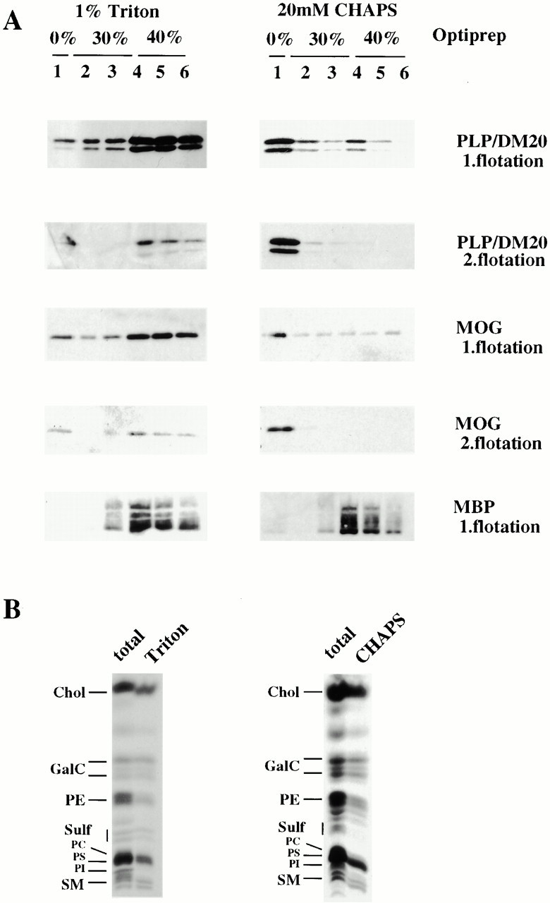
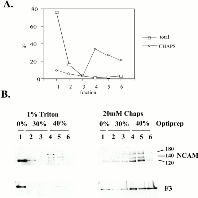
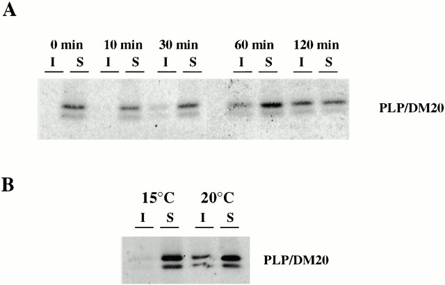
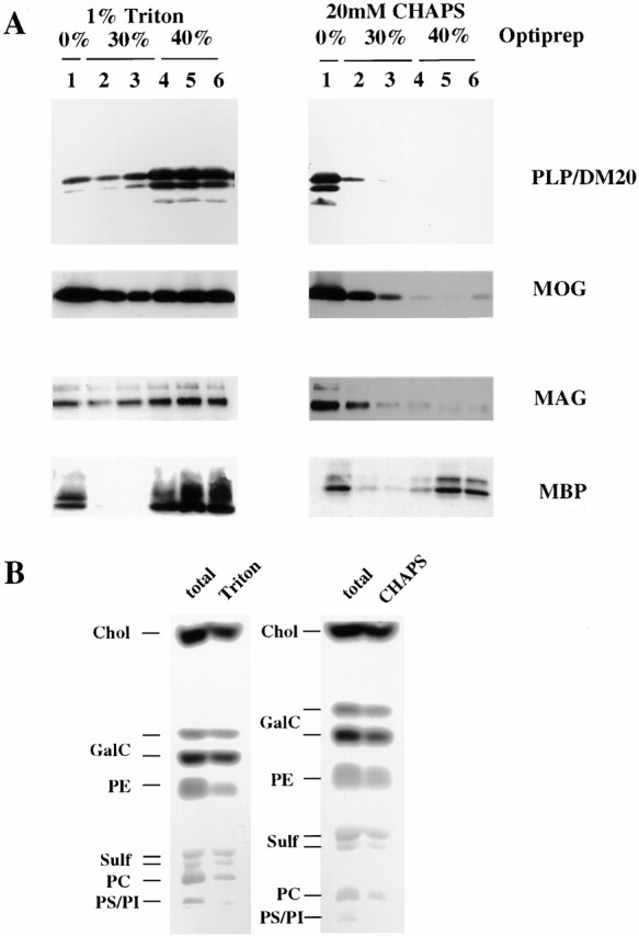
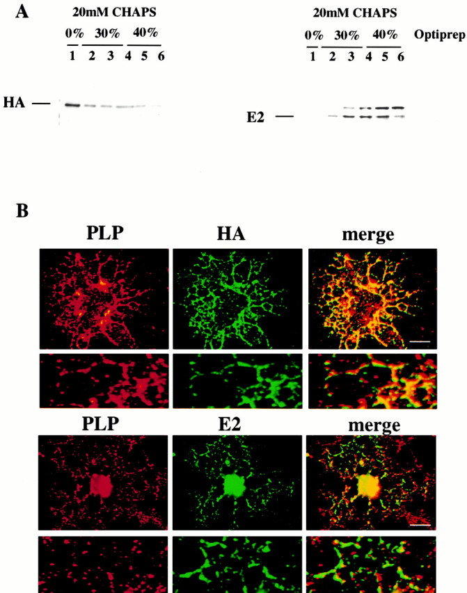
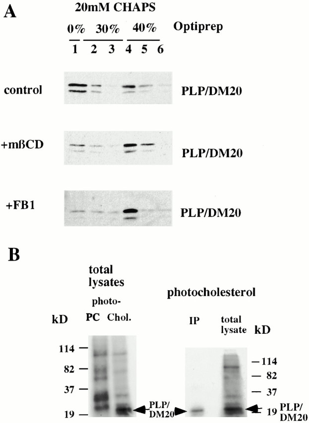
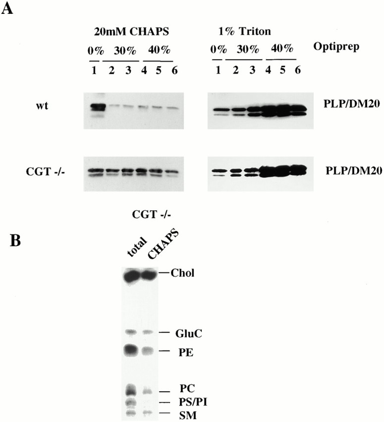
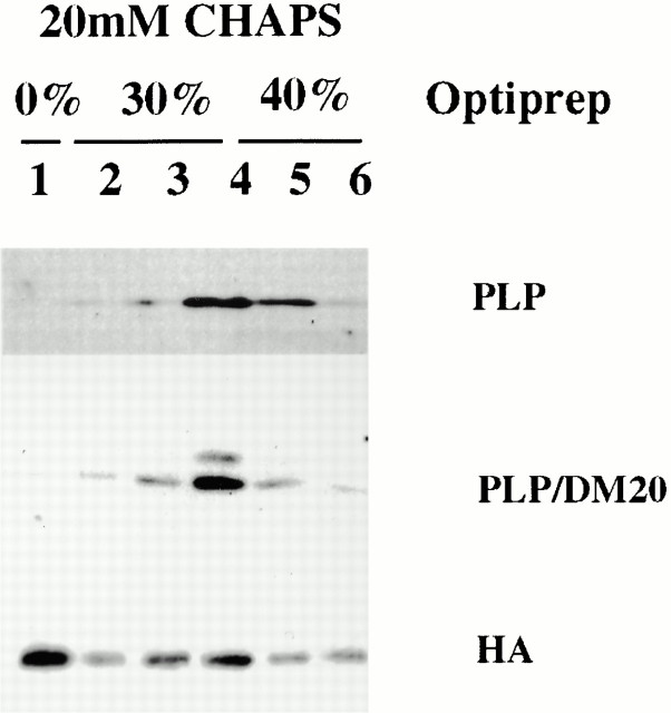
Myelin is a specialized membrane enriched in glycosphingolipids and cholesterol that contains a limited spectrum of proteins. We investigated the assembly of myelin components by oligodendrocytes and analyzed the role of lipid–protein interactions in this process. Proteolipid protein (PLP), the major myelin protein, was recovered from cultured oligodendrocytes from a low-density CHAPS-insoluble membrane fraction (CIMF) enriched in myelin lipids. PLP associated with the CIMF after leaving the endoplasmic reticulum but before exiting the Golgi apparatus, suggesting that myelin lipid and protein components assemble in the Golgi complex. The specific association of PLP with myelin lipids in CIMF was supported by the finding that it was efficiently cross-linked to photoactivable cholesterol, but not to phosphatidylcholine, which is underrepresented in both myelin and CIMF. Furthermore, depletion of cholesterol or inhibition of sphingolipid synthesis in oligodendrocytes abolished the association of PLP with CIMF. Thus, PLP may be recruited to myelin rafts, represented by CIMF, via lipid–protein interactions. In contrast to oligodendrocytes, after transfection in BHK cells, PLP is absent from isolated CIMF, suggesting that PLP requires specific lipids for raft association. In mice deficient in the enzyme ceramide galactosyl transferase, which cannot synthesize the main myelin glycosphingolipids, a large fraction of PLP no longer associates with rafts. Formation of a cholesterol- and galactosylceramide-rich membrane domain (myelin rafts) may be critical for the sorting of PLP and assembly of myelin in oligodendrocytes.
Overview
The claims and statistics of this paper haven’t been checked yet.
Run the check — free, no account needed. It reads the open-access text and checks the sources it cites.
Integrity signals
Registry
Cites
Statistics
Figures
Verification
Claims the paper makes
The claims and statistics of this paper haven’t been checked yet.
Run the check — free, no account needed. It reads the open-access text and checks the sources it cites.
Claims other papers make about this one
Each is checked against this paper’s own text: does it say what the citing paper says it does?
The source backs part of the claim, not all of it — or backs it for a different population, condition or comparison.
In the citing paper
“The extra cholesterol supply is not simply the building blocks for lipid content in the myelin sheet, but also the key for proper myelin protein assembly and sorting (Simons et al. 2000).”
From Apolipoprotein E ε4 disrupts oligodendrocyte differentiation by interfering with astrocyte‐derived lipid transport (2023) · cites this paper as Simons et al. 2000
Experimental — accuracy not yet measured
Self-check
Percentages, sums and reported differences re-computed from the paper’s own numbers.
The self-check needs the paper’s full text.
Citations
Cites
A RAPID METHOD OF TOTAL LIPID EXTRACTION AND PURIFICATION
Bligh, E. G., & Dyer, W. J. (1959) · Canadian Journal of Biochemistry and Physiology · doi:10.1139/o59-099 · Cited by 54,751
Functional rafts in cell membranes
Simons, K., & Ikonen, E. (1997) · Nature · doi:10.1038/42408 · Cited by 9,757
Srimaroeng, C., Cecile, J. P., Walden, R., & Pritchard, J. B. (2013) · Cellular Physiology and Biochemistry · doi:10.1159/000350077 · Cited by 2,989
Brown, D. A., & Rose, J. K. (1992) · Cell · doi:10.1016/0092-8674(92)90189-j · Cited by 2,921
Structure and Function of Sphingolipid- and Cholesterol-rich Membrane Rafts
Brown, D. A., & London, E. (2000) · Journal of Biological Chemistry · doi:10.1074/jbc.r000005200 · Cited by 2,344
Adams, R. D., & Victor, M. (1977) · Medical Entomology and Zoology · Cited by 1,718
ENDOCYTOSIS AND MOLECULAR SORTING
Mellman, I. (1996) · Annual Review of Cell and Developmental Biology · doi:10.1146/annurev.cellbio.12.1.575 · Cited by 1,552
Lipid sorting in epithelial cells
Simons, K., & Gerrit van, M. E. E. R. (1988) · Biochemistry · doi:10.1021/bi00417a001 · Cited by 1,373
GPI-anchored proteins are organized in submicron domains at the cell surface
Varma, R., & Mayor, S. (1998) · Nature · doi:10.1038/29563 · Cited by 1,209
Lipid Domain Structure of the Plasma Membrane Revealed by Patching of Membrane Components
Harder, T., Scheiffele, P., Verkade, P., & Simons, K. (1998) · The Journal of Cell Biology · doi:10.1083/jcb.141.4.929 · Cited by 1,186
Sphingolipid–Cholesterol Rafts Diffuse as Small Entities in the Plasma Membrane of Mammalian Cells
Pralle, A., Keller, P., Florin, E., Simons, K., & Hörber, J. K. H. (2000) · The Journal of Cell Biology · doi:10.1083/jcb.148.5.997 · Cited by 988
The oligodendrocyte and its many cellular processes
PFEIFFER, S., WARRINGTON, A., & BANSAL, R. (1993) · Trends in Cell Biology · doi:10.1016/0962-8924(93)90213-k · Cited by 737
Schroeder, R., London, E., & Brown, D. (1994) · Proceedings of the National Academy of Sciences · doi:10.1073/pnas.91.25.12130 · Cited by 692
Scheiffele, P., Roth, M. G., & Simons, K. (1997) · The EMBO Journal · doi:10.1093/emboj/16.18.5501 · Cited by 638
Coetzee, T., Fujita, N., Dupree, J. L., Shi, R., Blight, A. R., Suzuki, K., Suzuki, K., & Popko, B. (1996) · Cell · doi:10.1016/s0092-8674(00)80093-8 · Cited by 624
Caveolae: where incoming and outgoing messengers meet.
Anderson, R. G. W. (1993) · Proceedings of the National Academy of Sciences · doi:10.1073/pnas.90.23.10909 · Cited by 607
Kurzchalia, T. V., Dupree, P., Parton, R. G., Kellner, R., Virta, H., Lehnert, M., & Simons, K. (1992) · The Journal of Cell Biology · doi:10.1083/jcb.118.5.1003 · Cited by 559
Microdomains of GPI-anchored proteins in living cells revealed by crosslinking
Friedrichson, T., & Kurzchalia, T. V. (1998) · Nature · doi:10.1038/29570 · Cited by 551
Matlin, K. S., & Simons, K. (1983) · Cell · doi:10.1016/0092-8674(83)90154-x · Cited by 508
Cholesterol binds to synaptophysin and is required for biogenesis of synaptic vesicles
Thiele, C., Hannah, M. J., Fahrenholz, F., & Huttner, W. B. (2000) · Nature Cell Biology · doi:10.1038/71366 · Cited by 498
Self-citationKlein, U., Gimpl, G., & Fahrenholz, F. (1995) · Biochemistry · doi:10.1021/bi00042a009 · Cited by 481
Sphingolipid organization in biomembranes: what physical studies of model membranes reveal
Brown, R. E. (1998) · Journal of Cell Science · doi:10.1242/jcs.111.1.1 · Cited by 479
Monier, S., Parton, R. G., Vogel, F., Behlke, J., HENSKE, A., & Kurzchalia, T. V. (1995) · Molecular Biology of the Cell · doi:10.1091/mbc.6.7.911 · Cited by 479
Influenza Viruses Select Ordered Lipid Domains during Budding from the Plasma Membrane
Scheiffele, P., Rietveld, A. G., Wilk, T., & Simons, K. (1999) · Journal of Biological Chemistry · doi:10.1074/jbc.274.4.2038 · Cited by 448
Isolation and Characterization of Myelin
Norton, W. T. (1977) · Springer eBooks · doi:10.1007/978-1-4615-7514-6_5 · Cited by 389
Caveolin-3 Associates with Developing T-tubules during Muscle Differentiation
Parton, R. G., Way, M., Zorzi, N., & Stang, E. (1997) · The Journal of Cell Biology · doi:10.1083/jcb.136.1.137 · Cited by 354
Bosio, A., Binczek, E., & Stoffel, W. (1996) · Proceedings of the National Academy of Sciences · doi:10.1073/pnas.93.23.13280 · Cited by 347
Self-citationNucleotide sequences of two mRNAs for rat brain myelin proteolipid protein
Milner, R. J., Lai, C., Nave, K., Lenoir, D., Ogata, J., & Sutcliffe, J. G. (1985) · Cell · doi:10.1016/0092-8674(85)90289-2 · Cited by 337
Nave, K., Lai, C., Bloom, F. E., & Milner, R. J. (1987) · Proceedings of the National Academy of Sciences · doi:10.1073/pnas.84.16.5665 · Cited by 302
Braun, P. E., & Brostoff, S. W. (1977) · Springer eBooks · doi:10.1007/978-1-4615-7514-6_6 · Cited by 278
Axo-Glial Interactions Regulate the Localization of Axonal Paranodal Proteins
Dupree, J., Girault, J., & Popko, B. (1999) · Journal of Cell Biology · doi:10.1083/jcb.147.6.1145 · Cited by 278
Saraste, J., Palade, G. E., & Farquhar, M. G. (1986) · Proceedings of the National Academy of Sciences · doi:10.1073/pnas.83.17.6425 · Cited by 252
Isolation and Characterization of Myelin
Norton, W. T., & Cammer, W. (1984) · Springer eBooks · doi:10.1007/978-1-4757-1830-0_5 · Cited by 248
Glycosphingolipid-enriched, detergent-insoluble complexes in protein sorting in epithelial cells
Fiedler, K., Kobayashi, T., Kurzchalia, T. V., & Simons, K. (1993) · Biochemistry · doi:10.1021/bi00076a009 · Cited by 244
Krämer, E., Klein, C., Koch, T., Boytinck, M., & Trotter, J. (1999) · Journal of Biological Chemistry · doi:10.1074/jbc.274.41.29042 · Cited by 232
Self-citationWeimbs, T., & Stoffel, W. (1992) · Biochemistry · doi:10.1021/bi00164a002 · Cited by 228
Self-citationImmunohistochemical localization of the neural cannabinoid receptor in rat brain
Pettit, D. A. D., Harrison, M. P., Olson, J. M., Spencer, R. F., & Cabral, G. A. (1998) · Journal of Neuroscience Research · doi:10.1002/(sici)1097-4547(19980201)51:3 · Cited by 228
Morphological Differentiation of Oligodendrocytes Requires Activation of Fyn Tyrosine Kinase
Osterhout, D. J., Wolven, A., Wolf, R. M., Resh, M. D., & Chao, M. V. (1999) · The Journal of Cell Biology · doi:10.1083/jcb.145.6.1209 · Cited by 226
Lees, M. B., & Brostoff, S. W. (1984) · Springer eBooks · doi:10.1007/978-1-4757-1830-0_6 · Cited by 187
Inhibition of sphingolipid synthesis affects axonal outgrowth in cultured hippocampal neurons
Harel, R., & Futerman, A. H. (1993) · Journal of Biological Chemistry · doi:10.1016/s0021-9258(19)85263-8 · Cited by 186
Brm̈mendorf, T., Hubert, M. J., Treubert, U., Leuschner, R., Tárnok, A., & Rathjen, F. G. (1993) · Neuron · doi:10.1016/0896-6273(93)90172-n · Cited by 179
Cerneus, D. P., Ueffing, E., Posthuma, G., Strous, G. J., & ENDE, A. V. D. (1993) · Journal of Biological Chemistry · doi:10.1016/s0021-9258(18)53671-1 · Cited by 168
Ostermeyer, A. G., Beckrich, B. T., Ivarson, K. A., Grove, K., & Brown, D. A. (1999) · Journal of Biological Chemistry · doi:10.1074/jbc.274.48.34459 · Cited by 158
Trotter, J., Bitter-Suermann, D., & Schachner, M. (1989) · Journal of Neuroscience Research · doi:10.1002/jnr.490220402 · Cited by 157
Self-citationJung, M., Sommer, I., Schachner, M., & Nave, K. (1996) · Journal of Neuroscience · doi:10.1523/jneurosci.16-24-07920.1996 · Cited by 145
Molecular Organization of Myelin
Braun, P. E. (1984) · Springer eBooks · doi:10.1007/978-1-4757-1830-0_3 · Cited by 142
Major myelin proteolipid: The 4-α-helix topology
Popot, J., Pham-Dinh, D., & Dautigny, A. (1991) · The Journal of Membrane Biology · doi:10.1007/bf01868534 · Cited by 137
Ledesma, M. D., Brügger, B., Bünning, C., Wieland, F. T., & Dotti, C. G. (1999) · The EMBO Journal · doi:10.1093/emboj/18.7.1761 · Cited by 131
Krämer‐Albers, E., Koch, T., Niehaus, A., & Trotter, J. (1997) · Journal of Biological Chemistry · doi:10.1074/jbc.272.14.8937 · Cited by 122
Self-citationKim, T., & Pfeiffer, S. E. (1999) · Journal of Neurocytology · doi:10.1023/a:1007001427597 · Cited by 104
Bohuslav, J., Cinek, T., & Hořejšı́, V. (1993) · European Journal of Immunology · doi:10.1002/eji.1830230409 · Cited by 96
Vaz, W. L. C., & Almeida, P. F. F. (1993) · Current Opinion in Structural Biology · doi:10.1016/0959-440x(93)90071-r · Cited by 76
Pereyra, P. M., Horváth, E., & Braun, P. E. (1988) · Neurochemical Research · doi:10.1007/bf00973301 · Cited by 61
Kuhlmann-Krieg, S., Sommer, I., & Schachner, M. (1988) · Developmental Brain Research · doi:10.1016/0165-3806(88)90031-4 · Cited by 55
Polarity signals in epithelial cells
Rodriguez-Boulan, E. J., & Zurzolo, C. (1993) · Journal of Cell Science · doi:10.1242/jcs.1993.supplement_17.2 · Cited by 46
de Vries, H., Schrage, C., & Hoekstra, D. (1998) · Molecular Biology of the Cell · doi:10.1091/mbc.9.3.599 · Cited by 41
Linington, C., & Waehneldt, T. V. (1990) · Journal of Neurochemistry · doi:10.1111/j.1471-4159.1990.tb01969.x · Cited by 29
GPI-Anchored Proteins and Glycosphingolipid-Rich Rafts: Platforms for Adhesion and Signaling
Trotter, J., Klein, C., & Krämer, E. (2000) · The Neuroscientist · doi:10.1177/107385840000600410 · Cited by 19
Self-citation
Cited by
Sorted by citations
Biologically Active Molecules with a “Light Switch”
Mayer, G., & Heckel, A. (2006) · Angewandte Chemie International Edition · doi:10.1002/anie.200600387 · Cited by 1,113
Myelination of the Nervous System: Mechanisms and Functions
Nave, K.-A., & Werner, H. B. (2014) · Annual Review of Cell and Developmental Biology · doi:10.1146/annurev-cellbio-100913-013101 · Cited by 983
High cholesterol level is essential for myelin membrane growth
Saher, G., Brügger, B., Lappe-Siefke, C., Möbius, W., Tozawa, R.-I., Wehr, M. C., Wieland, F., Ishibashi, S., & Nave, K.-A. (2005) · Nature Neuroscience · doi:10.1038/nn1426 · Cited by 799
Myelin in the Central Nervous System: Structure, Function, and Pathology
Stadelmann, C., Timmler, S., Barrantes‐Freer, A., & Simons, M. (2019) · Physiological Reviews · doi:10.1152/physrev.00031.2018 · Cited by 799
Self-citationAxon-Glial Signaling and the Glial Support of Axon Function
Nave, K., & Trapp, B. D. (2008) · Annual Review of Neuroscience · doi:10.1146/annurev.neuro.30.051606.094309 · Cited by 664
Metadata
- Authors
- Simons, M., Krämer, E.-M., Thiele, C., Stoffel, W., & Trotter, J.
- Affiliations
- Heidelberg University (DE)
- aDepartment of Neurobiology, University of Heidelberg, 69120 Heidelberg, Germany
- University of Cologne (DE)
- Venue
- The Journal of Cell Biology
- Publisher
- Rockefeller University Press
- Published
- 2000-10-02
- Volume
- 151
- Issue
- 1
- Pages
- 143-154
- ISSN
- 0021-9525 (ISSN-L)
- 1540-8140
- PMC
- PMC2189802
- OpenAlex
- W2098744846
- Cited by
- 293
- References
- 58
- Citations by year
- 2026: 5 · 2025: 2 · 2024: 7 · 2023: 6 · 2022: 12 · 2021: 5 · 2020: 10 · 2019: 15 · 2018: 3 · 2017: 12 · 2016: 4 · 2015: 8 · 2014: 17 · 2013: 9 · 2012: 14
- FWCI
- 5.31
- Type
- journal article
- Language
- en
- Open access
- green
- www.ncbi.nlm.nih.gov
- Licence
- cc-by-nc-sa
- Locations
- The Journal of Cell Biology (publishedVersion)
- Europe PMC (PubMed Central) (publishedVersion, cc-by-nc-sa)
- PubMed Central (publishedVersion, cc-by-nc-sa)
- PubMed (publishedVersion)
- Topics
- Life Sciences › Biochemistry, Genetics and Molecular Biology › Molecular Biology › Lipid Membrane Structure and Behavior
- Life Sciences › Biochemistry, Genetics and Molecular Biology › Molecular Biology › Sphingolipid Metabolism and Signaling
- Life Sciences › Biochemistry, Genetics and Molecular Biology › Cell Biology › Caveolin-1 and cellular processes
- Keywords
- proteolipid protein, lipid rafts, oligodendrocytes, cholesterol, galactosylceramide, glycosphingolipids, protein-lipid interactions, Golgi apparatus, sphingolipid biosynthesis
- MeSH terms
- Animals, Brain Chemistry, Cell Fractionation, Cholesterol, Cholic Acids, Detergents, Endoplasmic Reticulum, Galactosylceramides, Galactosyltransferases, Golgi Apparatus, Cricetinae, Myelin Sheath, Oligodendroglia, Solubility, Glycosylphosphatidylinositols, Mice, Knockout, Myelin Proteolipid Protein, Hemagglutinin Glycoproteins, Influenza Virus, Membrane Microdomains, N-Acylsphingosine Galactosyltransferase, Mice
About these checks
Registry, retraction, Cites and statistics results are computed, not judged, with one exception: a printed reference that no index lists is read once by a model before it counts as a citation error. Claims are extracted by a model and kept only when their quote appears verbatim in the text; each cited source is then checked, with a model’s help, for whether it says what the claim relies on. Those verdicts can be wrong — read the claim’s quote and the cited source before relying on one.
Statistics and arithmetic are recomputed, not judged.
Confidence bands are shown only for fields calibrated on our annotated test set. None is calibrated yet, so no band is shown.
What counts as a claim ›How we measure confidence ›How we check this ›
Are you an author, or do you see a problem with this page? Request a review
