

The DOI matches its Crossref record; 53 of the 53 references it cites were found in a registry or an index.
Mmm2p, a mitochondrial outer membrane protein required for yeast mitochondrial shape and maintenance of mtDNA nucleoids
Open accessAbstract
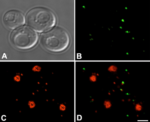
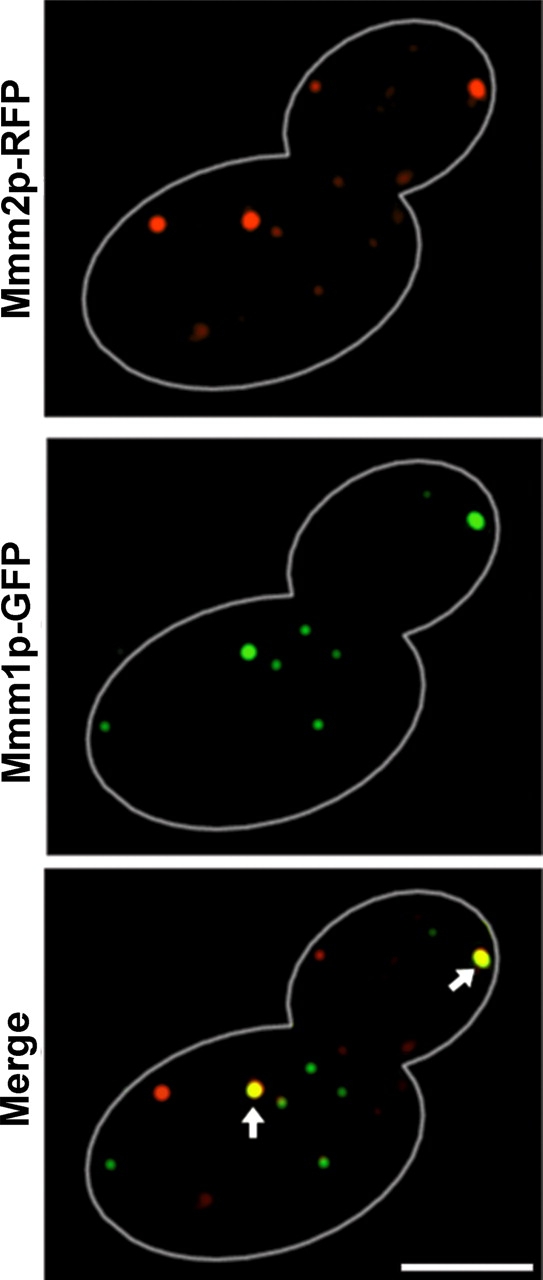
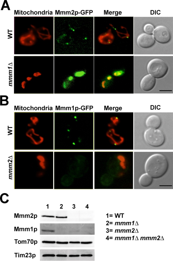
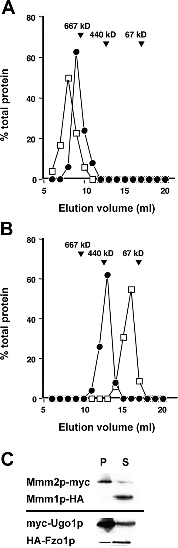
The mitochondrial outer membrane protein, Mmm1p, is required for normal mitochondrial shape in yeast. To identify new morphology proteins, we isolated mutations incompatible with the mmm1-1 mutant. One of these mutants, mmm2-1, is defective in a novel outer membrane protein. Lack of Mmm2p causes a defect in mitochondrial shape and loss of mitochondrial DNA (mtDNA) nucleoids. Like the Mmm1 protein (Aiken Hobbs, A.E., M. Srinivasan, J.M. McCaffery, and R.E. Jensen. 2001. J. Cell Biol. 152:401-410.), Mmm2p is located in dot-like particles on the mitochondrial surface, many of which are adjacent to mtDNA nucleoids. While some of the Mmm2p-containing spots colocalize with those containing Mmm1p, at least some of Mmm2p is separate from Mmm1p. Moreover, while Mmm2p and Mmm1p both appear to be part of large complexes, we find that Mmm2p and Mmm1p do not stably interact and appear to be members of two different structures. We speculate that Mmm2p and Mmm1p are components of independent machinery, whose dynamic interactions are required to maintain mitochondrial shape and mtDNA structure.
Overview
The claims and statistics of this paper haven’t been checked yet.
Run the check — free, no account needed. It reads the open-access text and checks the sources it cites.
Integrity signals
Registry
Cites
Statistics
Figures
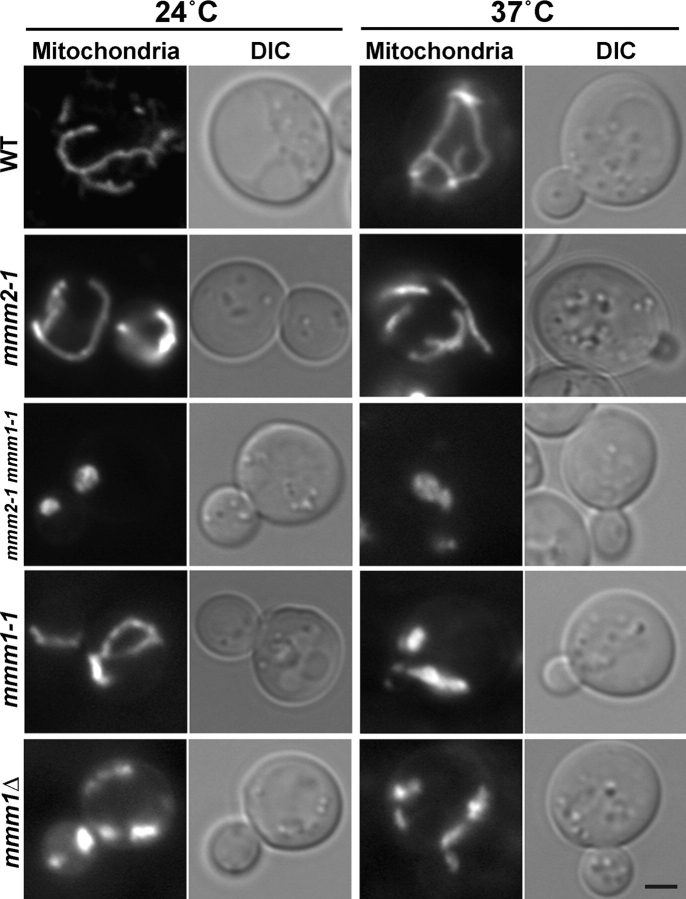
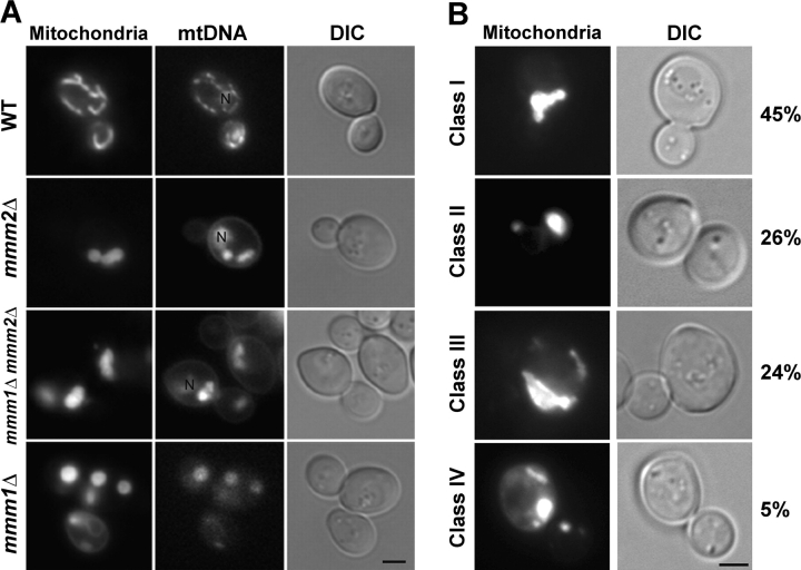
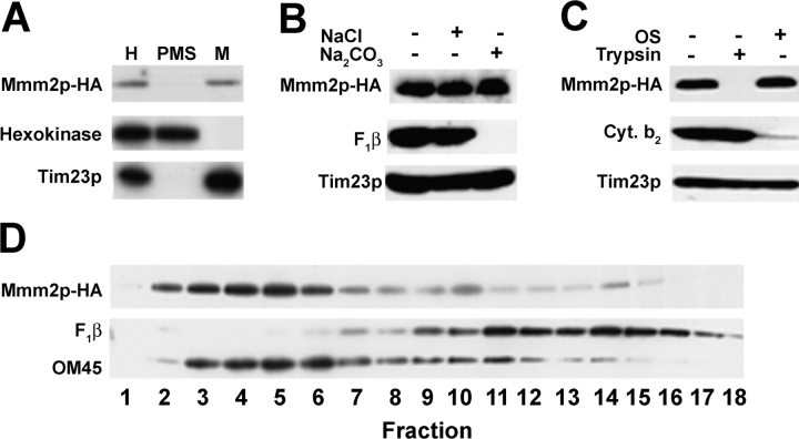
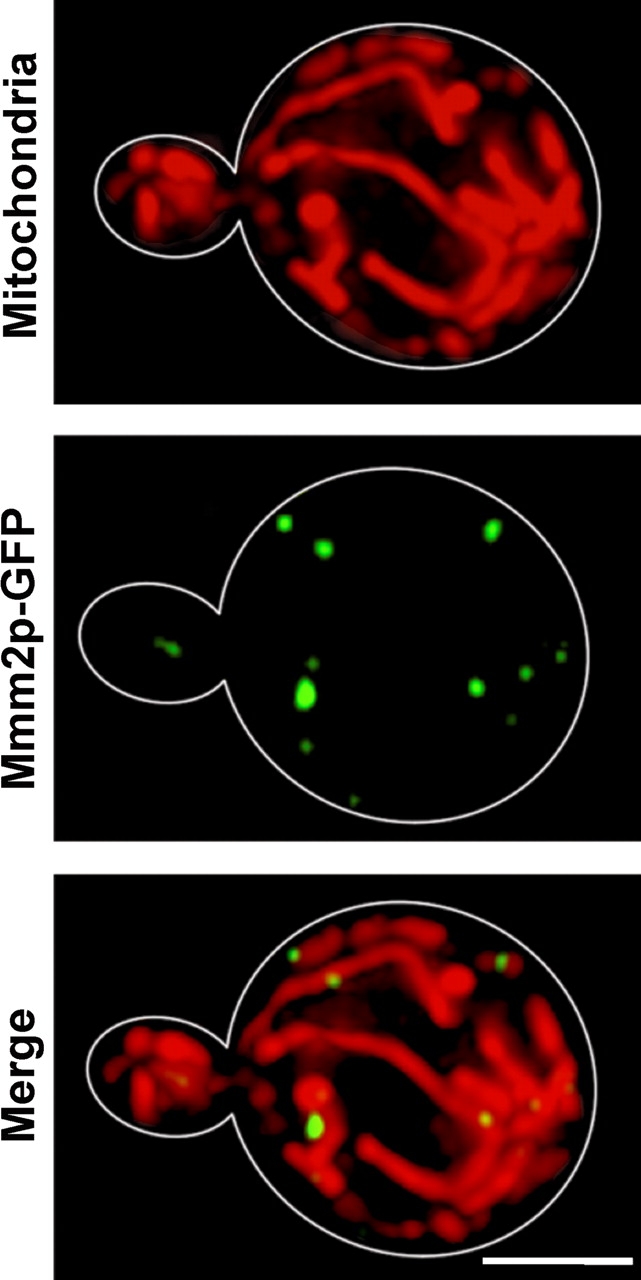
Verification
Claims the paper makes
The claims and statistics of this paper haven’t been checked yet.
Run the check — free, no account needed. It reads the open-access text and checks the sources it cites.
Claims other papers make about this one
Each is checked against this paper’s own text: does it say what the citing paper says it does?
The cited source doesn’t support the claim as written.
In the citing paper
“In particular, deletion mutants of the ERMES core complex display defects in the segregation of mtDNA into daughter cells, resulting in the loss of mitochondria over several generations of growth in S. cerevisiae [27,29,30,45].”
From In Candida glabrata, ERMES Component GEM1 Controls Mitochondrial Morphology, mtROS, and Drug Efflux Pump Expression, Resulting in Azole Susceptibility (2023) · cites this paper as [30]
Checked by a reasoning model — may be wrong.
Experimental — accuracy not yet measured
Self-check
Percentages, sums and reported differences re-computed from the paper’s own numbers.
The self-check needs the paper’s full text.
Citations
Cites
Sikorski, R. S., & Hieter, P. (1989) · Genetics · doi:10.1093/genetics/122.1.19 · Cited by 8,590
Sherman, F., Fink, G. R., & Hicks, J. B. (1979) · Cold Spring Harbor Laboratory eBooks · Cited by 7,430
Brachmann, C. B., Davies, A., Cost, G. J., Caputo, E. M., Li, J., Hieter, P., & Boeke, J. D. (1998) · Yeast · doi:10.1002/(sici)1097-0061(19980130)14:2 · Cited by 3,418
Hoffman, C. S., & Winston, F. (1987) · Gene · doi:10.1016/0378-1119(87)90131-4 · Cited by 2,492
Boeke, J. D., Lacroute, F., & Fink, G. R. (1984) · Molecular and General Genetics MGG · doi:10.1007/bf00330984 · Cited by 2,354
Bähler, J., Wu, J., Longtine, M. S., Shah, N. G., Mckenzie, A., Steever, A. B., Wach, A., Philippsen, P., & Pringle, J. R. (1998) · Yeast · doi:10.1002/(sici)1097-0061(199807)14:10 · Cited by 2,208
Attardi, G. M., & Schatz, G. (1988) · Annual Review of Cell Biology · doi:10.1146/annurev.cb.04.110188.001445 · Cited by 1,392
Daum, G., Böhni, P. C., & Schatz, G. (1982) · Journal of Biological Chemistry · doi:10.1016/s0021-9258(18)33617-2 · Cited by 1,123
Transformation in yeast: Development of a hybrid cloning vector and isolation of the can1 gene
Broach, J. R., Strathern, J. N., & Hicks, J. B. (1979) · Gene · doi:10.1016/0378-1119(79)90012-x · Cited by 1,073
The ATP synthase is involved in generating mitochondrial cristae morphology
Paumard, P., Vaillier, J., Coulary, B., Schaëffer, J., Soubannier, V., Mueller, D. M., Brèthes, D., di Rago, J., & Velours, J. (2002) · The EMBO Journal · doi:10.1093/emboj/21.3.221 · Cited by 809
Research works printed but not linked: 5
- 1991, Mol. Cell. Biol., 11, 1295
- 1996, Biol. Chem., 377, 535
- 1987, J. Cell Sci., 88(Pt 4), 431
- 1949, Arch. Biochem., 22, 331
- 1993, Mol. Cell. Biol., 13, 5418
Cited by
Sorted by citations
An ER-Mitochondria Tethering Complex Revealed by a Synthetic Biology Screen
Kornmann, B., Currie, E., Collins, S. R., Schuldiner, M., Nunnari, J., Weissman, J. S., & Walter, P. (2009) · Science · doi:10.1126/science.1175088 · Cited by 1,275
Mitochondrial Morphology and Dynamics in Yeast and Multicellular Eukaryotes
Okamoto, K., & Shaw, J. M. (2005) · Annual Review of Genetics · doi:10.1146/annurev.genet.38.072902.093019 · Cited by 737
Mitochondrial protein import: from proteomics to functional mechanisms
Schmidt, O., Pfanner, N., & Meisinger, C. (2010) · Nature Reviews Molecular Cell Biology · doi:10.1038/nrm2959 · Cited by 717
Making heads or tails of phospholipids in mitochondria
Osman, C., Voelker, D. R., & Langer, T. (2011) · The Journal of Cell Biology · doi:10.1083/jcb.201006159 · Cited by 599
Phosphatidylethanolamine Metabolism in Health and Disease
Calzada, E., Onguka, O., & Claypool, S. M. (2015) · International review of cytology · doi:10.1016/bs.ircmb.2015.10.001 · Cited by 558
The organization and inheritance of the mitochondrial genome
Chen, X. J., & Butow, R. A. (2005) · Nature Reviews Genetics · doi:10.1038/nrg1708 · Cited by 496
The Conserved GTPase Gem1 Regulates Endoplasmic Reticulum–Mitochondria Connections
Kornmann, B., Osman, C., & Walter, P. (2011) · Proceedings of the National Academy of Sciences · doi:10.1073/pnas.1111314108 · Cited by 335
Murley, A., Lackner, L. L., Osman, C., West, M., Voeltz, G. K., Walter, P., & Nunnari, J. (2013) · eLife · doi:10.7554/elife.00422 · Cited by 326
Multiple pathways for sorting mitochondrial precursor proteins
Bolender, N., Sickmann, A., Wagner, R., Meisinger, C., & Pfanner, N. (2008) · EMBO Reports · doi:10.1038/sj.embor.7401126 · Cited by 319
The Metabolic Theory of Pulmonary Arterial Hypertension
Paulin, R., & Michelakis, E. D. (2014) · Circulation Research · doi:10.1161/circresaha.115.301130 · Cited by 328
Determinants and Functions of Mitochondrial Behavior
Labbè, K., Murley, A., & Nunnari, J. M. (2014) · Annual Review of Cell and Developmental Biology · doi:10.1146/annurev-cellbio-101011-155756 · Cited by 324
Wang, Y., & Bogenhagen, D. F. (2006) · Journal of Biological Chemistry · doi:10.1074/jbc.m604501200 · Cited by 303
Interactions of mitochondria with the actin cytoskeleton
Boldogh, I. R., & Pon, L. A. (2006) · Biochimica et Biophysica Acta (BBA) - Molecular Cell Research · doi:10.1016/j.bbamcr.2006.02.014 · Cited by 270
Yeast Miro GTPase, Gem1p, regulates mitochondrial morphology via a novel pathway
Frederick, R. L., McCaffery, J. M., Cunningham, K. W., Okamoto, K., & Shaw, J. M. (2004) · The Journal of Cell Biology · doi:10.1083/jcb.200405100 · Cited by 247
Organelle-Based Aggregation and Retention of Damaged Proteins in Asymmetrically Dividing Cells
Zhou, C., Slaughter, B. D., Unruh, J. R., Guo, F., Yu, Z., Mickey, K., Narkar, A., Trimble, R., McClain, M. L., & Li, R. (2014) · Cell · doi:10.1016/j.cell.2014.09.026 · Cited by 241
Ott, C., Roß, K., Straub, S. P., Thiede, B., Götz, M., Goosmann, C., Krischke, M., Mueller, M. J., Krohne, G., Rudel, T., et al. (2012) · Molecular and Cellular Biology · doi:10.1128/mcb.06388-11 · Cited by 227
ER–mitochondrial junctions can be bypassed by dominant mutations in the endosomal protein Vps13
Lang, A. B., Peter, A. T. J., Walter, P., & Kornmann, B. (2015) · The Journal of Cell Biology · doi:10.1083/jcb.201502105 · Cited by 223
Kopec, K. O., Alva, V., & Lupas, A. N. (2010) · Bioinformatics · doi:10.1093/bioinformatics/btq326 · Cited by 212
ERMES-mediated ER-mitochondria contacts: molecular hubs for the regulation of mitochondrial biology
Kornmann, B., & Walter, P. (2010) · Journal of Cell Science · doi:10.1242/jcs.058636 · Cited by 210
Ubiquitin–Proteasome System and mitochondria — Reciprocity
Livnat‐Levanon, N., & Glickman, M. H. (2010) · Biochimica et Biophysica Acta (BBA) - Gene Regulatory Mechanisms · doi:10.1016/j.bbagrm.2010.07.005 · Cited by 197
Walther, D. M., Rapaport, D., & Tommassen, J. (2009) · Cellular and Molecular Life Sciences · doi:10.1007/s00018-009-0029-z · Cited by 178
Spelbrink, J. N. (2010) · IUBMB Life · doi:10.1002/iub.282 · Cited by 171
The cell-type specificity of mitochondrial dynamics
Kuznetsov, A. V., Hermann, M., Saks, V. A., Hengster, P., & Margreiter, R. (2009) · The International Journal of Biochemistry & Cell Biology · doi:10.1016/j.biocel.2009.03.007 · Cited by 184
Nguyen, T., Lewandowska, A., Choi, J., Markgraf, D. F., Junker, M., Bilgin, M., Ejsing, C. S., Voelker, D. R., Rapoport, T. A., & Shaw, J. M. (2012) · Traffic · doi:10.1111/j.1600-0854.2012.01352.x · Cited by 166
Role of Essential Genes in Mitochondrial Morphogenesis inSaccharomyces cerevisiae
Altmann, K., & Westermann, B. (2005) · Molecular Biology of the Cell · doi:10.1091/mbc.e05-07-0678 · Cited by 165
Role of MINOS in mitochondrial membrane architecture and biogenesis
van der Laan, M., Bohnert, M., Wiedemann, N., & Pfanner, N. (2012) · Trends in Cell Biology · doi:10.1016/j.tcb.2012.01.004 · Cited by 156
(De)constructing Mitochondria: What For?
Dimmer, K. S., & Scorrano, L. (2006) · Physiology · doi:10.1152/physiol.00010.2006 · Cited by 151
Structure–Function Insights into Direct Lipid Transfer between Membranes by Mmm1–Mdm12 of ERMES
Kawano, S., Tamura, Y., Kojima, R., Bala, S., Asai, E., Michel, A. H., Kornmann, B., Riezman, I., Riezman, H., Sakae, Y., et al. (2018) · Journal of Cell Biology · doi:10.1083/jcb.201704119 · Cited by 148
“, 1994); Mdm10, a β-barrel mitochondrial outer membrane (OM) protein (Sogo and Yaffe, 1994); Mdm34/Mmm2, an OM protein without an obvious transmembrane segment (Youngman et al., 2004), and Mdm12, a peripheral OM protein (Berger et al.”
BackgroundComposition and Topology of the Endoplasmic Reticulum–Mitochondria Encounter Structure
Stroud, D. A., Oeljeklaus, S., Wiese, S., Bohnert, M., Lewandrowski, U., Sickmann, A., Guiard, B., van der Laan, M., Warscheid, B., & Wiedemann, N. (2011) · Journal of Molecular Biology · doi:10.1016/j.jmb.2011.09.012 · Cited by 144
Positioning mitochondrial plasticity within cellular signaling cascades
Soubannier, V., & McBride, H. M. (2008) · Biochimica et Biophysica Acta (BBA) - Molecular Cell Research · doi:10.1016/j.bbamcr.2008.07.008 · Cited by 146
Biogenesis of β-barrel membrane proteins of mitochondria
Paschen, S. A., Neupert, W., & Rapaport, D. (2005) · Trends in Biochemical Sciences · doi:10.1016/j.tibs.2005.08.009 · Cited by 137
Making connections: interorganelle contacts orchestrate mitochondrial behavior
Klecker, T., Böckler, S., & Westermann, B. (2014) · Trends in Cell Biology · doi:10.1016/j.tcb.2014.04.004 · Cited by 137
Biogenesis of mitochondrial outer membrane proteins
Walther, D. M., & Rapaport, D. (2008) · Biochimica et Biophysica Acta (BBA) - Molecular Cell Research · doi:10.1016/j.bbamcr.2008.04.013 · Cited by 121
Inhibition of mitochondrial fission favours mutant over wild-type mitochondrial DNA
Malena, A., Loro, E., Di Re, M., Holt, I. J., & Vergani, L. (2009) · Human Molecular Genetics · doi:10.1093/hmg/ddp281 · Cited by 107
Visualizing multiple inter-organelle contact sites using the organelle-targeted split-GFP system
Kakimoto, Y., Tashiro, S., Kojima, R., Morozumi, Y., Endo, T., & Tamura, Y. (2018) · Scientific Reports · doi:10.1038/s41598-018-24466-0 · Cited by 107
Yeast Mitochondrial Division and Distribution Require the Cortical Num1 Protein
Cerveny, K. L., Studer, S. L., Jensen, R. E., & Sesaki, H. (2007) · Developmental Cell · doi:10.1016/j.devcel.2007.01.017 · Cited by 106
Self-citationThe mitochondrial outer membrane protein MDI promotes local protein synthesis and mtDNA replication
Zhang, Y., Chen, Y., Guček, M., & Xu, H. (2016) · The EMBO Journal · doi:10.15252/embj.201592994 · Cited by 108
Tamura, Y., Onguka, O., Hobbs, A. E. A., Jensen, R. E., Iijima, M., Claypool, S. M., & Sesaki, H. (2012) · Journal of Biological Chemistry · doi:10.1074/jbc.m111.338665 · Cited by 107
Self-citationUnderstanding aging: revealing order out of chaos
Dufour, É., & Larsson, N. (2004) · Biochimica et Biophysica Acta (BBA) - Bioenergetics · doi:10.1016/j.bbabio.2004.04.020 · Cited by 103
Dimmer, K. S., Jakobs, S., Vogel, F., Altmann, K., & Westermann, B. (2005) · The Journal of Cell Biology · doi:10.1083/jcb.200410030 · Cited by 101
Mcp1 and Mcp2, two novel proteins involved in mitochondrial lipid homeostasis
Tan, T., Özbalci, C., Brügger, B., Rapaport, D., & Dimmer, K. (2013) · Journal of Cell Science · doi:10.1242/jcs.121244 · Cited by 116
Protein import into plant mitochondria: signals, machinery, processing, and regulation
Murcha, M. W., Kmiec, B., Kubiszewski-Jakubiak, S., Teixeira, P. F., Glaser, E. G., & Whelan, J. (2014) · Journal of Experimental Botany · doi:10.1093/jxb/eru399 · Cited by 93
Becker, T., Wenz, L.-S., Thornton, N., Stroud, D., Meisinger, C., Wiedemann, N., & Pfanner, N. (2011) · Journal of Molecular Biology · doi:10.1016/j.jmb.2010.11.002 · Cited by 89
Mitochondrial inheritance in yeast
Westermann, B. (2013) · Biochimica et Biophysica Acta (BBA) - Bioenergetics · doi:10.1016/j.bbabio.2013.10.005 · Cited by 87
Ellenrieder, L., Opaliński, Ł., Becker, L., Krüger, V., Mirus, O., Straub, S. P., Ebell, K., Flinner, N., Stiller, S. B., Guiard, B., et al. (2016) · Nature Communications · doi:10.1038/ncomms13021 · Cited by 84
Multiple Pathways Influence Mitochondrial Inheritance in Budding Yeast
Frederick, R. L., Okamoto, K., & Shaw, J. M. (2008) · Genetics · doi:10.1534/genetics.107.083055 · Cited by 84
Lipid homeostasis in mitochondria
Tamura, Y., Kawano, S., & Endo, T. (2020) · Biological Chemistry · doi:10.1515/hsz-2020-0121 · Cited by 81
Assembly of β-barrel proteins in the mitochondrial outer membrane
Höhr, A. I. C., Straub, S. P., Warscheid, B., Becker, T., & Wiedemann, N. (2014) · Biochimica et Biophysica Acta (BBA) - Molecular Cell Research · doi:10.1016/j.bbamcr.2014.10.006 · Cited by 78
The role of Sec3p in secretory vesicle targeting and exocyst complex assembly
Luo, G., Zhang, J., & Guo, W. (2014) · Molecular Biology of the Cell · doi:10.1091/mbc.e14-04-0907 · Cited by 75
Phospholipid Transport via Mitochondria
Tamura, Y., Sesaki, H., & Endo, T. (2014) · Traffic · doi:10.1111/tra.12188 · Cited by 75
One ring to bring them all and in the darkness bind them: The trafficking of heme without deliverers
Chambers, I. G., Willoughby, M. M., Hamza, I., & Reddi, A. R. (2020) · Biochimica et Biophysica Acta (BBA) - Molecular Cell Research · doi:10.1016/j.bbamcr.2020.118881 · Cited by 77
Förtsch, J., Hummel, E., Krist, M., & Westermann, B. (2011) · The Journal of Cell Biology · doi:10.1083/jcb.201012088 · Cited by 73
Crystal structure of Mdm12 reveals the architecture and dynamic organization of the ERMES complex
Jeong, H., Park, J., & Lee, C. (2016) · EMBO Reports · doi:10.15252/embr.201642706 · Cited by 71
Hales, K. G. (2004) · Mitochondrion · doi:10.1016/j.mito.2004.05.007 · Cited by 70
Tucey, T. M., Verma-Gaur, J., Nguyen, J., Hewitt, V. L., Lo, T. L., Shingu-Vazquez, M., Robertson, A. A. B., Hill, J. R., Pettolino, F. A., Beddoe, T., et al. (2016) · mSphere · doi:10.1128/msphere.00074-16 · Cited by 66
Crosstalk between Mitochondria and Cytoskeleton in Cardiac Cells
Kuznetsov, A. V., Javadov, S. A., Grimm, M. F., Margreiter, R., Ausserlechner, M. J., & Hagenbuchner, J. (2020) · Cells · doi:10.3390/cells9010222 · Cited by 71
Bereiter‐Hahn, J., & Jendrach, M. (2010) · International review of cytology · doi:10.1016/s1937-6448(10)84001-8 · Cited by 67
Organization, dynamics and transmission of mitochondrial DNA: Focus on vertebrate nucleoids
Malka, F., Lombès, A., & Rojo, M. (2006) · Biochimica et Biophysica Acta (BBA) - Molecular Cell Research · doi:10.1016/j.bbamcr.2006.04.001 · Cited by 64
Coonrod, E. M., Karren, M. A., & Shaw, J. M. (2007) · Traffic · doi:10.1111/j.1600-0854.2007.00550.x · Cited by 62
Organelle contact zones as sites for lipid transfer
Tamura, Y., Kawano, S., & Endo, T. (2018) · The Journal of Biochemistry · doi:10.1093/jb/mvy088 · Cited by 60
Fusion, fission, and transport control asymmetric inheritance of mitochondria and protein aggregates
Böckler, S., Chelius, X., Hock, N., Klecker, T., Wolter, M., Weiß, M., Braun, R. J., & Westermann, B. (2017) · The Journal of Cell Biology · doi:10.1083/jcb.201611197 · Cited by 60
The plant mitochondrial protein import apparatus — The differences make it interesting
Murcha, M. W., Wang, Y., Narsai, R., & Whelan, J. (2013) · Biochimica et Biophysica Acta (BBA) - General Subjects · doi:10.1016/j.bbagen.2013.09.026 · Cited by 58
Endoplasmic reticulum–mitochondria junction is required for iron homeostasis
Xue, Y., Schmollinger, S., Attar, N., Campos, O. A., Vogelauer, M., Carey, M., Merchant, S., & Kurdistani, S. K. (2017) · Journal of Biological Chemistry · doi:10.1074/jbc.m117.784249 · Cited by 58
Role of membrane contact sites in protein import into mitochondria
Horvath, S. E., Rampelt, H., Oeljeklaus, S., Warscheid, B., van der Laan, M., & Pfanner, N. (2014) · Protein Science · doi:10.1002/pro.2625 · Cited by 61
Itoh, K., Tamura, Y., Iijima, M., & Sesaki, H. (2013) · Molecular Biology of the Cell · doi:10.1091/mbc.e13-03-0125 · Cited by 58
Mitochondrial contact sites as platforms for phospholipid exchange
Dimmer, K. S., & Rapaport, D. (2016) · Biochimica et Biophysica Acta (BBA) - Molecular and Cell Biology of Lipids · doi:10.1016/j.bbalip.2016.07.010 · Cited by 56
To, T., Fadul, M. J., & Shu, X. (2014) · Nature Communications · doi:10.1038/ncomms5072 · Cited by 54
Mitochondrial Fission: Regulation and ER Connection
Lee, H., & Yoon, Y. (2014) · Molecules and Cells · doi:10.14348/molcells.2014.2329 · Cited by 53
Mdm36 Is a Mitochondrial Fission-promoting Protein inSaccharomyces cerevisiae
Hammermeister, M., Schödel, K., & Westermann, B. (2010) · Molecular Biology of the Cell · doi:10.1091/mbc.e10-02-0096 · Cited by 57
Molecular machinery of mitochondrial dynamics in yeast
Merz, S., Hammermeister, M., Altmann, K., Dürr, M., & Westermann, B. (2007) · Biological Chemistry · doi:10.1515/bc.2007.110 · Cited by 50
Schnarwiler, F., Niemann, M., Doiron, N., Harsman, A., Käser, S., Mani, J., Chanfon, A., Dewar, C. E., Oeljeklaus, S., Jackson, C. B., et al. (2014) · Proceedings of the National Academy of Sciences · doi:10.1073/pnas.1404854111 · Cited by 49
Pro370Leu mutant myocilin impairs mitochondrial functions in human trabecular meshwork cells.
He, Y., Leung, K. W., Zhuo, Y., & Ge, J. (2009) · PubMed Central · Cited by 48
Lipin1 deficiency causes sarcoplasmic reticulum stress and chaperone‐responsive myopathy
Rashid, T., Nemazanyy, I., Paolini, C., Tatsuta, T., Crespin, P., de Villeneuve, D., Brodesser, S., Bénit, P., Rustin, P., Baraibar, M. A., et al. (2018) · The EMBO Journal · doi:10.15252/embj.201899576 · Cited by 49
ER–mitochondria contacts promote mitochondrial-derived compartment biogenesis
English, A. M., Schuler, M.-H., Xiao, T., Kornmann, B., Shaw, J. M., & Hughes, A. L. (2020) · The Journal of Cell Biology · doi:10.1083/jcb.202002144 · Cited by 48
Wideman, J. G., & Muñoz-Gómez, S. A. (2016) · Biochimica et Biophysica Acta (BBA) - Molecular and Cell Biology of Lipids · doi:10.1016/j.bbalip.2016.01.015 · Cited by 47
Ergosterol reduction impairs mitochondrial DNA maintenance in S. cerevisiae
Cirigliano, A., Macone, A., Bianchi, M. M., Oliaro‐Bosso, S., Balliano, G., Negri, R., & Rinaldi, T. (2018) · Biochimica et Biophysica Acta (BBA) - Molecular and Cell Biology of Lipids · doi:10.1016/j.bbalip.2018.12.002 · Cited by 48
Martinez-Guzman, O., Willoughby, M. M., Saini, A. G., Dietz, J. V., Bohovych, I., Medlock, A. E., Khalimonchuk, O., & Reddi, A. R. (2020) · Journal of Cell Science · doi:10.1242/jcs.237917 · Cited by 47
TAC102 Is a Novel Component of the Mitochondrial Genome Segregation Machinery in Trypanosomes
Trikin, R. M., Doiron, N., Hoffmann, A., Haenni, B., Jakob, M., Schnaufer, A. C., Schimanski, B., Zuber, B., & Ochsenreiter, T. (2016) · PLoS Pathogens · doi:10.1371/journal.ppat.1005586 · Cited by 46
Lipid synthesis and membrane contact sites: a crossroads for cellular physiology
Fernández-Murray, J. P., & McMaster, C. R. (2016) · Journal of Lipid Research · doi:10.1194/jlr.r070920 · Cited by 45
Eisenberg‐Bord, M., Tsui, H. S., Antunes, D., Fernández-del-Río, L., Bradley, M. C., Dunn, C. D., Nguyen, T. P. T., Rapaport, D., Clarke, C. F., & Schuldiner, M. (2019) · Contact · doi:10.1177/2515256418825409 · Cited by 46
Kondo‐Okamoto, N., Ohkuni, K., Kitagawa, K., McCaffery, J. M., Shaw, J. M., & Okamoto, K. (2006) · Molecular Biology of the Cell · doi:10.1091/mbc.e06-02-0145 · Cited by 43
Rinaldi, T., Hofmann, L., Gambadoro, A., Cossard, R., Livnat‐Levanon, N., Glickman, M. H., Frontali, L., & Delahodde, A. (2008) · Molecular Biology of the Cell · doi:10.1091/mbc.e07-07-0717 · Cited by 43
Mitochondrial protein import machineries and lipids: A functional connection
Gebert, N., Ryan, M. T., Pfanner, N., Wiedemann, N., & Stojanovski, D. (2010) · Biochimica et Biophysica Acta (BBA) - Biomembranes · doi:10.1016/j.bbamem.2010.08.003 · Cited by 38
Control of mitochondrial shape
Jensen, R. E. (2005) · Current Opinion in Cell Biology · doi:10.1016/j.ceb.2005.06.011 · Cited by 38
Self-citationEukaryote-wide sequence analysis of mitochondrial β-barrel outer membrane proteins
Imai, K., Fujita, N., Gromiha, M. M., & Horton, P. (2011) · BMC Genomics · doi:10.1186/1471-2164-12-79 · Cited by 37
Sakai, H., Shen, X., Koga, T., Park, B., Noskina, Y., TIBUDAN, M. L., & Yue, B. Y. J. T. (2007) · Journal of Cellular Physiology · doi:10.1002/jcp.21147 · Cited by 37
Kannan, M., Riekhof, W. R., & Voelker, D. R. (2014) · Traffic · doi:10.1111/tra.12236 · Cited by 36
Rasul, F., Zheng, F., Dong, F., He, J., Liu, L., Liu, W., Cheema, J. Y., Wei, W., & Fu, C. (2021) · Nature Communications · doi:10.1038/s41467-020-20866-x · Cited by 35
Connection of Protein Transport and Organelle Contact Sites in Mitochondria
Ellenrieder, L., Rampelt, H., & Becker, T. (2017) · Journal of Molecular Biology · doi:10.1016/j.jmb.2017.05.023 · Cited by 33
The mitochondrial Ras-related GTPase Miro: views from inside and outside the metazoan kingdom
Yamaoka, S., & Hara‐Nishimura, I. (2014) · Frontiers in Plant Science · doi:10.3389/fpls.2014.00350 · Cited by 30
Bacterial tail anchors can target to the mitochondrial outer membrane
Bal, G. L., Keskin, A., Seferoğlu, A. B., & Dunn, C. D. (2017) · Biology Direct · doi:10.1186/s13062-017-0187-0 · Cited by 28
Ota, K., Kito, K., Okada, S., & Ito, T. (2008) · Genes to Cells · doi:10.1111/j.1365-2443.2008.01228.x · Cited by 28
Geißel, B., Penka, M., Neubauer, M., & Wagener, J. (2017) · International Journal of Medical Microbiology · doi:10.1016/j.ijmm.2016.11.005 · Cited by 27
Peraza‐Reyes, L., Crider, D. G., & Pon, L. A. (2010) · BioEssays · doi:10.1002/bies.201000083 · Cited by 27
Wideman, J. G., Lackey, S. W. K., Srayko, M., Norton, K. A., & Nargang, F. E. (2013) · PLoS ONE · doi:10.1371/journal.pone.0071837 · Cited by 25
Daubois, L., Beaudet, D., Hijri, M., & de la Providencia, I. E. (2016) · BMC Microbiology · doi:10.1186/s12866-016-0627-5 · Cited by 25
Role of Intra- and Inter-mitochondrial Membrane Contact Sites in Yeast Phospholipid Biogenesis
Tamura, Y., & Endo, T. (2017) · Advances in experimental medicine and biology · doi:10.1007/978-981-10-4567-7_9 · Cited by 25
Stenger, M., Le, D. T., Klecker, T., & Westermann, B. (2020) · Microbial Cell · doi:10.15698/mic2020.09.729 · Cited by 27
Mitochondrial shape and function in trypanosomes requires the outer membrane protein, TbLOK1
Povelones, M. L., Tiengwe, C., Gluenz, E., Keith Gull, B., Englund, P. T., & Jensen, R. E. (2013) · Molecular Microbiology · doi:10.1111/mmi.12089 · Cited by 24
Self-citationGroß, L. E., Klinger, A., Spies, N., Ernst, T., Flinner, N., Simm, S., Ladig, R., Bodensohn, U., & Schleiff, E. (2021) · The Plant Cell · doi:10.1093/plcell/koab052 · Cited by 23
Metadata
- Authors
- Youngman, M. J., Hobbs, A. E. A., Burgess, S. M., Srinivasan, M., & Jensen, R. E.
- Affiliations
- Johns Hopkins University (US)
- Venue
- The Journal of Cell Biology
- Publisher
- Rockefeller University Press
- Published
- 2004-02-23
- Volume
- 164
- Issue
- 5
- Pages
- 677-688
- ISSN
- 0021-9525 (ISSN-L)
- 1540-8140
- PMC
- PMC2172170
- OpenAlex
- W1964155535
- Cited by
- 148
- References
- 53
- Citations by year
- 2026: 1 · 2025: 3 · 2024: 5 · 2023: 4 · 2022: 3 · 2021: 5 · 2020: 6 · 2019: 3 · 2018: 8 · 2017: 8 · 2016: 12 · 2015: 4 · 2014: 15 · 2013: 7 · 2012: 5
- FWCI
- 2.96
- Type
- journal article
- Language
- en
- Open access
- green
- www.ncbi.nlm.nih.gov
- Licence
- cc-by-nc-sa
- Locations
- The Journal of Cell Biology (publishedVersion)
- Europe PMC (PubMed Central) (publishedVersion, cc-by-nc-sa)
- PubMed Central (publishedVersion, cc-by-nc-sa)
- PubMed (publishedVersion)
- Funders
- National Institute of General Medical Sciences (R01GM054021, T32GM007445, R01-GM54021, 2T32-GM07445)
- Topics
- Life Sciences › Biochemistry, Genetics and Molecular Biology › Molecular Biology › Mitochondrial Function and Pathology
- Life Sciences › Biochemistry, Genetics and Molecular Biology › Cell Biology › Autophagy in Disease and Therapy
- Life Sciences › Biochemistry, Genetics and Molecular Biology › Molecular Biology › Fungal and yeast genetics research
- Keywords
- mitochondrial outer membrane proteins, mitochondrial morphology, mtDNA maintenance, Saccharomyces cerevisiae, protein-protein interaction, protein complex structure
- MeSH terms
- Cell Division, Cell Membrane, DNA, Mitochondrial, Membrane Proteins, Mitochondria, Nucleic Acid Conformation, Recombinant Fusion Proteins, Saccharomyces cerevisiae, Subcellular Fractions, Mitochondrial Proteins, Saccharomyces cerevisiae Proteins, Macromolecular Substances
About these checks
Registry, retraction, Cites and statistics results are computed, not judged, with one exception: a printed reference that no index lists is read once by a model before it counts as a citation error. Claims are extracted by a model and kept only when their quote appears verbatim in the text; each cited source is then checked, with a model’s help, for whether it says what the claim relies on. Those verdicts can be wrong — read the claim’s quote and the cited source before relying on one.
Statistics and arithmetic are recomputed, not judged.
Confidence bands are shown only for fields calibrated on our annotated test set. None is calibrated yet, so no band is shown.
What counts as a claim ›How we measure confidence ›How we check this ›
Are you an author, or do you see a problem with this page? Request a review
